Role of chemokines in promoting
instability of coronary atherosclerotic
plaques and the underlying
molecular mechanism
Z.X. Zhong, B. Li, C.R. Li, Q.F. Zhang, Z.D. Liu, P.F. Zhang, X.F. Gu,
H. Luo, M.J. Li, H.S. Luo, G.H. Ye and F.L. Wen
Department of Cardiology, Meizhou Hospital Affiliated to Zhongshan University, Meizhou, Guangdong, China
Abstract
Our aim was to investigate the role of chemokines in promoting instability of coronary atherosclerotic plaques and the underlying molecular mechanism. Coronary angiography and intravascular ultrasound (IVUS) were performed in 60 stable angina pectoris (SAP) patients and 60 unstable angina pectoris (UAP) patients. The chemotactic activity of monocytes in the 2 groups of patients was examined in Transwell chambers. High-sensitivity C-reactive protein (hs-CRP), monocyte chemoattractant protein-1 (MCP-1), regulated on activation in normal T-cell expressed and secreted (RANTES), and fractalkine in serum were examined with ELISA kits, and expression of MCP-1, RANTES, and fractalkine mRNA was examined with real-time PCR. In the SAP group, 92 plaques were detected with IVUS. In the UAP group, 96 plaques were detected with IVUS. The plaques in the UAP group were mainly lipid 51.04% (49/96) and the plaques in the SAP group were mainly fibrous 52.17% (48/92). Compared with the SAP group, the plaque burden and vascular remodeling index in the UAP group were significantly greater than in the SAP group (P,0.01). Chemotactic activity and the number of mobile monocytes in the UAP group were significantly greater than in the SAP group (P,0.01). Concentrations of hs-CRP, MCP-1, RANTES, and fractalkine in the serum of the UAP group were significantly higher than in the serum of the SAP group (P,0.05 or P,0.01), and expression of MCP-1, RANTES, and fractalkine mRNA was significantly higher than in the SAP group (P,0.05). MCP-1, RANTES, and fractalkine probably promote instability of coronary atherosclerotic plaque.
Key words: Atherosclerosis; Chemokine; Plaque; MCP-1; RANTES
Introduction
Atherosclerosis (AS), which is characterized by intima-based lipid accumulation, extracellular matrix accumulation, fibrosis and calcification, and the involvement of artery media, is the most common and significant pattern in arteriosclerosis (1). The main arteries involved in AS are the large and medium elastic muscular arteries, such as the aorta and coronary and cerebral arteries. Complications include occlusion, rupture, and hemorrhage (2,3). AS that involves the coronary artery is called coronary atherosclero-sis, which is the most common coronary artery disease. The reason that frequently involved lesions locate in the proximal part of coronary artery is because of closer proximity to the ventricle than other arteries and therefore it is burdened with the greatest systolic pressure (4). In addition, the orientation of the coronary artery changes due to the structure of the heart, so the coronary artery is burdened with the high shear stress of blood flow.
The mechanism underlying acute coronary syndrome (ACS) involves rupture of atherosclerotic plaques, platelet aggregation, and thrombosis (5,6). Several investigations have demonstrated that the pivotal link in the pathogen-esis of AS is migration of monocytes into the intima and ultimately into foam cells. Also, the migration of leuko-cytes is regulated by chemokines. It is suggested that monocyte chemoattractant protein-1 (MCP-1), regulated on activation normal T-cell expressed and secreted (RANTES), and fractalkine secreted by vascular endothe-lial cells, smooth muscle cells, and platelets, etc., are closely associated with the initiation and progression of AS (7).
The present study aimed to investigate the role of monocyte chemokines in atherosclerotic plaque and the underlying molecular mechanism by comparing the expres-sion of MCP-1, RANTES, and fractalkine in a stable angina
Correspondence: Z.X. Zhong:,zhongzhixiog@163.com..
pectoris (SAP) group with an unstable angina pectoris (UAP) group.
Patients and Methods
Patients
We studied 120 hospitalized patients with coronary artery disease (72 males and 48 females aged 48.5±9.8 years). Patients were assigned to 2 groups according to symptoms and diagnostic standards of the American Heart Association, namely 60 cases to the stable angina pectoris (SAP) group and 60 cases to the unstable angina pectoris (UAP) group. Coronary angiography (CAG) and intravas-cular ultrasound (IVUS) were performed in all patients. Patients with infectious diseases, tumors, and connective tissue diseases were excluded according to history, visible signs, and laboratory examination. All patients agreed to participate in this study and gave written informed consent. The protocol was approved by the Ethics Committee of Zhongshan University.
Blood specimen collection
Fasting blood samples from SAP patients were collected in the morning. The blood samples from UAP patients were collected once admitted to hospital. Under aseptic condi-tions, 10 mL of blood was collected from cubital veins as follows: 5 mL of blood was centrifuged for 15 min at about 300gand the serum preserved at ––706C under refrigeration; and 5 mL of blood was used to extract monocytes.
CAG and IVUS examination
A cardiovascular imaging instrument (Labnet Co., USA) was utilized to confirm the narrow lesions. After CAG, an IVUS instrument (Heraeus Co., Germany) was employed to examine the lesions. The IVUS probe was 3.2 F, 40 MHz. A guide wire (0.015 inch) was directed to the pathological vessels. After injecting 0.2 mg nitroglycerin into the coronary artery, the IVUS probe was inserted along the guide wire through the narrow lesions and into the distal part of the artery. The probe was then withdrawn at 0.3 mm/s, the proximal and distal images of the plaque were marked, and a video was recorded for analysis. The lesion and reference sites, located 8 mm away from the proximal and distal pathological parts, were examined in the same artery.
The IVUS measurement index was as follows: the external elastic membrane area (EEMA), lumen area (LA), plaque area (PA), maximal diameter of the plaque (Dmax), and minimal diameter of the plaque at the opposite side (Dmin) of the pathological site and reference site. The eccentricity index (EI), plaque burden (PB), and remodeling index (RI) were calculated according to the formulas: EI= (Dmax–Dmin)/Dmax, PB=PA/EEMA6100%, and RI=EEMA at the pathological site/mean EEMA of the proximal and distal parts of the reference site. EI.0.5 was defined as eccentric plaque; RI=0.95-1.05 was defined as nonremo-deling; RI.1.05 was defined as positive remodeling; and
RI,0.95 was defined as negative remodeling. The plaques were classified according to their echo characteristics as follows:1) lipid plaque, with a plaque echo less than that of the adventitia, has a low echo or anechoic area;2) fibrous plaque, with a plaque echo similar to that of adventitia;3) calcified plaque, with a plaque echo greater than that of adventitia, and accompanied with a sound shadow behind the plaque; and4) mixed plaque, with the characteristics of the plaques mentioned above. Plaque rupture is character-ized by a plaque fissure connected with the lumen and with a residual fibrous cap seen on the plaque. The ultrasound character of a thrombus is that of a crumbling echo in the lumen, which is stratiformed or leafy. When blood flows in the vessel, a thrombus appears as a shining crumbling echo (8). The IVUS images were observed by 2 operators independ-ently, and the combined results are reported in this study.
Assay of serum inflammatory factors
Serum hs-CRP, MCP-1, RANTES, and fractalkine were examined with kits according to manufacturer instructions. The hs-CRP kit was purchased from Sigma Co. (USA). The MCP-1, RANTES, and fractalkine kits were purchased from Beijing Rui Xiang Biotechnology Co., Ltd. (China).
Monocyte chemotactic function assay
Five milliliters of blood were drawn and heparin so-dium (30 U/mL) added. The blood was diluted with Hank’s solution (pH,7.0-7.2) in the ratio of 1:1. The diluted blood and lymphocyte separation solution were further diluted at a volume ratio of 3:1 and were centrifuged for 10 min at about 300g. A narrow belt of a white cloudy layer including lymphocytes and monocytes in the upper and middle interface was absorbed and was washed twice with Hank’s solution. Platelets were removed and 10% fetal bovine serum (0.5 mL/mL) was added to suspend the cells again. A cell suspension of 0.5 mL was removed and an equal amount of 0.1% trypan blue dye was added to detect the viability of the cells. In the experiments, the purity of mononuclear cells was 90% with a yield of 85% and a living cell rate of 90%.
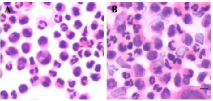
membrane was stained with hematoxylin and eosin and monocytes were counted by drawing a grid in the dyed membrane. After the mobile monocytes were detected under low power, the total cell number was counted in 5 random visual fields under high power and reported as a value divided by 10 high-power field values, and each sample was repeated 3 times.
Assay of MCP-1, RANTES, and fractalkine mRNA with real-time PCR
An iScript RNA extraction kit was employed (Fermentas Co., USA) to extract total RNA in the monocytes. Absorb-ance at 260 nm (A260) and A280 were examined with a UV spectrophotometer, and A260/A280 was calculated to evaluate the purity of the RNA. RNA concentration was calculated according to the formula: RNA concentration (mg/mL)=A2606dilution ratio640/1000 (mg/mL).
An iScript cDNA synthesis kit was employed to carryout RNA reverse transcription. Two micrograms of RNA were added to diethyl pyrocarbonate (DEPC) water to a volume of 20mL, and 3mL of 56supermix including dNTP, oligo(dT), and 2mL transcriptase was added, so the total volume
was 25mL. In the PCR amplifier, reverse transcription was
performed at 256C for 10 min, 426C for 10 min, and 256C for 10 min.
Three microliters of cDNA were taken from the total 25mL cDNA for RT-PCR. Amplification was performed in
an RT-PCR amplifier, under the following cycling condi-tions: 906C 45 s, 506C 60 s, 656C 20 s repeated 30 times. The primer sequence was the following: MCP-1 upstream primer 59-CCCGAAGGGTTACCTTTCGGC-39, down-stream primer 59-GGATTAGGGCGTTACCGGTCG-39; RANTE upstream primer 59-CCCGAAGGGTTACCTTTCG GC-39, downstream primer 59-CCCCGGCGAAGGAAGTT TCCTT-39; fractalkine upstream primer 59-TTCGGGTTGG GTCCAATACCT-39, downstream primer 59-TTGGTTCGA GTGCTTGGAACC-39;b-actin upstream primer 59-CGTGA CCCGTTACCTTAACGC-39, downstream primer 59-CAG GCCCTCAGGGTAATCGGC-39. mRNA expression was determined by the cycling threshold (Ct) rectified by b -actin, namely 2DCt(DCt=Ct ofb-actin––Ct of target gene).
Statistical analysis
A normal distribution test was applied to all measure-ment data, and the data accorded with normal distribution are reported as means±SE; skewness data are reported as median. The two groups were compared using at-test. Skewness data of the two groups were compared using a rank sum test. Enumeration data of the two groups were compared using ax2test. A value of P,0.05 was considered to be statistically significant.
Results
Results of CAG
In the SAP group, 20 cases showed one branch of
artery occlusion, 31 cases showed two branches of artery occlusion, and 9 cases showed three branches of artery occlusion. In the UAP group, 22 cases had one branch of artery occlusion, 30 cases had two branches of artery occlusion, and 8 cases had three branches of artery occlusion. Comparison between the two groups was not statistically different (P.0.05).
Results of IVUS
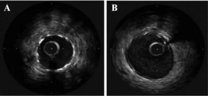
In the SAP group, 92 plaques were detected. In the UAP group, 90 plaques were detected. The lesions of the SAP patients were mainly fibrous plaques (52.17%) while those of the UAP group were mainly lipid plaques (51.04%). Comparison between the two groups was statistically different (Figure 1, Table 1). Comparison of EEMA, PA, EI, and PB between the two groups was statistically different (P,0.05 or P,0.01), but the compar-ison of LA was not statistically different. Comparcompar-ison of RI between the two groups was statistically different. The SAP group appeared as negative remodeling while the UAP group appeared as positive remodeling (Table 2). The ratio of ruptured plaques in the UAP group was about 70.83%, which was statistically different when compared with the SAP group (P,0.01; Table 1).
Figure 1. Intravascular ultrasound imaging of the stable (SAP) and unstable (UAP) angina pectoris groups. A, SAP group. Eccentrically-distributed fibrous plaques appeared with enhanced echo. B, UAP group. Eccentrically-distributed lipid plaques ap-peared with low echo.
Table 1. Plaque type of the lesions in the stable (SAP) and unstable (UAP) angina pectoris groups of patients.
Type SAP (n=60) UAP (n=60)
Number 92 96
Lipid plaque 12 (13.04) 49 (51.04)*
Fibrous plaque 48 (52.17) 12 (12.50)* Calcified plaque 15 (16.30) 17 (17.71)
Mixed plaque 17 (18.48) 18 (18.75)
Ruptured plaque 34 (36.96) 68 (70.83)*
Level of inflammatory factors in serum
Serum levels of hs-CRP, MCP-l, RANTES, and fractalk-ine in the UAP group were significantly higher than those in the SAP group (P,0.01; Table 3).
Monocyte chemotactic activity
Monocyte chemotactic activity in the UAP group was remarkably enhanced and the mobile number was greater than that in the SAP group. The total number of monocytes in all 5 high-power fields was 137±26 and 58±11, respec-tively, which was statistically different (P,0.01; Figure 2).
mRNA expression of MCP-1, RANTES, and fractalkine in monocytes
mRNA expression of MCP-1, RANTES, and fractalkine in the UAP group was significantly greater than in the SAP group (P,0.05; Table 3).
Discussion
In the present study, IVUS demonstrated that the plaques in UAP patients have the characteristics of
vulnerable plaques, i.e., eccentrically distributed lipid plaque. PB increased significantly and the pathological arteries showed positive remodeling. Plaque rupture rate increased significantly. Comparing the chemotactic activ-ity of monocytes in UAP patients with that in SAP patients, we found that chemotactic activity and the quantity of monocytes in the former group increased significantly compared with the SAP patients. This result indicated that chemotactic activity of monocytes was closely related to the stability of the plaques. The activity of inflammatory factors in the serum indicated that hs-CRP, MCP-1, RANTES, and fractalkine in UAP patients increased significantly, which was statistically different compared with the SAP patients. This result suggested that there was active inflammation in the plaques of UAP patients. MCP-1, RANTES, and fractalkine are not only the factors that enhance chemotactic activity in monocytes, but are also markers of plaque instability. Using molecular biological detection of MCP-1, RANTES, and fractalkine, we found that MCP-1, RANTES, and fractalkine mRNA expression in the UAP group was remarkably higher than in the SAP group, which further indicated that chemokines played a key role in plaque instability.
Chemokines played a pivotal role in the pathogenesis of AS. The subendothelial invasion of leukocytes in the blood is regulated by chemokines, and increasing atten-tion has been paid to the role of chemokines in the inflammation of plaques (9,10). Chemokines are grouped into 4 types: CXC chemokines (a-chemokines), CC chemokines (b-chemokines), C chemokines (c -chemo-kines), and CX3C chemokines (d-chemokines) (11,12). The rupture of vulnerable plaques in AS and thrombosis is the main mechanism of ACS, and the vulnerability of the plaques is the initiating factor of those processes. Further investigation of the mechanism of plaque vulnerability, to transform the vulnerable plaque into a stable state, would be an important way to prevent ACS. A recent study suggested that MCP-1 (CCL2/CCR2), RANTES (CCL5/ CCR5), and fractalkine (CX3CLl/CX3CRl) secreted by vascular endothelial cells, smooth muscle cells, and platelets, etc., are most closely associated with the
Table 2.Intravascular ultrasound examination results of the stable (SAP) and unstable (UAP) angina pectoris groups of patients.
SAP (n=60) UAP (n=60)
Number 92 96
EEMA (mm2) 9.67 ± 1.62 13.14 ± 1.84*
LA (mm2) 4.61 ± 0.86 4.57 ± 0.79
PA (mm2) 6.76 ± 1.29 9.07 ± 1.67#
PB (%) 58.13 ± 5.19 69.38 ± 7.26#
EI 0.48 ± 0.12 0.61 ± 0.13*
RI 0.95 ± 0.16 1.27 ± 0.21#
EEMA: external elastic membrane area; LA: lumen area; PA: plaque area; PB: plaque burden; EI: eccentricity index; RI: remodeling index. * P,0.05,#P,0.01vsSAP group (t-test).
Table 3.Serum high-sensitivity C-reactive protein (hs-CRP) and chemokine levels and chemokine mRNA expression in the stable (SAP) and unstable (UAP) angina pectoris groups of patients.
SAP (n=60) UAP (n=60) Serum levels
hs-CRP (mg/L) 1.98 ± 0.86 5.01 ± 1.13* MCP-l (ng/L) 41.06 ± 6.64 92.15 ± 9.28* RANTES (ng/L) 31.24 ± 5.32 49.67 ± 6.26#
Fractalkine (ng/L) 29.84 ± 5.66 57.39 ± 6.91# mRNA expression
MCP-l 32.15 ± 6.28 46.92 ± 7.71*
RANTES 31.64 ± 6.13 43.92 ± 7.68*
Fractalkine 21.56 ± 4.94 34.81 ± 6.21*
* P,0.05,#P
,0.01vsSAP group (t-test).
initiation and progression of AS plaque (13).
The stability of AS plaques has been investigated by researchers around the world using IVUS. Sillesen et al. (14) reported that plaques with thin fibrous caps and large lipid cores were easy to rupture when using IVUS. Purushothaman et al. (15) compared ruptured and non-ruptured plaques in the same artery in ACS patients with IVUS and found that the ruptured plaques showed obvious irregularity and the artery appeared to have positive remodeling. Ueda (16) found that the plaques in unstable angina were mainly irregularly distributed lipid plaques with positive vascular remodeling. By comparing IVUS, serol-ogy, and molecular biology of SAP patients with those of UAP patients, we evaluated the general mechanisms underlying vulnerable plaques: 1) the levels of hs-CRP, MCP-1, RANTES, and fractalkine in the serum are sensitive markers for observing the conditions in UAP patients; 2) unstable plaques show irregularly distributed lipid plaques with low echo and have a larger PA and obvious positive remodeling; and3) mRNA expression of MCP-l, RANTES, and fractalkine in UAP patients increases significantly, which promotes inflammation and further leads to the formation of vulnerable plaques (17,18).
MCP-1 is the first CC chemokine identified by cloning. A study suggests that the binding of MCP-1 to its receptor CCR2 plays an important role in the initiation and progres-sion of AS (19). Lee et al. (20) fed mice deficient in LDL receptors and MCP-1 with a high-cholesterol diet and reported that the level of total cholesterol and every kind of cholesterol were the same in LDL receptor/MCP-1 gene-deficient mice and LDL receptor gene-gene-deficient/wild MCP-1 mice. However, the lipid disposition in the former group was 83% of that in the latter group, and very little macrophage infiltration was observed in LDL receptor/MCP-1 gene-deficient mice. Apart from regulating monocyte adhesion
and migration into the arterial wall, MCP-1 regulates chemotaxis and promotes proliferation of smooth muscle cells. It also triggers tissue factor generation and is involved in the development of AS directly or indirectly.
Fractalkine belongs to the group of CX3C chemokines and binds to its receptor CX3CRl to play a significant role in inflammation. A recent study suggested that fractalkine/ CX3CRl was closely related to the initiation and progression of AS. Feig et al. (21) found that gene expression of fractalkine existed in human atherosclerotic plaques and was highly associated with the macrophages within the plaques. Correa et al. (22) reported that fractalkine led to vascular smooth muscle cell dysfunction by promoting chemotaxis and migration of monocytes, inducing superoxide anion generation and reducing NO secretion, which predisposed to AS. Nozue et al. (23) found that fractalkine gene expression in patients with coronary artery disease increased dramati-cally, which could be attenuated by employing statin drugs. RANTES belongs to the CC chemokine group and binds to CCR5, acts on monocytes, T-cells, and eosino-phils, and drives them into the endothelium to promote AS. Zernecke et al. (24) found that RANTES mRNA expression existed in human atherosclerotic plaques. Sun et al. (25) showed that RANTES mRNA expression in monocytes increased dramatically in patients with hyperhomocystei-nemia and that RANTES was the important chemokine in promoting AS.
In summary, the present study suggested that chemo-kines such as MCP-1, RANTES, and fractalkine promoted the instability of coronary atherosclerotic plaques. These findings provided further insight into the role of chemokines in regulating the stability of atherosclerotic plaques and suggested that inhibiting the activity of chemokines such as MCP-1, RANTES, and fractalkine might be of therapeutic value for the treatment of atherosclerosis.
References
1. Roy SK, Cespedes A, Li D, Choi TY, Budoff MJ. Chronic kidney disease is associated with increased coronary artery atherosclerosis as revealed by multidetector computed tomo-graphic angiography.Tex Heart Inst J2012; 39: 811-816. 2. Thej MJ, Kalyani R, Kiran J. Atherosclerosis in coronary
artery and aorta in a semi-urban population by applying modified American Heart Association classification of atherosclerosis: An autopsy study.J Cardiovasc Dis Res
2012; 3: 265-271, doi: 10.4103/0975-3583.102692. 3. Khan R, Jang IK. Evaluation of coronary allograft
vasculo-pathy using multi-detector row computed tomography: a systematic review.Eur J Cardiothorac Surg2012; 41: 415-422, doi: 10.1016/j.ejcts.2011.06.033.
4. Babic S, Nezic D, Radak D. Is the routine screening for significant atherosclerotic renal artery stenosis during cor-onary angiography/intervention indispensable? J Zhejiang Univ Sci B2013; 14: 83, doi: 10.1631/jzus.B1200240. 5. Andrassy M, Volz HC, Schuessler A, Gitsioudis G, Hofmann
N, Laohachewin D, et al. HMGB1 is associated with
atherosclerotic plaque composition and burden in patients with stable coronary artery disease. PLoS One 2012; 7: e52081, doi: 10.1371/journal.pone.0052081.
6. La Grutta L, Galia M, Gentile G, Lo Re G, Grassedonio E, Coppolino F, et al. Comparison of iodinated contrast media for the assessment of atherosclerotic plaque attenuation values by CT coronary angiography: observations in anex vivomodel.Br J Radiol2013; 86: 20120238, doi: 10.1259/ bjr.20120238.
7. Karshovska E, Weber C. Atherosclerosis: cell biology and lipoproteins - new mechanistic links in atherosclerosis: chemokines mediating the effects of lipids, platelets and dendritic cells. Curr Opin Lipidol 2012; 23: 400-401, doi: 10.1097/MOL.0b013e3283555a9c.
doi: 10.1016/j.jcmg.2011.05.005.
9. Nie P, Li D, Hu L, Jin S, Yu Y, Cai Z, et al. Atorvastatin improves plaque stability in ApoE-knockout mice by regulating chemokines and chemokine receptors. PLoS One2014; 9: e97009, doi: 10.1371/journal.pone.0097009. 10. Recio C, Oguiza A, Lazaro I, Mallavia B, Egido J,
Gomez-Guerrero C. Suppressor of cytokine signaling 1-derived peptide inhibits Janus kinase/signal transducers and activa-tors of transcription pathway and improves inflammation and atherosclerosis in diabetic mice. Arterioscler Thromb Vasc Biol2014; 34: 1953-1960, doi: 10.1161/ATVBAHA.114. 304144.
11. Koenen RR, Weber C. Chemokines: established and novel targets in atherosclerosis.EMBO Mol Med 2011; 3: 713-725, doi: 10.1002/emmm.201100183.
12. Tellez A, Schuster DS, Alviar C, Lopez-Berenstein G, Sanguino A, Ballantyne C, et al. Intramural coronary lipid injection induces atheromatous lesions expressing proin-flammatory chemokines: implications for the development of a porcine model of atherosclerosis.Cardiovasc Revasc Med
2011; 12: 304-311, doi: 10.1016/j.carrev.2011.03.007. 13. Rayner KJ, Moore KJ. The plaque ‘‘micro’’ environment:
microRNAs control the risk and the development of athero-sclerosis. Curr Atheroscler Rep 2012; 14: 413-421, doi: 10.1007/s11883-012-0272-x.
14. Sillesen H, Muntendam P, Adourian A, Entrekin R, Garcia M, Falk E, et al. Carotid plaque burden as a measure of subclinical atherosclerosis: comparison with other tests for subclinical arterial disease in the High Risk Plaque BioImage study. JACC Cardiovasc Imaging 2012; 5: 681-689, doi: 10.1016/j.jcmg.2012.03.013.
15. Purushothaman KR, Purushothaman M, Levy AP, Lento PA, Evrard S, Kovacic JC, et al. Increased expression of oxidation-specific epitopes and apoptosis are associated with haptoglobin genotype: possible implications for plaque progression in human atherosclerosis. J Am Coll Cardiol
2012; 60: 112-119, doi: 10.1016/j.jacc.2012.04.011. 16. Ueda Y. [Evaluation of atherosclerosis by IVUS and
angioscopy].Nihon Rinsho2011; 69 (Suppl 1): 496-502. 17. Poupel L, Combadiere C. [Atherosclerosis: on the trail of
chemokines]. Biol Aujourdhui 2010; 204: 285-293, doi: 10.1051/jbio/2010026.
18. Koenen RR, Weber C. Platelet-derived chemokines in vascular remodeling and atherosclerosis. Semin Thromb Hemost2010; 36: 163-169, doi: 10.1055/s-0030-1251500. 19. Koenen RR, von Hundelshausen P, Nesmelova IV,
Zernecke A, Liehn EA, Sarabi A, et al. Disrupting functional interactions between platelet chemokines inhibits athero-sclerosis in hyperlipidemic mice.Nat Med2009; 15: 97-103, doi: 10.1038/nm.1898.
20. Lee CH, Tai BC, Lim GH, Chan MY, Low AF, Tan KC, et al. Correlation between high density lipoprotein-cholesterol and remodeling index in patients with coronary artery disease: IDEAS (IVUS diagnostic evaluation of atherosclerosis in Singapore)-HDL study.Int J Cardiovasc Imaging 2012; 28: 33-41.
21. Feig JE, Vengrenyuk Y, Reiser V, Wu C, Statnikov A, Aliferis CF, et al. Regression of atherosclerosis is characterized by broad changes in the plaque macrophage transcriptome.PLoS One2012; 7: e39790, doi: 10.1371/journal.pone.0039790. 22. Correa CR, Dias-Melicio LA, Calvi SA, Lastoria S, Soares
AM. Activation of monocytes and cytokine production in patients with peripheral atherosclerosis obliterans. J Inflamm2011; 8: 23, doi: 10.1186/1476-9255-8-23. 23. Nozue T, Yamamoto S, Tohyama S, Fukui K, Umezawa S,
Onishi Y, et al. Impact of diabetes mellitus on coronary atherosclerosis and plaque composition under statin therapy - subanalysis of the TRUTH study.Circ J2012; 76: 2188-2196, doi: 10.1253/circj.CJ-11-1303.
24. Zernecke A, Weber C. Chemokines in the vascular inflammatory response of atherosclerosis.Cardiovasc Res
2010; 86: 192-201, doi: 10.1093/cvr/cvp391.