Excitato ry amino acid re ce pto r blo ckade
within the caudal pre sso r are a and
ro stral ve ntro late ral me dulla alte rs
cardio vascular re spo nse s to nucle us
raphe o bscurus stimulatio n in rats
1Departamento de Ciências Fisiológicas, Centro Biomédico,
Universidade Federal do Espírito Santo, Vitória, ES, Brasil
2Associação Educacional de Vitória (FAESA), Vitória, ES, Brasil
N.F. Silva2, J.G.P. Pires1,
M.A. Dantas1 and
H.A. Futuro Neto1
Abstract
Pressor responses elicited by stimulation of the nucleus raphe obscu-rus (NRO) depend on the integrity of the rostral ventrolateral medulla (RVLM). Therefore, to test the participation of excitatory amino acid (EAA) receptors in the cardiovascular responses evoked by NRO stimulation (1 ms, 100 Hz, 40-70 µA, for 10 s), the EAA antagonist kynurenic acid (Kyn) was microinjected at different sites in the ventrolateral medullar surface (2.7 nmol/200 nl) of male Wistar rats (270-320 g, N = 39) and NRO stimulation was repeated. The effects of NRO stimulation were: hypertension (DMAP = +43 ± 1 mmHg, P<0.01), bradycardia (DHR = -30 ± 7 bpm, P<0.01) and apnea. Bilateral microinjection of Kyn into the RVLM, which did not change baseline parameters, almost abolished the bradycardia induced by NRO stimulation (DHR = -61 ± 3 before vs -2 ± 3 bpm after Kyn, P<0.01, N = 7). Unilateral microinjection of Kyn into the CVLM did not change baseline parameters or reduce the pressor response to NRO stimulation (DMAP = +46 ± 5 before vs +48 ± 5 mmHg after Kyn, N = 6). Kyn bilaterally microinjected into the caudal pressor area reduced blood pressure and heart rate and almost abolished the pressor response to NRO stimulation (DMAP = +46 ± 4 mmHg before vs +4 ± 2 mmHg after Kyn, P<0.01, N = 7). These results indicate that EAA receptors on the medullary ventrolateral surface play a role in the modulation of the cardiovascular responses induced by NRO stimula-tion, and also suggest that the RVLM participates in the modulation of heart rate responses and that the caudal pressor area modulates the pressor response following NRO stimulation.
Co rre spo nde nce
H.A. Futuro Neto Av. Marechal Campos, 1468 29040-090 Vitória, ES Brasil
Fax: + 55-27-3335-7330 E-mail: futuront@ npd.ufes.br
Research supported by CNPq and FINEP.
Received August 7, 2001 Accepted June 24, 2002
Ke y words
·Raphe nuclei ·Electrical stimulation ·Ventrolateral medulla ·Caudal pressor area ·Excitatory amino acid
receptors
·Kynurenic acid ·Blood pressure
Intro ductio n
The participation of the nucleus raphe obscurus (NRO) in cardiovascular regula-tion using electrical stimularegula-tion has been demonstrated in various species. For instance,
predomi-nant. In anesthetized rats, electrical or chem-ical stimulation of the NRO and other raphe nuclei frequently causes increases in arterial BP (3,6) with wide heart rate (HR) variabil-ity. Dreteler et al. (7) demonstrated a rise in BP as a result of 5-HT1A receptor agonist
microinjection into the NRO, and proposed that the related bradycardia was a barore-flex-mediated response. In contrast, Coleman and Dampney (8) observed sympathoinhibi-tion and hypotension evoked by microinjec-tion of L-glutamate into sites within the NRO in anesthetized rabbits.
Anatomical studies of rats have indicated that caudal raphe neurons project either di-rectly to the intermediolateral (IML) cell column (9-11) or by sending axons to sites in the ventrolateral medulla (VLM) (12-14). Therefore, there is no conclusive evidence concerning NRO projections or their precise role in cardiovascular regulation. Campos Jr. et al. (15) showed that the pressor effects caused by stimulating the NRO require an intact rostral ventrolateral medulla (RVLM), an important area in the VLM involved in autonomic control (16), which directly projects to the IML (9). Preliminary results from our laboratory (17) failed to demon-strate direct functional connections between NRO and RVLM. We therefore hypothesized that the NRO might not have monosynaptic connections to the RVLM, and that the re-sponses to NRO stimulation may be medi-ated by indirect projections to the RVLM.
At least two other areas in the VLM that are involved in autonomic control can influ-ence RVLM activity: the caudal ventrolat-eral medulla (CVLM) (18,19), a sympatho-inhibitory area with projections to the RVLM (20), and the caudal pressor area (CPA) (21), originally described as a hypertensive area in deeply anesthetized or decerebrate animals (22-24). The functional connections between the CPA and the RVLM (23,25) possibly oc-cur via CVLM glutamatergic neurons (26).
The various cardiovascular and respira-tory responses evoked by NRO stimulation
have been credited to activation of seroto-nergic neurons (7). However, other neuro-transmitters are present in NRO neurons, including excitatory amino acids (EAA; mainly L-glutamate). Since the blockade of 5-HT1-3 receptors in the VLM did not block
the cardiovascular responses evoked by NRO stimulation (17), the present study was de-signed to test the possible role of EAA-mediated NRO projections to the RVLM, CVLM and CPA in these responses.
Mate rial and Me thods
Male Wistar rats weighing 270-320 g were anesthetized with urethane (1.2 g/kg, after induction with ether) injected into the left femoral vein cannulated with a polyeth-ylene catheter (PE50). Supplementary doses of anesthetic were administered whenever necessary, as indicated by the paw pinch reflex and/or by unstable cardiovascular re-cordings. Arterial BP was recorded through a heparinized (40 U/ml) saline-filled cath-eter (PE50) inserted into the left femoral artery and connected to a pressure trans-ducer (Statham P23 Db, Valley View, OH, USA). Pulsatile BP, mean arterial BP (MAP) and HR were recorded with a Gould RS 3400 polygraph. A tracheotomy was per-formed in all animals and respiration was recorded with a pneumotachograph (Fleish 0000) and a low-pressure transducer (Vala-dine) connected to the polygraph. Whenever necessary the rats were artificially ventilated (Harvard Rodent Ventilator, South Natick, MA, USA). All cardiovascular and respira-tory activity data were digitalized (Biopac MP 100) and stored on a PC hard disk for later processing. Rectal temperature was maintained between 36-37ºC with a homeo-thermic blanket (Harvard).
bregma, 8 mm depth) was performed with stainless steel electrodes insulated through-out except at the tip (tip size 30-40 µm, resistance 15-30 kW), the anode being a clip attached to a neck muscle. The dorsal sur-face of the brainstem was exposed by retrac-tion of the skin and muscles at the base of the skull, followed by the removal of the atlanto-occipital membrane and portions of the oc-cipital bone. Kynurenic acid (Kyn; 2.7 nmol/ 200 nl) and artificial cerebrospinal fluid (CSF) containing 122 mM NaCl, 25.7 mM NaHCO3, 1 mM MgSO4, and 1 mM CaCl2,
with pH adjusted to 7.2-7.4, were microin-jected bilaterally into the RVLM (+3 mm AP in relation to the obex, 1.5 mm L), CVLM (+1 mm AP in relation to the obex, 1.5 mm L) or CPA (-1 mm AP in relation to the obex, 1.5 mm L). In a separate group of four ani-mals, Kyn was microinjected unilaterally into the CVLM. Vertical positioning was obtained by slowly lowering both micropi-pettes placed inside the guide cannulae (which were 6-8 mm shorter than the mi-cropipettes) until a slight displacement be-tween them was observed (the displacement occurs when the tip of the micropipette touches the pia mater adjacent to the ventral medullar surface; the depth was 0.79 mm for the RVLM in relation to the dorsal surface of the cerebellum, 0.59 mm for the CVLM in relation to the dorsal surface of the cerebel-lum and 0.33 mm for the CPA in relation to the dorsal surface of the medulla. Therefore the microinjections were applied to the ven-tral layers of the nuclei).
At least three electrical stimulations of the NRO were performed at 3-min intervals and the means of the measured parameters were considered as controls. Kyn microin-jection was performed ~1.5 min after the last control NRO stimulation and after a period of ~1.5 min a new series of three NRO stimulations was performed. A separate group of CSF-treated animals was used as time-matched controls.
At the end of each experiment the
posi-tion of the stimulus or of the microinjecposi-tion was determined by passing a DC current of 5-20 µA for 1 min (stainless steel electrode anode) or by microinjecting 200 nl 2% Evan’s blue dye. The animals were then heparinized and transcardially perfused with 200 ml sa-line solution (0.9% NaCl) and a solution of 1% potassium ferrocyanide in 10% formal-dehyde. The brain was removed and placed in formaldehyde for 7 days. Sites of microin-jections or electrical NRO stimulation were identified and plotted on a schematic dia-gram.
Data are reported as means ± SEM. Paired or unpaired t-tests were used whenever
ap-propriate for statistical comparisons of the means. Two-way ANOVA was used for com-parisons between groups. In all cases, the level of significance was set at P£0.05.
Re sults
Effe ct of e le ctrical stimulation of the NRO on arte rial blood pre ssure , he art rate and
re spiration of ane sthe tize d rats
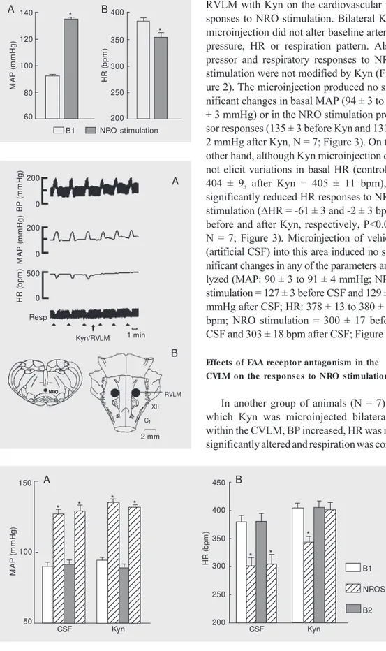
In the 39 anesthetized rats studied, basal MAP and HR were 92 ± 1 mmHg and 384 ± 7 bpm, respectively. Electrical stimulation of a site within the NRO produced an in-crease of BP (MAP = 135 ± 2, DMAP = +43 ± 1 mmHg, P<0.01, N = 39; Figure 1). HR changes during NRO stimulation were vari-able, but bradycardia was observed in most animals (HR = 353 ± 9, DHR = -30 ± 7 bpm, P<0.01, N = 39; Figure 1). The stimulus frequently elicited apnea. These animals were divided into subgroups to test the effects of bilateral microinjections of CSF and Kyn within the RVLM, CVLM or CPA on the responses to NRO stimulation.
Effe cts of EAA re ce ptor antagonism in the
RVLM on the re sponse s to NRO stimulation
RVLM with Kyn on the cardiovascular re-sponses to NRO stimulation. Bilateral Kyn microinjection did not alter baseline arterial pressure, HR or respiration pattern. Also, pressor and respiratory responses to NRO stimulation were not modified by Kyn (Fig-ure 2). The microinjection produced no sig-nificant changes in basal MAP (94 ± 3 to 89 ± 3 mmHg) or in the NRO stimulation pres-sor responses (135 ± 3 before Kyn and 131 ± 2 mmHg after Kyn, N = 7; Figure 3). On the other hand, although Kyn microinjection did not elicit variations in basal HR (control = 404 ± 9, after Kyn = 405 ± 11 bpm), it significantly reduced HR responses to NRO stimulation (DHR = -61 ± 3 and -2 ± 3 bpm, before and after Kyn, respectively, P<0.01, N = 7; Figure 3). Microinjection of vehicle (artificial CSF) into this area induced no sig-nificant changes in any of the parameters ana-lyzed (MAP: 90 ± 3 to 91 ± 4 mmHg; NRO stimulation = 127 ± 3 before CSF and 129 ± 5 mmHg after CSF; HR: 378 ± 13 to 380 ± 15 bpm; NRO stimulation = 300 ± 17 before CSF and 303 ± 18 bpm after CSF; Figure 3).
Effe cts of EAA re ce ptor antagonism in the
CVLM on the re sponse s to NRO stimulation
In another group of animals (N = 7) in which Kyn was microinjected bilaterally within the CVLM, BP increased, HR was not significantly altered and respiration was
com-Figure 1. M ean arterial blood pressure (M AP; panel A) and heart rate (HR; panel B) before and after nucleus raphe obscu-rus (NRO) stimulation. B1, base-line before stimulation. Data are reported as means ± SEM for all 39 rats. * P<0.01 compared to B1 (Student t-test).
Figure 3. Effect on mean arterial blood pressure (M AP; panel A) and heart rate (HR; panel B) of bilateral microinjections of cere-brospinal fluid (CSF, 200 nl) or kynurenic acid (Kyn, 2.7 nmol/ 200 nl) w ithin the rostral ventro-lateral m edulla and effect of electrical nucleus raphe obscu-rus st im ulat ion (NROS). B1, baseline values; B2, post-drug baseline values. Data are re-ported as means ± SEM for 7 rats. * P<0.01 compared to the preceding baseline value (Stu-dent t-test).
M A P ( m m H g ) 140 H R ( b p m ) 400 A B
B1 NRO stimulation 120 100 80 60 350 300 250 200 * *
Figure 2. A, Tracings from one rat show ing the cardiovascular and respiratory responses to nucleus raphe obscurus (NRO) stimulation (10 s at 100 Hz, 60 µA, and 1-ms pulse duration) be-fore and after bilateral microin-jection of kynurenic acid (Kyn, 2.7 nmol/200 nl) w ithin the ros-tral ventrolateral medulla (RVLM ). BP, pulsatile blood pressure; M AP, m ean art erial BP; HR, heart rate; Resp, respiration. The triangles denote electrical stimu-lations, and the arrow indicates microinjection. B, The left repre-sentative section show s the lo-cation of the NRO stimulation site. The illustration on the right show s the injection site w ithin the RVLM .
RVLM 2 mm 1 min Kyn/RVLM Resp H R ( b p m ) 500 0 XII C1 NRO NRO NRO NRO NRO A B M A P ( m m H g ) 150 H R ( b p m ) 450 123 123 123 123 123 123 123 123 123 123 123 123 123 123 123 123 123 123 123 123 123 123 123 123 123 123 123 123 123 123 123 123 123 123 123 123 123 123 123 123 123 123 123 1234 1234 1234 1234 1234 1234 1234 1234 1234 1234 1234 1234 1234 1234 1234 1234 1234 1234 1234 1234 1234 1234 1234 123 123 123 123 123 123 123 123 123 123 123 123 123 123 123 123 123 123 123 123 123 123 123 123 123 123 123 123 123 123 123 123 123 123 123 123 123 123 123 123 123 123 123 123 123 123 123 123 123 123 123 123 123 123 123 123 123 123 123 123 123 123 123 123 123 123 123 123 123 123 123 123 123 123 123 123 123 123 123 123 123 123 123 123 123 123 * * * * * * * B1 NROS B2 100 50 400 350 300 250 200
CSF Kyn CSF Kyn
pletely blocked. HR responses were found to be variable; for instance, in Figure 4 one such tracing is shown, where an attenuation of HR was observed.
Statistical analysis of the pooled data (Figure 5)showed that Kyn injected bilater-ally into the CVLM provoked an increase in MAP (89 ± 3 to 124 ± 4 mmHg, P<0.01, N = 7), and that the pressor response to NRO stimulation was slightly attenuated (DMAP = +48 ± 4 to +43 ± 3 mmHg after Kyn, P<0.05, N = 7).
Nonsignificant changes in cardiovascu-lar and respiratory parameters to NRO stim-ulation were induced by CSF microinjec-tions into the CVLM (baseline: MAP = 91 ± 4 to 92 ± 5 mmHg; NRO stimulation: DMAP = +41 ± 4 to +41 ± 5 mmHg; baseline: HR = 367 ± 31 to 369 ± 29 bpm; NRO stimulation:
DHR = -19 ± 26 to -25 ± 23 bpm, before and after CSF, respectively, N = 6).
Bilateral Kyn microinjection into the CVLM induced an increase in basal BP and a statistically significant reduction of the pressor response to NRO stimulation. There-fore, a separate group of rats with unilateral Kyn microinjection was used to determine the influence of changes in basal BP on the reduction of NRO responses. After unilat-eral Kyn injection, which induced a smaller increase in basal BP and did not block respi-ration, no significant effects were observed on the pressor responses to NRO stimulation
1 min Resp B P ( m m H g ) 200 0 H R ( b p m ) 200 2 mm Kyn/CVLM NRO NRO NRO NRO NRO 0 500 0 M A P ( m m H g ) CVLM XII C1 A B
Figure 4. A, Tracings from one rat show ing the cardiovascular and respiratory responses to nucleus raphe obscurus (NRO) stimulation (10 s at 100 Hz, 60 µA, and 1-ms pulse duration) before and after bilateral micro-injection of kynurenic acid (Kyn, 2.7 nmol/200 nl) w ithin the cau-dal vent rolat eral m edulla (CVLM ). BP, pulsat ile blood pressure; M AP, mean arterial BP; HR, heart rate; Resp, respi-rat ion. The t riangles denot e stimulations, and the arrow indi-cates microinjection. B, The left representative section show s the location of the NRO stimula-tion site. The illustrastimula-tion on the right show s the injection site w ithin the CVLM .
(DMAP = +46 ± 5 before Kyn and +48 ± 5 after Kyn, N = 6; data not shown). The use of this second group proved that the reduction in the pressor response was due to alteration in basal BP and not to a real blockade of the NRO response.
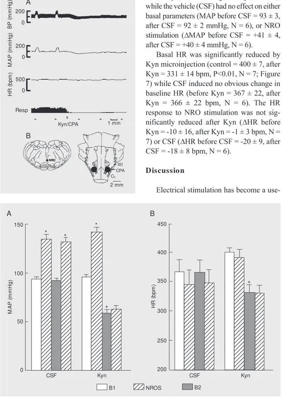
Effe cts of EAA re ce ptor antagonism in the CPA on the re sponse s to NRO stimulation
The cardiovascular and respiratory changes
Figure 5. Effect on mean arterial blood pressure (M AP; panel A) and heart rate (HR; panel B) of bilateral microinjections of cere-brospinal fluid (CSF, 200 nl) or kynurenic acid (Kyn, 2.7 nmol/ 200 nl) w ithin the caudal ventro-lateral medulla and effects of electrical nucleus raphe obscu-rus st im ulat ion (NROS). B1, baseline values; B2, post-drug baseline values. Data are re-ported as means ± SEM for 7 rats. +P<0.05 for B2 compared
to B1 and * P<0.01 compared to preceding baseline value, B1 (ANOVA). M A P ( m m H g ) 200 H R ( b p m ) 450 123 123 123 123 123 123 123 123 123 123 123 123 123 123 123 123 123 123 123 123 123 123 123 123 123 123 123 123 123 123 123 123 123 123 123 123 123 123 123 123 123 123 123 123 123 123 123 123 123 123 123 123 123 123 123 123 123 123 123 123 123 123 123 123 123 123 123 12 12 12 12 12 12 12 12 12 12 12 12 12 12 12 12 123 123 123 123 123 123 123 123 123 123 123 123 123 12 12 12 12 12 12 12 12 12 12 12 12 12 12 12 12 12 12 12 12 123 123 123 123 123 123 123 123 123 123 123 123 123 123 123 123 123 123 123 1234 1234 * * + * B1 NROS B2 150 50 400 350 300 250 200
CSF Kyn CSF Kyn
A B
croinjections of Kyn within the CPA signifi-cantly reduced both basal MAP (before Kyn = 96 ± 2, after Kyn = 59 ± 4 mmHg, P<0.01, N
= 7; Figure 7), and the pressor response to NRO stimulation (DMAP = +46 ± 4 mmHg before Kyn, +4 ± 2 mmHg after Kyn, P<0.01), while the vehicle (CSF) had no effect on either basal parameters (MAP before CSF = 93 ± 3, after CSF = 92 ± 2 mmHg, N = 6), or NRO stimulation (DMAP before CSF = +41 ± 4, after CSF = +40 ± 4 mmHg, N = 6).
Basal HR was significantly reduced by Kyn microinjection (control = 400 ± 7, after Kyn = 331 ± 14 bpm, P<0.01, N = 7; Figure 7) while CSF induced no obvious change in baseline HR (before Kyn = 367 ± 22, after Kyn = 366 ± 22 bpm, N = 6). The HR response to NRO stimulation was not sig-nificantly reduced after Kyn (DHR before Kyn = -10 ± 16, after Kyn = -1 ± 3 bpm, N = 7) or CSF (DHR before CSF = -20 ± 9, after CSF = -18 ± 8 bpm, N = 6).
D iscussio n
Electrical stimulation has become a
use-Figure 6. A, Tracings from one rat show ing the cardiovascular and respiratory responses to nucleus raphe obscurus (NRO) stimulation (10 s at 100 Hz, 60 µA, and 1-ms pulse duration) be-fore and after bilateral microin-jection of kynurenic acid (Kyn, 2.7 nmol/200 nl) w ithin the cau-dal pressor area (CPA). BP, pul-sat ile blood pressure; M AP, mean arterial BP; HR, heart rate; Resp, respiration. The triangles denote stimulations, and the ar-row indicates microinjection. B, The left representative section show s the location of the NRO stimulation site. The illustration on the right show s the injection site w ithin the CPA.
1 min Resp B P ( m m H g ) 200 0 H R ( b p m ) 200 2 mm Kyn/CPA NRO NRONRO NRONRO 0 500 0 M A P ( m m H g ) XII C1 A B CPA M A P ( m m H g ) 150 100 0 50 1234 1234 1234 1234 1234 1234 1234 1234 1234 1234 1234 1234 1234 1234 1234 1234 1234 1234 1234 1234 1234 1234 1234 1234 1234 1234 1234 1234 1234 1234 1234 1234 1234 1234 1234 1234 1234 1234 1234 1234 1234 1234 1234 1234 1234 1234 1234 1234 1234 1234 1234 1234 1234 1234 1234 1234 1234 1234 1234 1234 1234 1234 1234 1234 1234 1234 1234 1234 1234 1234 1234 1234 1234 1234 1234 1234 1234 1234 1234 1234 1234 1234 1234 1234 1234 1234 1234 1234 1234 1234 1234 1234 1234 1234 1234 1234 1234 1234 1234 1234 1234 1234 1234 1234 1234 1234 1234 1234 1234 1234 1234 1234 1234 1234 1234 1234 1234 1234 1234 1234 1234 1234 1234 1234 1234 1234 1234 1234 1234 1234 1234 1234 1234 1234 1234 1234 1234 1234 1234 1234 1234 1234 1234 1234 1234 1234 1234 1234 1234 1234 1234 1234 1234 1234 1234 1234 1234 1234 1234 1234 1234 1234 1234 1234 1234 1234 1234 1234 1234 1234 1234 1234 1234 1234 1234 1234 1234 1234 1234 1234 1234 1234 1234 1234 1234 1234 1234 1234 1234 1234 1234 1234 1234 1234 1234 1234 1234 1234 1234 1234 1234 1234 1234 1234 1234 1234 1234 1234 1234 1234 1234 1234 1234 1234 1234 1234 H R ( b p m ) 450 400 200 350 300 250 + + CSF Kyn 1234 1234
B1 NROS B2
CSF Kyn
Figure 7. Effect on mean arterial blood pressure (M AP; panel A) and heart rate (HR; panel B) of bilateral microinjections of cere-brospinal fluid (CSF, 200 nl) or kynurenic acid (Kyn, 2.7 nmol/ 200 nl) w ithin the caudal pressor area, and effects of electrical nucleus raphe obscurus stimula-tion (NROS). B1, baseline ues; B2, post-drug baseline val-ues. Data are reported as means ± SEM for 6 rats. +P<0.01
com-paring B2 w ith B1 and * P<0.01 compared to preceding baseline values (ANOVA).
after NRO stimulation mediated by bilateral Kyn microinjections within the CPA are shown in Figure 6. BP fell, HR was reduced and Kyn also induced respiratory arrest. Bilateral
mi-A B
* *
ful tool to study the participation of the NRO in autonomic control in various species (2,3) by permitting a larger sampling rate in each experiment. The weakness of this technique is that it excites not only cell bodies but also fibers passing through the region. In con-trast, neurochemical stimulation preferen-tially stimulates cell bodies. To avoid this problem the stimulation parameters were chosen so as to minimize current spread and activation of fibers of passage. These same stimulation parameters were previously used in the rat and other species in which they were paired with neurochemical stimulation, with the two different techniques yielding similar results (3-5). The present experimen-tal protocol requires a large number of NRO stimulations separated by a short time inter-val so as to capture the full result of EAA receptor blockade in the different areas of the ventral medulla. These requirements make it difficult to use neurochemical stim-ulation of the NRO since tachyphylaxis will limit the number of successive doses of EAA that can be administered and will also force the experimenter to markedly increase the time interval between doses. In view of these limitations, we decided to use electrical stim-ulation in our experimental protocol.
As stated before, the present study was designed to evaluate the role of EAA recep-tors of the VLM in the cardiovascular re-sponses to electrical NRO stimulation in the rat. Previous results have suggested that func-tional integrity of the RVLM is essential to obtain pressor responses evoked by electri-cal NRO stimulation (15) and that the NRO does not project directly to the IML (12,13). A particularly dense anatomical projection from the NRO to RVLM has been demon-strated (14,27). The cited authors suggest that these projections are mainly serotoner-gic but do not provide evidence for their function.
EAA receptors within the RVLM do not participate in the pressor responses to NRO stimulation since bilateral microinjections
of Kyn (an antagonist of ionotropic EAA receptors) into the RVLM did not block the effects of NRO stimulation activity, although they blocked the HR responses to NRO stim-ulation, demonstrating that these receptors were probably involved in cardiac regula-tion following NRO stimularegula-tion. It was not clear if HR responses were due to direct NRO cell stimulation or to a secondary re-flex response evoked by the barorere-flex or the chemoreflex. Dreteler et al. (7) observed mild bradycardia in response to electrical NRO stimulation, which was often associ-ated with the baroreflex. On the other hand, Amano et al. (28) and Koshiya et al. (29) demonstrated that bilateral microinjection of Kyn into the RVLM blocked the sympa-thetic chemoreflex response but left the sym-pathetic baroreflex response intact. Also, glutamate microinjection into the NRO re-sulted in a decrease in BP and HR in rabbits (8). Therefore it is possible that NRO neu-rons can inhibit cardiac sympathetic activity directly or indirectly within the RVLM with-out affecting the pressor responses. This dif-ferential activation has been previously dem-onstrated when the NRO is stimulated (30). Nevertheless, the changes in HR observed could also be due to activation of the nucleus ambiguus and or the peri-ambiguus region by NRO stimulation.
stimula-tion. These results agree with other studies showing that the CPA is involved in cardio-vascular control in anesthetized rats (22,24, 26). However, the precise nature of this par-ticipation is not clear. One such possibility was proposed by Natarajan and Morrison (26), who demonstrated a pathway from CPA
to RVLM involving sympathoexcitatory neu-rons in the vicinity of the CVLM. The pres-ent study suggests the existence of a pathway from CPA to RVLM mediating the pressor response elicited by NRO activation, but the precise nature of these projections has not been fully identified.
Re fe re nce s
1. Adair JR, Hamilton BL, Scappaticcika KA, Helke CJ & Gillis RA (1977). Cardiovascu-lar responses to electrical stimulation of the medullary raphe area of the cat. Brain Research, 128: 141-145.
2. Yen CT, Blum PS & Spath JA (1983). Con-trol of cardiovascular function by electri-cal stimulation w ithin the medullary raphe region of the cat. Experimental Neurol-ogy, 79: 666-679.
3. Futuro Neto HA, Silva SR, Dantas M A & Gilbey M P (1990). A comparison of the effects of stimulation w ithin the raphe nuclei on arterial BP in the anesthetized rabbit and rat. Acta Physiologica et Phar-macologica Latinoamericana, 40: 175-184. 4. Faria M GC, Dantas M A & Futuro-Neto HA (1996). Pressor effects elicited by stimu-lation w ithin the medullary raphe nuclei of the golden hamster (M esocricetus aura-tus). Brazilian Journal of M edical and Bio-logical Research, 29: 533-540.
5. Almada GL, Pires JGP, Dantas M A & Futuro Neto HA (1997). Pressor effects elicited by stimulation w ithin the medul-lary raphe nuclei of the guinea pig (Cavia porcellus). Acta Physiologica, Pharmaco-logica et Therapeutica Latinoamericana, 47: 229-236.
6. Kuhn DM , Wolf WA & Lovenberg W (1980). Pressor effects of electrical stimu-lation of the dorsal and median raphe nu-clei in anesthetized rats. Journal of Phar-macology and Experimental Therapeutics, 214: 403-409.
7. Dreteler GA, Wouters W, Saxena PR & Ramage AG (1991). Pressor effects fol-low ing microinjection of 5-HT1A receptor
agonists into the raphe obscurus of the anaesthetized rat. British Journal of Phar-macology, 102: 317-322.
8. Coleman M J & Dampney RA (1995). Pow -erful depressor and sympathoinhibitory effects evoked from neurons in the cau-dal raphe pallidus and obscurus. Ameri-can Journal of Physiology, 268: R1295-R1302.
9. Amendt K, Czachurski J, Dembow sky K & Seller H (1979). Bulbospinal projections to the intermediolateral cell column: a neu-roanatomical study. Journal of the Auto-nomic Nervous System, 1: 103-117. 10. Loew y AD (1981). Raphe pallidus and
raphe obscurus projections to the inter-mediolateral cell column in the rat. Brain Research, 222: 129-133.
11. Gilbey M P, Futuro Neto HA & Zhou SY (1995). Respiratory-related discharge pat-terns of caudal raphe neurons projecting to the upper thoracic spinal cord in the rat. Journal of the Autonomic Nervous Sys-tem, 50: 263-273.
12. Bacon SJ, Zagon A & Smith AD (1990). Electron microscopic evidence of a mono-synaptic pathw ay betw een cells in the caudal raphe nuclei and sympathetic pre-ganglionic neurons in the rat spinal cord. Experimental Brain Research, 79: 589-602.
13. Zagon A (1993). Innervation of serotoner-gic medullary raphe neurons from cells of the rostral ventrolateral medulla in rats. Neuroscience, 55: 849-867.
14. Zagon A (1995). Internal connections in the rostral ventromedial medulla of the rat. Journal of the Autonomic Nervous System, 53: 43-56.
15. Campos Jr RR, Futuro Neto HA & Guert-zenstein PG (1993). Role of the rostral ventrolateral medulla in the pressor re-sponse to stimulation of the nucleus raphe obscurus. Brazilian Journal of M edi-cal and Biologiedi-cal Research, 26: 623-631. 16. Guertzenstein PG & Silver A (1974). Fall in BP produced from discrete regions of the ventral surface of the medulla by glycine and lesions. Journal of Physiology, 242: 489-503.
17. Futuro Neto HA, Dantas M A, Gilbey M P & Pires JGP (1996). A study of the nature of the projections from nucleus raphe ob-scurus to the ventrolateral medulla. Soci-ety for NeuroscienceAbstracts, 22: 954. 18. Bisset GW, Feldberg W, Guertzenstein
PG & Rocha e Silva Jr M (1975). Vaso-pressin release by nicotine: the site of action. British Journal of Pharmacology, 54: 463-474.
19. Feldberg W & Guertzenstein PG (1976). Vasodepressor effects obtained by drugs acting on the ventral surface of the brain stem. Journal of Physiology, 258: 337-355.
20. Cravo SL & M orrison SF (1993). The cau-dal ventrolateral medulla is a source of tonic sympathoinhibition. Brain Research, 621: 133-136.
21. Feldberg W & Guertzenstein PG (1986). Blood pressure effects of leptazol applied to the ventral surface of the brainstem of cats. Journal of Physiology, 372: 445-456. 22. Gordon FJ & M cCann LA (1988). Pressor responses evoked by microinjections of L-glutamate into the caudal ventrolateral medulla of the rat. Brain Research, 457: 251-258.
23. Possas OS, Campos Jr RR, Cravo SL, Lopes OU & Guertzenstein PG (1994). A fall in arterial BP by blockade of the caudalmost ventrolateral medulla: the caudal pressor area. Journal of the Auto-nomic Nervous System, 49: 235-245. 24. Campos Jr RR, Possas OS, Cravo SL,
Lopes OU & Guertzenstein PG (1994). Putative pathw ays involved in responses evoked from the caudal pressor area. Bra-zilian Journal of M edical and Biological Research, 27: 2467-2479.
25. Campos Jr RR & M cAllen RM (1999). Tonic drive to sympathetic premotor neu-rons of rostral ventrolateral medulla from caudal pressor area neurons. American Journal of Physiology, 276: R1209-R1213. 26. Natarajan M & M orrison SF (2000). Sym-pathoexcitatory CVLM neurons mediate responses to caudal pressor area stimula-tion. American Journal of Physiology, 279: R364-R374.
catechol-amine neurons in the rat. Neuroscience Letters, 108: 22-28.
28. Amano M , Asari T & Kubo T (1994). Exci-tatory amino acid receptors in the rostro-ventrolateral medulla mediate hyperten-sion induced by carotid body chemore-cept or st im ulat ion. Naunyn-Schm ie-deberg’s Archives of Pharmacology, 349: 549-554.
29. Koshiya N, Huangfu D & Guyenet PG (1993). Ventrolateral medulla and sympa-thetic chemoreflex in the rat. Brain Re-search, 609: 174-184.
30. Futuro Neto HA & Coote JH (1982). Desynchronised sleep-like pattern of sym-pathetic activity elicited by electrical stim-ulation of sites in the brainstem. Brain Research, 252: 269-276.