It’s my pleasure to introduce Professor Eric Liou, from Taipei, Taiwan. He is one of the most important members of the world new generation of orthodontists, who will be in Brazil for the irst time in October 2009, for a speech at the 7th Brazilian Association of Orthodontists Meeting that will be held in Brasília-DF. We irst met in Dallas, in 1998, during the American Association of Orthodontists annual meeting. At that time he had just won the Dewel Award, given to the best clinical article of the year published at the AJODO. Since there I’ve followed his brilliant work, where he has accomplished several clinical and experimental researches whose results have given rise to some controversial and have generated a lot of interest at the international orthodontic community for its originality and vanguard. His ields of interest involve osteogenic distraction, skeletal anchorage and mechanisms to accelerate orthodontic move-ment. However, without doubt, one of the most important topics, frequently present in his presentations is the new protocol for maxillary protraction called Alt-RAMEC, used both for patients with or without cleft lip and palate. This is the subject of this interview conducted by me and by three other important Brazilian orthodontists, who have a lot of experience in rapid maxillary expansion and in the treatment of cleft lip and palate patients: Dr. Omar Gabriel da Silva Filho, Dr. Daniela Gamba Garib and Dr. Gerson Ulema Ribeiro.
Enjoy the reading!!!!
Ricardo Machado Cruz
Eric Liou
• Undergraduate degree in Dentistry from the School of Dentistry, Taipei Medical University, Taipei, Taiwan.
• Graduate degree from the Department of Orthodontics and Craniofacial Dentistry at Chang Gung Memorial Hospital, Taipei, Taiwan.
• Master in Surgery, University of Illinois, Chicago, USA.
• Assistant Professor and Director of the Research and Development Committee of the Faculty of Dentistry at Chang Gung Memorial Hospital, Taipei, Taiwan.
• Orthodontist at the Department of Orthodontics and Craniofacial Dentistry at Chang Gung Memorial Hospital, Taipei, Taiwan.
• President, Taiwan Association of Cleft Lip and Palate. • Winner, Best Clinical Article, AJODO, 1998.
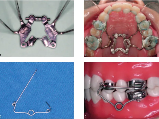
double-hinged expander (US Patent No. 6334771 B1) (Fig. 1A, B) and the intraoral β-Ti maxillary protraction springs (US patent 6273713 B1) (Fig. 1C, D). The complete protocol involves 7 weeks of Alt-RAMEC (Tab. 1). It should always keep the expander opened at the end of the protocol because an expanded maxilla allows a greater amount of maxillary protraction than an unex-panded maxilla, and the exunex-panded space between the central incisors could be saved for relieving anterior crowding or for compensating dental ef-fects, such as the proclined maxillary incisors due to the protraction. In comparison between the growing patients with and without cleft lip and palate, the eficacy of this protocol for the maxil-lary orthopedic protraction is similar and has no signiicant difference (Tab. 2).
You state that the Alt-RAMEC protocol opens the circumaxillary sutures and shows an ex-treme orthopedic effect in patients with cleft lip and palate, where you had an average of 5.8mm of maxillary protraction. This protocol could be used with the same eficacy in
pa-tients without issures? Daniela Gamba Garib
and Omar Gabriel da Silva ilho
The protocol of alternate rapid maxillary ex-pansions and constrictions (Alt-RAMEC) is ei-ther effective in patients with or without cleft lip and palate. The Alt-RAMEC was developed in 2005 for the growth of a hypoplastic maxilla not only for the growing patients with cleft lip and palate18, but also for those without cleft15,16. The clinical devices and protocol are exactly the same for both groups of patients. The devices are the
FIGURE 1 - A, B) Double-hinged expander, C, D) intraoral β-Ti maxillary protraction spring.
A
C
B
Could your technique of effective maxillary orthopedic protraction be implemented us-ing conventional screws, like those used in Haas or Hyrax appliances? What is the main difference between the double-hinged ex-pander used in your protocol and the
clas-sical one? Daniela Gamba Garib and Ricardo
Machado Cruz
The Hyrax and the double-hinged expanders could be both effective for the opening of circum-axillary sutures under the protocol of Alt-RAMEC. The key is the protocol of Alt-RAMEC rather than the types of expander. However, in terms of the extent of anterior displacement after Alt-RAMEC, the double-hinged expander has been revealed superior to the other types of expander. Several types of rapid maxillary expander have been used for the purpose of maxillary protraction. They are the fan-typed14,25 or Hyrax-typed built with two acrylic resin halves7, splints20, or in a hygienic
design2. These appliances expand and rotate the maxilla outward in a V-shaped manner28.
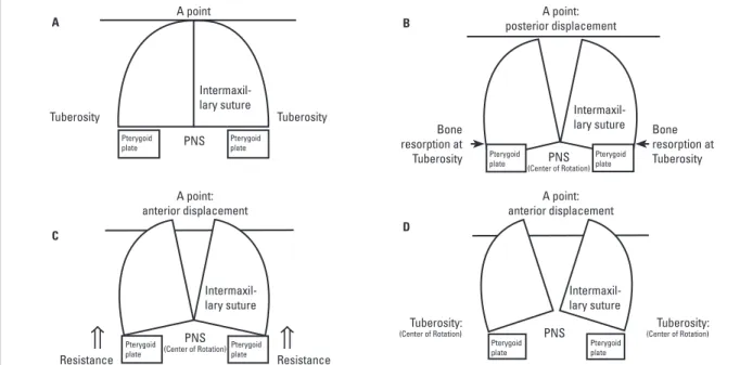
The center of rotation is located around the posterior nasal spine3,13. The expansion force is distributed not only in the maxilla but also ex-tends into the circumaxillary structures4,12. It is postulated that this entails bone resorption behind the maxilla and consequently results in posterior displacement of maxilla2 (Fig. 2A, B). On the contrast, it is postulated as well that this entails the circumaxillary structures such as pterygpoid plates to displace the maxilla forward8,9 (Fig. 2C). These two assumptions explain why some of the clinical studies on Hyrax-typed expanders reported an anterior displacement of maxilla1,7,30, while some others reported no signiicant displacement22,24 or even a posterior displacement of maxilla5,23. The posterior displacement of maxilla compromises the maxillary protraction in Class III patients.
The double-hinged rapid maxillary expander is developed for a greater anterior displacement of maxilla17,18. Its coniguration is similar to a W-appliance and has 2 hinges of rotation. It consists of a jackscrew in the center, two bolts holding the screw, a body holding the bolts at the anterior and two hinges of rotation at the posterior (Fig. 1A, B). It expands and rotates each half of the max-illa outward through the two hinges of rotation. This model of expansion entails forward rotation of maxilla with less chance of bone resorption behind the maxillary tuberosities (Fig. 2D), and this has been veriied in an experimental study in 14 cats that the double–hinged expander signii-cantly displaced the maxilla more anteriorly than the Hyrax expander10 (Tab. 3). It is therefore, in
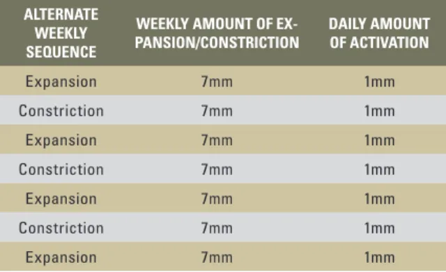
TABLE 1 - Clinical protocol for alternate rapid maxillary expansions and constrictions (Alt-RAMEC).
AlternAte weekly sequenCe
weekly Amount of ex-pAnsion/ConstriCtion
DAily Amount of ACtivAtion
Expansion 7mm 1mm
Constriction 7mm 1mm
Expansion 7mm 1mm
Constriction 7mm 1mm
Expansion 7mm 1mm
Constriction 7mm 1mm
Expansion 7mm 1mm
TABLE 2 - Comparison of the amount of maxillary protraction at A point between the growing patients with cleft lip and palate and those without (regular Class III growing patients).
growing pAtients with Cleft
lip AnD pAlAte (n=10)
regulAr ClAss iii growing pAtients (n=13)
stAtistiCs
maxillary protraction at
A point (mm)
5,8 ± 2,3 5,3 ± 1,1 n.s.
n.s. = no signiicant difference.
hyrAx expAnDer
(n=7)
DouBle-hingeD ex-pAnDer (n=7)
stAtistiCs
Anterior displacement
of maxilla (mm)
terms of the anterior displacement of maxilla, a double-hinged maxillary expander is superior to the other types of expander for the treatment of a hypoplastic maxilla in growing Class III or cleft patient.
To clinically setup the double-hinged expand-er in a patient, maxillary irst premolars and mo-lars are banded and maxillary impression is taken. The double-hinged expander is then placed and oriented on the maxillary cast so that its screw is perpendicular to the intermaxillary suture no matter it is for a patient with cleft lip and palate or regular Class III patient. The expander is than soldered to the molar and premolar bands. Two anterior extension arms (0.051-inch stainless steel wires) extending bilaterally from the premolar bands toward the central incisors are soldered to the premolar bands (Fig. 1B). The premolar and molar bands and the anterior extension arms are sandblasted before cementation. After
cementa-tion of the expander, the anterior extension arms are bonded to the anterior teeth with composite resin (Fig. 1B). One day after cementation, the double-hinged expander is activated according to the protocol of Alt-RAMEC.
Is it possible to install facial masks instead of the pair of TMA intraoral springs or do you utilize both methods simultaneously for max-illary protraction? Do you think the results in maxillary protraction would be the same? Do you have any clinical or experimental
evi-dences comparing the two methods? Ricardo
Machado Cruz
The eficiency of maxillary protraction depends mostly on the opening of circumaxillary sutures. An adequate opening of the circuumaxillary sutures is the prerequisite for a good amount of maxil-lary protraction, no matter it is a facemask, β-Ti maxillary protraction spring, or combination of a FIGURE 2 - Schematic illustrations of the postulated maxillary displacement after rapid maxillary expansion. A) The maxilla before expansion: the semi-circles represent the right and left maxillae; the rectangles represent the pterygoid plates. B) Posterior displacement of the maxilla after expansion by a Hyrax expander: each half of the maxilla rotates outward and backward around the posterior nasal spine (PNS), which entails bone resorption behind the maxillary tuberosities and results in posterior displacement of maxilla. C) Anterior displacement of the maxilla after expansion by a Hyrax expander: each half of the maxilla rotates outward and backward around the PNS, which entails the circumaxillary structures to displace the maxilla forward and results in anterior displacement of maxilla. D) Anterior displacement of the maxilla after expansion by a double-hinged expander: each half of the maxilla rotates outward and forward around the maxillary tuberosities, which geometrically results in anterior displacement of maxilla and less chance of bone resorption behind the maxillary tuberosities.
A
C
B
D
A point A point:
posterior displacement
A point: anterior displacement
A point: anterior displacement
Intermaxil-lary suture
Intermaxil-lary suture Tuberosity
Resistance Tuberosity
Resistance
Bone resorption at Tuberosity
Tuberosity:
(Center of Rotation)
PNS
(Center of Rotation)
PNS
(Center of Rotation)
PNS
Bone resorption at Tuberosity
Tuberosity:
(Center of Rotation) Pterygoid
plate
Pterygoid plate
PNS
Pterygoid plate
Pterygoid plate
Pterygoid plate
Pterygoid plate Pterygoid
plate
Pterygoid plate
⇑
⇑
Intermaxil-lary suture
to deliver a protraction force through the center of resistance of the maxilla due to its intraoral design. The orthopedic mechanics of the β-Ti maxillary protraction spring is by breaking down the biting force into a forward component of force that pro-tracts the maxilla, and an upward component of force that rotates the mandible backward. But at the same time it also tilts the palatal plane upward and opens the bite at the anterior. The upward tilting of palatal plane and bite opening are not usual in the application of facemask or modiied protraction headgear.
Therefore the β-Ti maxillary protraction spring is indicated in cases with overclosure of mandible and anterior overbite. The maxillary protraction spring is contraindicated in cases with a high mandibular plane angle or anterior open bite Class III patients. Facemask or modiied protraction headgear could be a more suitable device for such cases. In comparison of the eficiency between the
β-Ti maxillary protraction spring and facemask under the same protocol of Alt-RMEC, our clini-cal studies (Tab. 4) for growing patients with cleft lip and palate have revealed that the maxillary protraction by using facemask was 5.01 ± 1.48mm in 6 months26, whereas it was 5.8 ± 2.3mm in 3 months18 (Fig. 3).
Have you observed any nasal bone fracture during the execution of the alt-RAMEC
proto-col? Omar Gabriel da Silva Filho
Until now, we have not seen or observed ex-perimentally and clinically any nasal bone or other facemask and β-Ti maxillary protraction spring.
Facemask, modiied protraction headgear, or heavy Class III elastics are the most frequent used devices for maxillary protraction. Their eficiency in maxil-lary protraction has been well documented7,20,21,27. By applying the protraction force 30o down ante-riorly, they are able to direct a forward and down-ward protraction force so that the maxilla could be protracted through the center of resistance of the maxilla. However, they are extraoral devices and dependent largely on patient’s compliance for their eficiency.
On the other hand, the β-Ti maxillary protrac-tion spring18 has been the only intraoral device that delivers reasonable orthopedic force for maxillary orthopedic protraction. Its eficiency is independent of patient’s compliance. However, it is a custom-made device and has the inherent disadvantages that most of the intraoral springs have. It breaks sometimes during treatment due to the material fatigue, and mechanically it is almost impossible
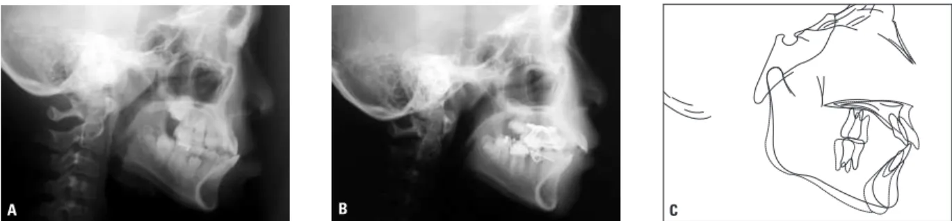
FIGURE 3 - The maxilla was protracted for 6.5mm by using intraoral β-Ti maxillary protraction springs. A) Lateral cephalogram before protraction, B) lateral cephalogram after the protraction, C) cephalometric superimposition.
A B C
β-ti mAxillAry protrACtion spring (n=10)
fACemAsk
(n=20) stAtistiCs
maxillary protraction
at A point (mm)
5,8 + 2,3 5,01 ± 1,48 n.s. TABLE 4 - Comparison of the amount of maxillary protraction at A point between the β-Ti maxillary protraction spring 18 and facemask26 under the same protocol of Alt-RMEC in growing patients with cleft lip and palate.
facial bone fracture during or after the protocol of Alt-RAMEC. Anatomically, the nasal bones articu-late with the frontonasal processes of maxilla and the frontal bones. The sutures surrounding nasal bones are the internasal nasal, frontonasal, and the nasomaxillary sutures. Sutures are the tissues absorbing stress and strain whenever forces are applied on bones. Cracks or fractures could only happen whenever the sutures surrounding the nasal bones have fused.
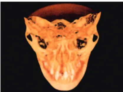
In our experimental study, we grossly examined the skull and facial bone specimen of 6 cats under-went 5 weeks of Alt-RAMEC29. We detected the sutures surrounding the nasal bones were opened 100% quantitatively in the internasal and nasomax-illary sutures and 58.3% in the nasofrontal suture. No crack or fracture line in the nasal bone or other facial bones was detected (Fig. 4).
Clinically, we have revealed in a clinical cepha-lometric study that, in addition to the maxilla, the nasal bones were displaced after Alt-RAMEC and then protracted anteriorly by the maxillary protraction springs18. Among those cases, some reported short term discomforts in the regions of nasal bones and zygomatic key-ridges during the period of Alt-RAMEC but not in the period of maxillary orthopedic protraction. The discomfort and displacement indicated that the sutures sur-rounding nasal bones were being disarticulated, or the nasal bones cracked or even fractured. How-ever the clinical evidences indicated no cracks or fractures in the nasal bones. No compression pain, skin bruise, or discoloration, has been observed or reported in any of the patients. More direct and further clinical evidences also have been revealed in our current prospective cone beam CT study in 14 patients who had undergone 7 weeks of Alt-RAREC, in which no fracture or crack line was observed in any of the circumaxillary facial bones, including the nasal bones (Fig. 5).
However, the concerns of nasal or other facial bones fracture or crack during the period of Alt-RAMEC could be a serious issue, especially in cases whose circumaxillary sutures have begun to fuse or are fused.
The alt-RAMEC technique disarranges and weakens all maxillary sutures, requiring al-ways the maintenance of the integrity of the periosteum in order to prevent foreign cells to colonize the bone sutural space. If this problem occurs, there could be the forma-tion of an osteoarthrosis, a pseudo-articula-tion formed by ibrous tissue. What would be your approach to this problem
(pseudo-ar-ticulation) if this happens? Gerson Luiz Ulema
Ribeiro
This is a question under the assumption if the circumaxillary sutures are over expanded and the integrity of a suture or periosteum is ruptured so that foreign cells invaginate and colonize in the suture resulting in osteoarthrosis or pseudo-articu-lation. This could be a serious concern whenever a suture is intentionally over expanded and exceeded the biological and physiological limitations of a suture. However, to the limit of my knowledge, the biological and physiological limitations of a suture have not been reported before.
FIGURE 4 - No crack or fracture line in the nasal bone or other facial bones after 5 weeks of Alt-RAMEC was detected in an experimental study in cats29.
The suture is an osteogenic and osteolytic tissue that allowed for a certain degree of expansion and constriction. Within the biological and physiologi-cal limitations, expansion of a suture leads to bone formation, and constriction leads to bone resorp-tion. This phenomenon also has been referred as the sutural distraction osteogenesis and osteolysis, respectively, that resembles the callus distraction osteogenesis or osteolysis in the long bone19. For the callus distraction osteogenesis in long bone, it has been revealed that the optimal (biological and physiological) rate of distraction is 1mm/day, more than 1mm/day of distraction results in osteoarthro-sis, and less than 1mm/day results in premature
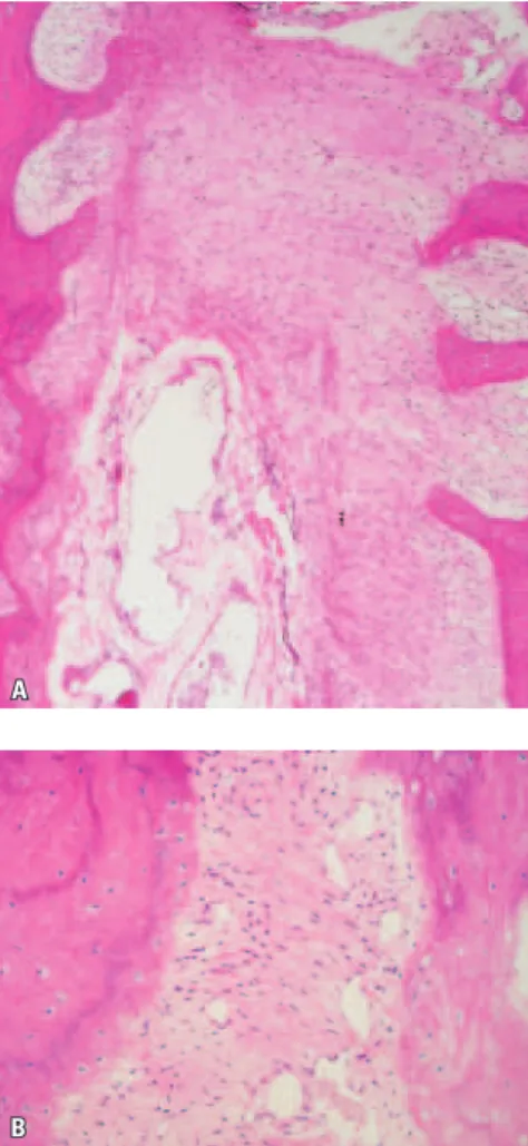
consolidation of the distraction site11. It also has been considered that 1mm/day is the biological, physiological, and optimal rate for rapid maxillary suture expansion7,8,9. The optimal rate of expansion or distraction for any osteogenetic tissue, such as suture or callus, is 1mm/day. In our experimental study on Alt-RAMEC29, we opened and constricted the maxilla 1mm/day and we did not observed foreign cells invagination, colonization, and osteo-arthrosis in any of the circummaxillary sutures (Fig. 6). The daily expansion or constriction of a suture should be biologically and physiologically conined within 1mm/day.
How long does it take to correct the sagittal maxillary deiciency after the alt-RAMEC
pro-tocol? Omar Gabriel da Silva Filho
No matter it is for growing patients with cleft lip and palate or regular Class III growing patients, it takes 1 to 2 months to correct the sagittal maxil-lary deiciency by using the intraoral β-Ti maxil-lary protraction springs15,16,18. The total treatment period is 6 months, including 3 steps in sequence:
1) 7 weeks of Alt-RAMEC to loosen the maxilla. 2) 1 to 2 months of active maxillary protraction by using the intraoral maxillary protraction springs.
3) 2 to 3 months of maintenance by keeping the maxillary protraction springs intraorally without adding extra force.
Your protocol advocated 9 weeks, than 7 weeks of alternate rapid maxillary expansions and contractions with the aim of disarticulate the maxilla before its protraction. The same procedure with 3 or 5 weeks of expansion/
contraction would be less eficient? Daniela
Gamba Garib and Ricardo Machado Cruz To open the sagittally running circumaxillary sutures quantitatively enough for maxillary pro-traction, 3 or 5 weeks of Alt-RAMEC might be enough. However, at least 7 weeks of Alt-RAMEC would be necessary to open the coronally running circumaxillary sutures quantitatively enough for maxillary protraction.
It remains controversial to what width the ex-FIGURE 6 - No foreign cells invagination, colonization, steoarthrosis,
and over growth of osteoid in the circummaxillary sutures after 5 weeks of Alt-RAMEC in experimental study in cats 29. A) intermaxillary suture, B) zygomaticomaxillary suture.
pansion should reach to disarticulate the circumax-illary sutures. Some report that 5mm of expansion is well enough, while some others report at least 12 to 15mm6. It seems that a greater amount of expan-sion will disarticulate the circumaxillary sutures more effectively than lesser expansion. However, to expand the maxilla beyond 15mm is neither clinically practical nor acceptable to patients. It is dificult to accommodate a jackscrew longer than 15mm across the palate without irritating palatal mucosa. After such an expansion, the expanded maxillary dental arch may be too wide to coordi-nate transversely with the mandibular dental arch. It is usually not necessary to increase the maxillary transverse dimension when the maxilla has been repositioned anteriorly. Rapid maxillary expander should be used to displace maxilla anteriorly and to disarticulate circumaxillary sutures rather than to expand maxilla transversely.
To disarticulate circumaxillary sutures without over-expansion of maxilla, the protocol of Alt-RAMEC is much more practical and feasible for such a performance. This is similar to the simple tooth extraction in which we repeatedly rock the tooth buccally and lingually until the tooth is disarticulated from the alveolar socket. However, the question remains controversial to how many expansions and constrictions are needed for a suf-icient disarticulation of the circumaxillary sutures. It was 9 weeks of Alt-RAMEC when we irst developed the protocol for growing patients with cleft lip and palate18, then we shifted the protocol to 7 weeks for regular Class III growing patients15,16. Clinically, it has been our observation and experi-ence that the effects of 9 weeks and 7 weeks of Alt-RAMEC are similar for the subsequent maxillary protraction. Experimentally, we have revealed in 12 cats that, after 5 weeks of Alt-RAMEC, the circum-axillary sutures running sagittally were signiicantly opened more quantitatively (94.4-100.0%) than those running coronally (56.9-58.3%), no matter they articulated directly or indirectly to the maxilla. The coronally running circumaxillary sutures were not opened quantitatively enough (56.9 to 58.3%) after 5 weeks of Alt-RAMEC. It is the sutures
running coronally rather than the sutures running sagittally needed to be well opened quantitatively for maxillary protraction. To open the coronal running sutures quantitatively enough for maxil-lary protraction, it is therefore at least 7 weeks of Alt-RAMEC would be necessary.
To examine whether the maxilla is clinically loosened enough for the protraction and to indi-vidualize the period of Alt-RAMEC needed for different patient, one could hold the maxilla with his/her ingers and rock the maxilla up and down. The patient would feel the mobility of maxilla or you could see the up and down movement of the maxilla once the Alt-RAMEC has loosened the maxilla enough.
Considering that successive expansions and contractions could lead to a healing process that will form a very dense new bone in the suture, what would you do if it would be nec-essary another rapid maxillary expansion in
the future? Gerson Luiz Ulema Ribeiro
The formation of a very dense new bone or osteoarthrosis (no new bone formation) in an expanded suture is the two opposite extremities. We did not observe these two extremities in our experimental studies in cats. All the circumaxillary sutures were intact grossly after 5 weeks of Alt-RAMEC29. No suture disruption or overgrowth of osteoid has been observed histologically (Fig. 6). The ibrous tissue and ibroblasts were stretched in the direction of expansion and constriction, and the blood vessels dilated, and the osteoid and rest-ing lines lay along the edges of the sutures (Fig. 6). The bone remodeling (osteogenesis by osteoblasts, and osteolysis by osteoclast) in a suture was kept within the biological and physiological limitation under 1mm/day of suture expansion or constric-tion, although the circumaxillary sutures became signiicantly wider after 5 weeks of Alt-RAMEC.
protraction due to the growth of mandible. Our policy to avoid the second maxillary protraction is to protract the maxilla as late as possible so that the adverse effects of mandibular growth could be the least. The optimal timing therefore is right before the fusion of circumaxillary sutures. Alt-RAMEC is a technique developed for this purpose. Our clinical studies have revealed that the optimal timing for maxillary protraction, either for a growing patient with cleft lip and palate or regular Class III patient, is at the cervical vertebrate stage 2 (CVS 2). It is the beginning of puberty, around the ages of 11~13 in girls and ages of 13~15 in boys.
Is the result of this alt-RAMEC protocol sta-ble, independently of the issure, or there is a difference when this protocol is applied in
patients without issure? Omar Gabriel da
Sil-va Filho
The treatment results by Alt-RAMEC and maxillary protraction have been revealed stable. Orthodontic tooth movement relapses so does the maxillary orthopedic protraction relapses. It
is therefore better to over protract the maxilla as much as possible in any of the maxillary protrac-tion techniques.
It was our clinical study that the maxilla in growing Class III patients was protracted forward 5.2±1.3mm under the protocol of Alt-RAMEC and the subsequent maxillary orthopedic protraction, and the percentage of post-protraction relapse was 20 to 30% of the protraction amount. Our further study on the long term results revealed that the maxilla, including the amount of relapse, grew 3.5±1.6mm further forward 5 years after the protraction. This meant the Alt-RAMEC did not jeopardize the suture growth of maxilla and the post-treatment maxillary growth compensated the amount of relapse. The total amount of maxillary growth in the horizontal direction, including the protraction, relapse, and post-treatment growth was 8.7 mm (5.2+3.5 mm)in 5.5 years, which would be good enough for maintaining the treat-ment results and covering the growth of mandible in most of the growing Class III or cleft patients through their puberty.
1. AKKAYA, S.; LORENZON, S.; ÜÇEM, T. T. A comparison of sagittal and vertical effects between bonded rapid and slow maxillary expansion procedures. Eur. J. Orthod., Oxford, v. 21, no. 2, p. 175-180, Apr. 1999.
2. BIEDERMAN, W.; CHEM, B. Rapid correction of Class III malocclusion by midpalatal expansion. Am. J. Orthod., St. Louis, v. 63, no. 1, p. 47-55, 1973.
3. BRAUN, S.; BOTTREL, J. A.; LEE, K. G.; LUNAZZI, J. J.; LEGAN, H. L. The biomechanics of rapid maxillary sutural expansion. Am. J. Orthod. Dentofacial Orthop., St. Louis, v. 118, no. 3, p. 257-261, Sept. 2000.
4. CHACONAS, S. J.; CAPUTO, A. A. Observation of orthopedic force distribution produced by maxillary orthodontic appliances. Am. J. Orthod., St. Louis, v. 82, no. 6, p. 492-501, Dec. 1982.
5. COZZA, P.; GIANCOTTI, A.; PETROSINO, A. Rapid palatal expansion in mixed dentition using a modiied expander: A cephalometric investigation. J. Orthod., London, v. 28, no. 2, p. 129-134, June 2001.
6. HAAS, A. J. Long-term posttreatment evaluation of rapid palatal expansion. Angle Orthod., Appleton, v. 50, no. 3, p. 189-217, July 1980.
7. HAAS, A. J. Palatal expansion: Just the beginning of
dentofacial orthopedics. Am. J. Orthod., St. Louis, v. 57, no. 3, p. 219-255, 1970.
8. HAAS, A. J. Rapid expansion of the maxillary dental arch and nasal cavity by opening the midpalatal suture. Angle Orthod., Appleton, v. 31, no. 2, p. 73-90, Apr. 1961.
REFERENCES
9. HAAS, A. J. The treatment of maxillary deiciency by opening the midpalatal suture. Angle Orthod., Appleton, v. 35, no. 3, p. 200-217, July 1965.
10. HUANG, C. T.; WANG, Y. C.; HUANG, C. S.; LIOU, E. J. Maxillary displacement after rapid maxillary expansions: An animal study. J. Taiwan Assoc. Orthod., Taipei,
v. 20, no. 2, p. 19-31, 2008.
11. ILIZAROV, G. A.; LEDIOV, V. L.; SHITIN, V. P. The course of compact bone reparative regeneration in distraction osteosynthesis under different conditions of bone fragment ixation (experimental study). Eksp. Khir. Anesteziol., Moscou, v. 14, no. 6, p. 3-12, Nov./Dec. 1969.
12. ITOH, T. Photoelastic effect of maxillary protraction on the craniofacial complex. Am. J. Orthod., St. Louis, v. 88, p. 117-124, 1985.
13. LEE, K. G.; RYU, Y. K.; PARK, Y. C.; RUDOLPH, D. J. A study of holographic interferometry on the initial reaction of maxillofacial complex during protraction. Am. J. Orthod. Dentofacial Orthop., St. Louis, v. 111, no. 6, p. 623-632, 1997. 14. LEVRINI, L.; FILIPPI, V. A fan-shaped maxillary expander.
J. Clin. Orthod., Boulder, v. 33, no. 11, p. 642-643, 1999. 15. LIOU, E. J. An innovative technique for maxillary protraction in
Class III growing patients: The effective maxillary orthopedic protraction. J. Clin. Orthod., Boulder, v. 39, p. 68-75, 2005. 16. LIOU, E. J. Effective maxillary orthopedic protraction for
Daniela Gamba Garib
- Doctorate Professor of Orthodontics - HRAC/FOB-USP. - Post-Doctorate, Harvard School of Dental Medicine,
Boston, USA
- Doctorate in Orthodontics, FOB-USP. - Master/specialist in Orthodontics, FOB-USP.
Gerson Luiz Ulema Ribeiro
- Professor, Specialization course in Orthodontics and Facial Orthopedics, UFSC.
- Professor, undergraduate and graduate programs, UFSC.
- Doctor in Orthodontics, UFRJ. - Master in Orthodontics, UFRJ.
- Licensed by the Brazilian Board of Orthodontics and Facial Orthopedics.
Omar Gabriel da Silva Filho
- Orthodontist at the Hospital for Rehabilitation of Craniofacial Anomalies (USP), Bauru.
- Master in Orthodontics, Unesp/Araçatuba. - Coordinator, refresher course in Interceptive
Orthodontics, Society for the Social Promotion of Cleft Lip and Palate Patients (PROFIS).
- Faculty, specialization course in Orthodontics, PROFIS. - Editor, Dental Press Orthodontics Clinical Magazine.
Ricardo Machado Cruz
- Full Professor, Orthodontics, UNIP/Brasília. - Doctor in Animal Biology/Genetics, UnB.
- Master in Orthodontics and Facial Orthopedics, FO-UFRJ.
- Licensed by the Brazilian Board of Orthodontics and Facial Orthopedics.
Contact:
Eric Liou
E-mail: [email protected]
17. LIOU, E. J.; CHEN, P. K. T. New orthodontic and orthopaedic managements on the premaxillary deformities in patients with bilateral cleft before alveolar bone grafting. Ann. R. Coll. Surg. Engl., London, v. 7, no. 3, p. 73-82, Aug. 2003. 18. LIOU, E. J.; TSAI, W. C. A new protocol for maxillary protraction
in cleft patients: Repetitive weekly protocol of alternate rapid maxillary expansions and constrictions. Cleft Palate Craniofac. J., Pittsburgh, v. 42, no. 2, p. 121-127, Mar. 2005.
19. LIU, C.; SONG, R.; SONG, Y. Sutural expansion osteogenesis for management of the bony-tissue defect in cleft palate repair: Experimental studies in dogs. Plast. Reconstr. Surg., Hagerstown, v. 105, no. 6, p. 2012-2025, 2000.
20. McNAMARA JR., J. A. An orthopedic approach to the treatment of Class III malocclusion in young patients. J. Clin. Orthod., Boulder, v. 21, no. 9, p. 598-608, Sep. 1987. 21. NANDA, R. Biomechanical and clinical considerations of a
modiied protraction headgear. Am. J. Orthod., St. Louis, v. 78, no. 2, p.125-139, 1980.
22. PANGRAZIO-KULBERSH, V.; BERGER, J.; KERSTEN, G. Effects of protraction mechanics on the midface. Am. J. Orthod. Dentofacial Orthop., St. Louis, v. 114, no. 5, p. 484-491, Nov. 1998.
23. SARVER, D. M.; JOHNSTON, M. W. Skeletal changes in vertical and anterior displacement of the maxilla with bonded rapid palatal expansion appliances. Am. J. Orthod. Dentofacial Orthop., St. Louis, v. 95, no. 6, p. 462-466, June 1989.
24. SILVA FILHO, O. G.; BOAS, M. C.; CAPELOZZA FILHO, L. Rapid maxillary expansion in the primary and mixed dentitions: A cephalometric evaluation. Am. J. Orthod. Dentofacial Orthop., St. Louis, v. 100, no. 2, p. 171-179, Aug. 1991. 25. SUZUKI, A.; TAKAHAMA, Y. A jointed fan-type expander: A
newly designed expansion appliance for the upper dental arch of patients with cleft lip and or palate. Cleft Palate J., Pittsburgh, v. 26, no. 3, p. 239-241, 1989.
26. TSAI, W. C.; HUANG, C. S.; LIN, C. T.; LIOU, E. J. Dentofacial changes of combined double-hinged rapid maxillary expansion and protraction facemask therapy. J. Taiwan Assoc. Orthod., Taipei, v. 20, no. 2, p. 5-18, 2008.
27. TURLEY, P. K. Orthopedic correction of Class III malocclusion with palatal expansion and custom protraction headgear.
J. Clin. Orthod., Boulder, v. 22, no. 5, p. 314-325, May 1988. 28. VARDIMON, A. D.; BROSH, T.; SPIEGLER, A.; LIEBERMAN,
M.; PITARU, S. Rapid palatal expansion: Part 1. Mineralization pattern of the midpalatal suture in cats. Am. J. Orthod. Dentofacial Orthop., St. Louis, v. 113, no. 4, p. 371-378, Apr. 1998.
29. WANG, Y. C.; CHANG, P. M. S.; LIOU, E. J. Opening of circumaxillary sutures by alternate rapid maxillary expansions and constrictions. Angle Orthod., Appleton, v. 79, no. 2, p. 230-234, Mar. 2009.