Ionic Fragmentation of Methyl Methacrylate Induced by
Synchrotron Radiation and Multiphoton Ionization
Cristina M. Quintellaa, G. Gerson B. de Souzab, and Joselito B. Macielb
a
Instituto de Química, Universidade Federal da Bahia, Campus de Ondina,
40.170-290 BA, Brasil; e-mail: [email protected]
b
Center for Advanced Microstructures and Devices, Louisiana State University, LA,
USA and Instituto de Química, Universidade Federal do Rio de Janeiro, RJ, Brazil#
Received: February 26, 1998
Este trabalho estuda a fotofragmentação iônica do metilmetacrilato induzida por radiação síncrotron e por multifotoionização laser. As espécies iônicas foram identificadas por um espec-trômetro de massa por tempo de vôo recentemente desenvolvido. No caso da radiação síncrotron tanto fótons com baixa energia (12.1 eV) como fótons com alta energia (287.9 eV) foram utilizados. Apesar da intensidade da fragmentação aumentar com a energia de 12.1 eV até 287.9 eV, o seu padrão permanece basicamente o mesmo e em ambos os casos: o íon pai é facilmente identificável e os fragmentos predominantes são m/q = 15,39,41 e 69. A ionização multifotônica produz bem mais alto grau de fragmentação sendo que o íon pai não foi observado e o espectro é dominado por C+. Os íons m/q = 15, 39, 41 e 69 são detectados tanto utilizando radiação síncrotron como laser evidenciando seu alto grau de estabilidade. Íons com carga dupla não foram detectados.
Ionic fragmentation of methylmethacrylate has been observed using synchrotron radiation and laser excitation. A recently developed time-of-flight mass spectrometer was used for the ionic identification. In the case of synchrotron radiation, both low energy (12.1 eV) and high energy (287.9 eV) photons were used. Although a definite increase in fragmentation was observed while moving from 12.1 to 287.9 eV, the fragmentation pattern remained basically the same in both cases. The parent peak stays clearly visible and intense fragments, associated with m/q = 15, 39, 41 and 69 dominate both synchrotron radiation-induced spectra. Multiphoton ionization causes much exten-sive fragmentation, the parent ion could not be observed, and C+ ion becomes the most intense peak in the spectrum. Ions at m/q = 15, 39, 41 and 69 are observed using laser and synchrotron radiation, which demonstrates their high stability. Doubly or multiply-charged ions have not been observed.
Keywords: methylmethacrylate, synchrotron, multiphoton ionization
Introduction
Methylmethacrylate (MMA) [H2C=C(CH3)CO2CH3] is considered nowadays as the most important methacrylic acid ester commercially available, with a world consump-tion valued at over $2.1 billion in 19961. It is in particular used as a monomer for the fabrication of polymethyl-methacrylate (PMMA). This polymer is in its turn widely used as a contact lens material and as a resist in microelec-tronics2, among other applications.
Despite its relevance, and the importance of the inter-action of ultraviolet and X-ray photons with PMMA for microlithography techniques, not much information is available concerning the interaction of high energy photons with PMMA, particularly regarding its electronic structure and fragmentation pattern.
It is well known, on the other hand, that compared to the corresponding polymers, monomers usually show simi-lar electronic photoabsorption spectrum, with some blue shifting of the observed bands3. Study of the MMA excita-Article
#permanent address
J. Braz. Chem. Soc., Vol. 9, No. 6, 521-524, 1998. © 1998 Soc. Bras. Química
tion and fragmentation may accordingly bring new insights on the kinetics and dynamics of the PMMA fragmentation process, pointing out perhaps to new pathways for indus-trial procedures and applications.
The electronic excitation of MMA has been recently studied by Rocco et al.4 using the electron energy-loss technique. Detailed calculations of its electronic structure, using high quality wavefunctions have also been performed by Hollauer et al5.
The present paper is dedicated to the analysis of the ionic fragmentation of MMA, induced by synchrotron ra-diation (12.1 and 287.9 eV) and laser light (2.35 eV), the latter operating in a multiphotonic regime. It is part of a study on the light source dependence of the photofragmen-tation kinetics and dynamics6,7. Identification of the result-ing ions is based on the use of a recently developed time-of-flight mass spectrometer.
Experimental Setup
The experimental setup is discussed in detail else-where8 and consequently only a brief description will be given here. The molecular workstation basically consists of a time-of-flight mass spectrometer (TOFMS), a gas inlet and pumping system, attached to a high vacuum chamber (base pressure ≈ 1x10⁻⁹ Torr). The entire system was in its turn coupled to the Brazilian beamline9 installed at the storage ring from the Center for Advanced Microstructures and Devices (CAMD). An optical window is also available for the laser light input.
The gas inlet consists of a tube connecting the reservoir and a precision leak valve. Liquid MMA was vaporized through a hypodermic needle (inner diameter,0.4 mm) and expanded effusively into the vacuum chamber (working pressure ranged from1.8 x 10-7 to 1.7 x 10⁻⁶ Torr). The entire system is mounted on a XYZ manipulator to allow precise positioning at the interaction region. The stated purity of the MMA sample was 99% (Aldrich, catalog number M5,590-9).
Synchrotron radiation (SR) at selected wavelengths (102.5 nm or 12.1 eV and 4.31 nm or 287.9 eV) is admitted
into the chamber through a capillary (0.8 mm inner diame-ter) in order to reduce the beam spot size and intensity at the interaction region. A Nd:YAG laser (Spectra-Physics, Quanta-Ray GCR) at 2.35 eV was used for the multipho-tonic (MP) measurements. The MP spectra were acquired with the laser frequency of 10 Hz (Table 1) and power density in the order of 109 W/cm2. Under our experimental conditions the laser power was not raised over 85 mW to avoid saturation of the detection system. Decrements down to 17 mW did not change the overall mass spectra pattern. The time-of-flight mass spectrometer basically consists of an efficient electrostatic ion extraction system (extensive ion optics simulation with the SIMION program shows that an efficiency close to 100% should be attained in the extraction of ions with up to 40 eV kinetic energy), a drift tube (25 cm) and a microchannel detection system. After extraction from the ionizing region the positive ions travel through three metallic grids (each with a nominal 90% transmission), before reaching the microchannel plate de-tector. Taking 40% as a reasonable value for the detector efficiency10, we estimate the positive ion collection effi-ciency as approximately 30%. Immediately after the ioni-zation/fragmentation event, ions and the corresponding electrons are extracted by a 500 V cm-1 field and acceler-ated to opposing directions, perpendicular to the gas beam. A time-to-digital converter is used in the determination of the ions time-of-flight with the start provided by a pulse generated by the photoelectrons. The count rates were kept below 10 Hz for MP and at 300 Hz for SR measurements.
Results and Discussion
Synchrotron radiation and laser light may induce quite different ionization and fragmentation processes11-13. In fact, their temporal structure, light intensity and photon energy differ strongly (Table 1) and may consequently induce different fragmentation/ionization pathways.
Synchrotron radiation is a tunable source of photons with a very broad energy distribution. In the present case the photons were energy selected by a grazing incidence, toroidal grating monochromator, and the minimum energy
522 Quintella et al. J. Braz. Chem. Soc.
Table 1. Characteristics of the laser light and synchrotron light as used in this work.
Characteristic Laser Light Synchrotron Light
Photons per Pulse 108 102 - 104
Pulse Duration 10 ns 10-2 - 10-1 ns
Pulse Frequency 10 Hz 500 MHz
Photons energy 2.35 eV 12.1 eV
Energy Resolution 0.01 eV 0.1 eV and high harmonics contamination
Predominant Processes Multiphotonic Monophotonic
Beam focalization Excellent Good
available was 12.0 eV. This minimum energy is already larger then the first ionization potential of MMA, which is calculated to occur at approximately 8.5 eV4,5. The short pulse duration and relative low intensity induces single photon processes.
In the case of laser light, photons of much lower energy than the molecular ionization potential, long pulse duration and high intensity were available, inducing the absorption of several photons.
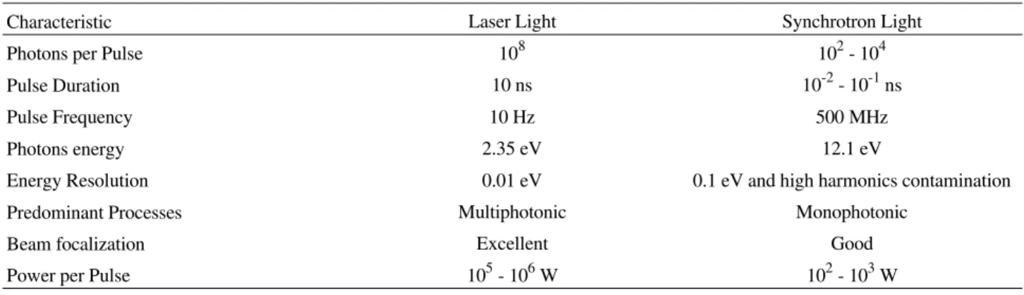
Typical mass spectra for SR (12.1 and 287.9 eV) can be seen in Fig. 1. A spectrum obtained through MP ionization (MPI) is shown in Fig. 2. It becomes immediately clear that MPI leads to a more extensive fragmentation of the mole-cule. In particular, the parent peak (m/q = 100), clearly defined in the synchrotron-radiation spectrum, cannot be seen in the laser spectrum. Small mass fragments, like C+, CH+, CH
2+ and CH3+ become, on the other hand, much more observable in the laser-excited spectrum. It is rele-vant, in this context, to mention that the parent MMA peak can still be easily observed even in the mass spectrum obtained at a much larger synchrotron photon energy (287.9 eV) as shown in Fig. 1b.
At 12.1 eV the synchrotron spectrum (Fig. 1a) is domi-nated by m/q = 15, 39, 41, 69 and 100. The first and the last peaks can be easily assigned to CH3+ and to the parent ion, C5O2H8+, respectively. Breakage of the only single C-C bond yields the most intense peak, C3H5+ (m/q = 41). Peak m/q = 39 is probably associated with the formation of the same ion, but with two hydrogen atoms less, C3H3+. Peak m/q = 69 results from the loss of the methoxy group. The ethylenic and carbonyl double bonds seem to be reasonably stable at this photon energy.
As we move on to287.9 eV, a definite increase in fragmentation occurs, but the parent peak is still prominent in the spectrum. The C3H3+ peak becomes the most intense, and a definite increase in intensity can also be observed, associated with the m/q = 15 (CH3+) and m/q = 1 (H+) fragments. 287.9 eV corresponds to a C1s → π* (C=C) transition. At this very high energy, the observation of doubly (or even multiply charged) ions may be expected, resulting from either a direct or a resonant Auger process14. The fact that these ions cannot be observed points out to their unstable nature, at least within the present time scale (a few microseconds). Experimental evidence of their ex-istence has been obtained using a different experimental scheme (Photoion-Photoion Coincidence technique or PIPICO)6.
MPI shows more extensive fragmentation than single photon ionization (SPI). As a result the more massive fragments m/q = 82 to 101 are absent from the MPI spec-trum. The SPI fragments are preferentially concentrated in m/q = 33 to 51 (49% branching ratio), whilst the MPI fragments, although being maximum in the same mass range (34%), also have high branching ratio for m/q = 12 to 18 (25%) and m/q = 24 to 32 (25%).
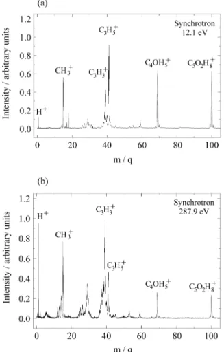
The complete absence of the parent ion in the MPI spectrum (Fig. 2) reveals the existence of very effective fragmentation processes. In particular m/q = 12 (C+) be-comes the most intense peak. A simple thermodynamic calculation shows that, to yield it, at least approximately
Vol. 9, No. 6, 1998 Ionic Fragmentation of Methyl Methacrylate 523
Figure 2. MMA mass spectrum obtained through laser MPI at 2.35 eV.
25 eV are required to produce this ionic fragment corre-sponding to an eleven photon process. To induce complete atomization of the MMA molecule 63 eV would be neces-sary15,16 and would correspond to the absorption of 27 photons. From the present experimental results it is not possible to point out whether this level of atomization would have proceeded either through a sequential mecha-nism, or through a concerted, true multi-photon absorption. In a sequential mechanism the first step would involve ionization (which would require the absorption of at least four photons), and the resulting fragments (neutral or charged) would absorb additional photons and suffer more extensive fragmentation. It is already known, for instance, that with high laser intensity extensive skeletal fragmenta-tion is readily produced and that in the extreme limit even multiply charged ions can be observed12. In the present case only singly-charged ions have been observed.
The laser spectrum shows, on the other hand, that ionic fragments associated with the breakage of the single carb-on-carbon or carbon-oxygen bonds (m/q = 41 and m/q = 69) are particularly stable, as they show a significant inten-sity even in the presence of such strong fragmentation processes.
Conclusions
Ionic fragmentation of MMA has been observed using synchrotron radiation and laser excitation, coupled to a time-of-flight technique. In the case of synchrotron radia-tion both low energy (12.1 eV) and high energy (287.9 eV) photons were used. Taking into consideration that the first ionization potential for MMA occurs at 8.5 eV, the lower energy is only capable of ionizing valence-shell electrons, while the higher energy corresponds to the excitation of core electrons (C 1s). Although we do observe a definite increase in fragmentation while moving from 12.1 to 287.9 eV, the fragmentation pattern remains basically the same in both cases. The parent peak is clearly visible and intense fragments, associated with m/q = 15, 39, 41 and 69 dominate both spectra. Doubly or multiply-charged ions are extremely unstable (at least in a microsecond scale) and have not been observed in the mass spectra.
Multiphoton ionization causes much extensive frag-mentation, and the parent ion could not be observed in the laser spectrum. As a demonstration of the severe degree of atomization induced on the molecule, the C+ ion becomes the most intense peak on the spectrum (as for a comparison, using synchrotron radiation this ion was barely observable at 287.9 eV and not observed at all at 12.1 eV). Very stable ions are observed at m/q = 15, 39, 41 and 69.
Acknowledgments
The authors would like to thank the Center for Advan-ced Microstructures and Devices (CAMD, Baton Rouge, LA, USA) and the Laboratório Nacional de Luz de Síncro-tron (LNLS, Campinas,Brazil) for support and assistance. LNLS is also acknowledged for the use of the Toroidal Grating Monochromator and related beamline. The Fun-dação Universitária José Bonifácio (FUJB, Brazil) and the Conselho Nacional de Desenvolvimento Científico e Tec-nológico (CNPq, Brazil) are gratefully acknowledged for financial assistance.
References
1. http://www-cmrc.sri.com.
2. Choi, J.O.; Moore, J.A.; Corelli, J.C.; Silverman, J.P.; Bakhu, H. J. Vac. Sci. Technol. 1988, B6, 2286. 3. Onari, S. J. Phys. Soc. Japan1969, 26, 500.
4. Rocco, M.L.M.; Lopes, M.C.A.; Hollauer, E.; Mon-teiro, E.E.; de Souza, G.G.B. Chem. Phys.1997, 223, 15.
5. Hollauer, E.; Rocco, M.L.M.; Lopes, M.C.A.; de Souza, G.G.B. Chem. Phys.1998 (submitted). 6. Quintella, C.M.; de Souza, G.G.B.; Mundim, M.S.P.
In Laser Techniq. for State Select. and State-to-State Chem. IV; Hepburn, J.W.; Continetti, R.E.; Johnson, M.A., eds, SPIE, v. 3271, 227-335, 1998.
7. Quintella, C.M.; de Souza, G.G.B.; Mundim, M.S.P. J. Braz. Chem. Soc. 1998, (to be submitted).
8. Maciel, J.B.; Morikawa, E.; de Souza, G.G.B. In Syn-chrotron Radiation InstrumentationNational Confer-ence; Am. Inst. Phys. Conference Proceedings, Ernest Fontes; 1997.
9. Fonseca, P.T.; Pacheco, J.G.; d’A Samogin, E.; de Castro, A.R.B. Rev. Sci. Instrum.1992, 63, 1256. 10. Reagan, N.R.; Frees, L.C.; Gray, J.W. J. Vac. Sci.
Technol. 1987, A5, 2389.
11. Baer, T. Annu. Rev. Phys. Chem.1989, 40, 637. 12. Gedanken, A.; Robin, M.J.; Kuebler, N.A. J. Phys.
Chem.1982, 86, 4096.
13. Reisler, H.; Wittig, C. In Photodissociation and Pho-toionization, Lawley, K.P., ed, John Wiley & Sons Ltd.; 1985.
14. Morin, P.; de Souza, G.G.B.; Nenner, I.; Lablanquie, P. Phys. Rev. Lett.1986, 56, 131.
15. Atkins, P.W. Physical Chemistry; Oxford University Press; Frome, 1994; 5th Edition.
16. Winn, J.S. Physical Chemistry; Harper Collins Col-lege Publishers, 1995.