www.mech-sci.net/5/1/2014/ doi:10.5194/ms-5-1-2014
©Author(s) 2014. CC Attribution 3.0 License.
Mechanical
Sciences
Open Access
Devices for accurate placement of epidural Tuohy needle
for Anaesthesia administration
N. Vaughan1, V. N. Dubey1, M. Y. K. Wee2, and R. Isaacs2 1
Bournemouth University, School of Design Engineering & Computing, Bournemouth, UK 2
Poole Hospital NHS Foundation Trust, Poole, UK
Correspondence to:N. Vaughan ([email protected])
Received: 17 April 2013 – Revised: 28 August 2013 – Accepted: 23 September 2013 – Published: 2 January 2014
Abstract. The aim of this project is to design two sterile devices for epidural needle insertion which can mea-sure in real time (i) the depth of needle tip during insertion and (ii) interspinous presmea-sure changes through a pressure measurement device as the epidural needle is advanced through the tissue layers. The length measure-ment device uses a small wireless camera with video processing computer algorithms which can detect and measure the moving needle. The pressure measurement device uses entirely sterile componenets including a pressure transducer to accurately measure syringe saline in mm Hg. The data from these two devices accurately describe a needle insertion allowing comparison or review of insertions. The data was then cross-referenced to pre-measured data from MRI or ultrasound scan to identify how ligemant thickness correlates to our measured depth and pressure data. The developed devices have been tested on a porcine specimen during insertions per-formed by experienced anaesthetists. We have obtained epidural pressures for each ligament and demonstrated functionality of our devices to measure pressure and depth of epidural needle during insertion. This has not previously been possible to monitor in real-time. The benefits of these devices are (i) to provide an alternative method to identify correct needle placement during the procedure on real patients. (ii) The data describing the speed, depth and pressure during insertion can be used to configure an epidural simulator, simulating the needle insertion procedure. (iii) Our pressure and depth data can be compared to pre-measured MRI and ultrasound to identify previously unknown links between epidural pressure and depth with BMI, obesity and body shapes.
1 Introduction
Epidural is commonly used as a form of pain relief during childbirth, for the treatment of chronic back pain or as a means to provide anaesthesia or analgesia during specific op-erations. The procedure consists of a long Tuohy needle be-ing carefully inserted in the lumbar spine through layers of skin, tissue and ligament into the epidural space. Monitoring pressure and the needle depth during an epidural insertion is crucial because it indicates when the needle is placed pre-cisely into the epidural space. If the needle is advanced too far it will puncture the dural sac and cause leakage of cere-brospinal fluid. Post dural puncture headaches may result and can be extremely disabling for the patient. Other risks include bleeding or nerve damage which may rarely lead to paralysis. If the needle is not within the epidural space, the pain relief
may be ineffective or inadequate due to improper placement of the catheter.
The most important aspect of needle insertion is to judge the correct depth of the needle, because it has to pass through on average 42 m of tissue layers although this increases in obese patients, before arriving at the small 6 mm epidural space (Jenkins, 2005). This skill requires precise knowledge of the feeling as the needle passes through various tissue lay-ers. The operator must judge the depth of the needle just from feeling. The point at which the needle arrives in epidu-ral space is identified by a characteristic loss-of-resistance (LOR), with corresponding drop of pressure.
ligament, interspinous ligament, ligamentum flavum and into the target epidural space. Each ligament has different density and texture, which affects the feel of the needle and the resis-tance to insertion (Vaughan et al., 2013). When the epidural space is reached, the resistance to insertion drops to zero and this indicates that the target has been reached so the device should show sudden drop in pressure. The depth can be pre-cisely monitored from the pre-determined value from MRI or ultrasound scan for each patient before carrying out the procedure therefore ensuring the accurate placement of the needle.
Currently there is no way to electronically measure epidu-ral needle depth or monitor epiduepidu-ral saline pressure for loss of resistance (LOR). These two novel devices developed in this paper provide solutions which can achieve these mea-surements in a sterile environment. The devices will provide several benefits to (i) provide a method alternative to oper-ators opinion to confirm correct needle placement during an in-vivo procedure (ii) collect pressure and depth data from patients to incorporate into an epidural simulator (iii) cross-reference measured data to MRI to identify how pressure and depth changes in patients with various BMI, tissue thick-nesses and densities.
2 Competing solutions
Epidural needle insertion is essentially a ‘blind’ procedure, but utilizes a well-known technique referred to as “loss of re-sistance” (LOR). During an epidural insertion, the operator tries to perceive which tissue layer the needle tip is pass-ing through by feelpass-ing the resistances on the needle. Cur-rent epidural precedure relies upon the operators judgement to identify LOR as the epidural needle travels through the various ligaments and potential spaces of the lumbar verte-bral column. There are many risks involved with the epidural procedure due to its complexity. Since the actual insertion is ‘blind’ and the end-point cannot be visualized, the anaes-thetist has only his/her perception from sensing and feeling of the needle resistance through the force stimuli in the fin-gertips and joints. There can be errors of judgment as to the location of the needle, exacerbated by individual patient characteristics of height, weight and body shape.
Our devices provide useful clues to the location of epidu-ral needle which could be used in combination with operators judgement and may increase chance of successfully finding the epidural space. In our camera based depth measurement system, the needle depth is visually monitored using brown and silver markings on the needle shaft at 10 mm intervals. The manual method of depth monitoring may not be reli-able since the anaesthetist has to pay attention to other as-pects of the procedure such as pressure and angle of inser-tion, besides this it is believed that there may potentially be errors of±5 mm depending on the skill and perception of the anaesthetist. Attempts have been made in the past to measure
the needle depth in real time by using metal calipers with an electronic sensor between the skin and the syringe barrel (Tran et al., 2009). Metal calipers are hard to sterilize for use in human trials and may interfere with the operator’s stan-dard procedure and would therefore not be ethically permit-ted in-vivo. Our novel sterile pressure measurement device captures the resultant pressures as the epidural needle is ad-vanced through the tissue layers and the novel image process-ing technique which measures the insertion depth of the nee-dle in real-time and transmits data wirelessly. Our pressure device can help to identify the sudden pressure drop at LOR, which identifies the needle is correctly in the epidural space, reducing risk of patient injury. Also the measured pressure data is very useful to (i) incorporate into a haptic epidural simulator and (ii) cross-reference data to pre-measured MRI and ultrasound to identify links between obesity and needle insertion pressure.
3 Description of the design
Both of our devices, the pressure monitor and the camera depth measurement device, include a novel wireless setup which has not been achieved before. The core component which enables our pressure device to obtain interspinous pressure measurements is a medical pressure transducer, or-dinarily used for blood pressure and pulse monitoring, which is entirely sterile. This provides an electronic pressure read-ing, which we then amplify and this goes through a micro-controller, and is then transmitted wirelessly. The receiver can be located in a separate hospital room, which minimises disturbance, up to one mile away. Our method uses a novel three-way-tap, enabling the transducer to be placed between the needle and the syringe, so that any pressure changes will be detected by the device.
algorithm is custom made, based around a core of OpenCV library.
A wireless data transmitter is utilized to minimize the equipment and disruption in the labour room. This uses
XBeeTM Pro 60 mW with a PCB antenna at 2.4 GHz
fre-quency and 250kbps data transfer rate that allows a maxi-mum of 100 m outdoor range. A computer receives the data remotely with a wireless receiver and displays a real-time graph on screen and simultaneously records the data to a file. The advantage of using a wireless transmitter is that the equipment is not only less cumbersome but also much less intrusive.
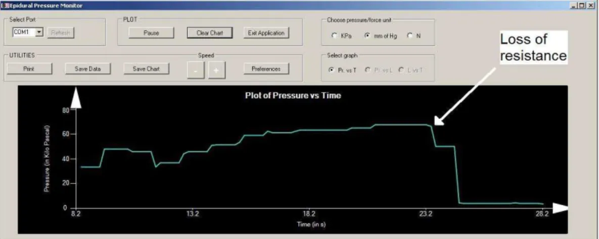
In obstetric practice, the LOR technique is performed with a saline-filled syringe connected directly onto the Tuohy needle, which maintains the plunger pressure until needle reaches the epidural space. Our design aims to minimize changes to this standard setup (Fig. 1). A small standard ster-ile three-way tap (BD ConnectaTM) is connected between the needle and syringe. The tap is connected to the pres-sure transducer via a one metre length of saline-filled sterile manometer tubing. The transducer’s electrical plug is con-nected by a short electrical cable to the wireless transmitter box which measures only 12 cm×5 cm×3 cm. The box con-tains a 5 V DC power supply, amplifier and the data trans-mitter. At the remote site, a wireless receiver is connected via Universal Serial Bus (USB) to the computer. Our custom designed software displays a real-time graph on screen and writes the data to a text file for later analysis; data retrieval speed can be varied in the software. The software clearly identifies loss-of-resistance as needle enters epidural space (Fig. 2) on the output from our pilot trial on a porcine ca-daver. The data recorded into a file was plotted as a line graph shown in Fig. 3.
On needle depth measurement there were several problems associated with needle detection that makes measurement hard to accomplish: (i) the needle is a thin object with small width of only a few pixels; (ii) the needle is reflective stain-less steel material which reflects colours from nearby objects, light sources and shadows; (iii) the needle is circular in cross section with different colours all around, the top is often illu-minated while the underside is often shadowed; (iv) there are camera noise and blur from the wireless camera; (v) tilting the needle up/down causes colour change with only a small tilt of the needle; (vi) tilting towards/away from the camera causes the visible length to decrease, which may be confused with reduced length due to insertion; (vii) the needle is not the only object in the foreground, due to the operator’s hands and the patient’s back; (viii) lighting conditions vary from room to room; (ix) there is no time to configure the camera each time, due to the short notice given when an epidural is going to be performed and the anaesthetist cannot wait be-cause the patient is often in pain; (x) the length is not fixed, but changes dynamically as needle is inserted. Solutions to these problems could involve controlling the environment with the use of fixed backgrounds, controlled light sources,
Figure 1.The disposable epidural pressure measurement system.
or restricting the movement. These precautions may not be feasible since they interfere with the operator’s procedure and may not be ethically permissible. We developed custom software which detects the needle in the image, measures the length of the visible needle externally and transmits the depth data of the needle wirelessly in real-time. The algorithm de-sign is able to solve the ten imaging problems listed by using the standard marking on the needle including blue handle and the silver 10 mm markings on the needle to accurately keep track of needle position, orientation and length in the major-ity of frames. The algorithmic implementation of the tech-nique and the software output is shown in Figs. 4 and 5. This uses a wireless camera measuring (20×20×20 mm) manu-factured by Ajoka that transmits a 640×480 pixel image as PAL AV in full colour over a 20 MHz wireless link. The AV receiver is connected to a 2.6 GHz Desktop PC with 2 GB RAM. The developed software uses features from OpenCV for image retrieval, memory handling and display.
4 Evaluation and testing
A porcine trial was conducted on a section of a cadaver for measurement of syringe pressure during insertion. The porcine tissue specimen was a double loin saddle cut. The ca-daver was obtained from a livestock farm within 24 hours of slaughter without being frozen or modified in any way after slaughter to preserve the integrity of the spinal tissues. The pig was a standard hybrid Large White cross Saddleback. The specimen contained the entire back in one piece, with the whole spine, and all tissue layers from external skin, through to the thoracic cavity. The porcine tissue was mounted verti-cally against a wooden support to mimic sitting position so that the cadaver would not move when pressure was applied during insertion.
Figure 2.Software output clearly showing LOR.
Figure 3.Line graph showing the KPa pressure measurement data.
therefore, needle patterns are easily discernible against the background. A saline drip bag was connected by tubing and flushed to fill the pressure transducer system. The vertical position of the transducer was compensated for the eleva-tion difference between the transducer and the insertion point throughout to cancel any gravitational effect on the pressure readings. Epidural insertions were performed by two experi-enced anaesthetists.
The recordings of pressure were then started and contin-uously recorded throughout needle insertion until after the LOR had been experienced. The majority of insertions lo-cated the epidural space in the first attempt and the corre-sponding data is presented in Table 1, these values were av-eraged from all pressure values throughout each tissue and by the maximum pressure peak prior to LOR. Force was cal-culated from pressure, multiplying by the known area of the syringe barrel. Table 2 shows the mean value of the data col-lected and the standard deviation from this trial. This data compared quite well with similar previous studies (Holton, 2001; Tran et al., 2009), length data was verified by the ac-tual measurement. The total insertion took about fifteen sec-onds with 10 frames per second. The maximum error from
the camera based length measurement was ±3 mm which
was determined by comparison of each frame to a manual review of the video. During testing, failures were determined
Figure 4.Implemented image processing algorithm.
as frames when needle markings were not identified by the algorithm so measurement did not work. The failure rate was 3 frames out of 150 which gave an overall 97.8 % reliability during this test.
5 Conclusions
Figure 5.Software output measuring needle length.
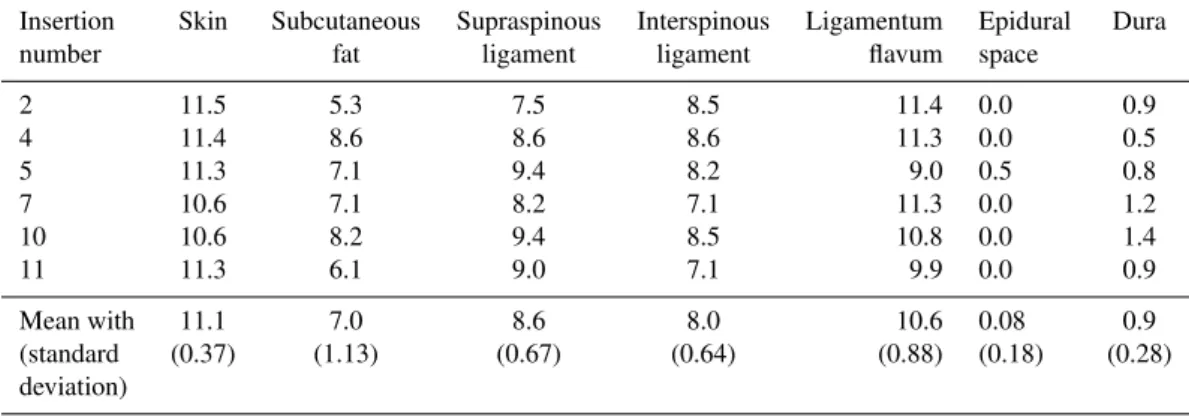
Table 1.Force (N) estimates for each tissue during needle insertions into porcine cadaver.
Insertion Skin Subcutaneous Supraspinous Interspinous Ligamentum Epidural Dura
number fat ligament ligament flavum space
2 11.5 5.3 7.5 8.5 11.4 0.0 0.9
4 11.4 8.6 8.6 8.6 11.3 0.0 0.5
5 11.3 7.1 9.4 8.2 9.0 0.5 0.8
7 10.6 7.1 8.2 7.1 11.3 0.0 1.2
10 10.6 8.2 9.4 8.5 10.8 0.0 1.4
11 11.3 6.1 9.0 7.1 9.9 0.0 0.9
Mean with 11.1 7.0 8.6 8.0 10.6 0.08 0.9
(standard (0.37) (1.13) (0.67) (0.64) (0.88) (0.18) (0.28)
deviation)
Table 2.Data from porcine trial (tissue thickness of each layer was measured at L2/L3).
Tissue Tissue Needle
layer thickness depth
(mm) (mm)
Skin 3 0
Subcutaneous fat 6 3
Supraspinous ligament 4 9 Interspinous ligament 26 13
Ligamentum flavum 3 39
Epidural space 6 42
Dura 15 48
on its metal shaft is clinically useful as there is currently no other way to measure the needle or monitor the depth in real-time during insertion. This could potentially be useful for precise location of Tuohy needle during epidurals to im-prove safety of the procedure. The image processing method is more ethically safe and less intrusive than using physical measuring devices which touch the needle (Tran et al., 2009). It also ensures sterility which is essential for the prevention
of infection during epidural insertion. A further study with labouring women has now begun after obtaining ethical ap-proval from the National Research Ethics Service in the UK, using our device and we hope that this will improve the safety of the procedure and thus reducing the morbidity and cost burden to the health services.
Acknowledgements. The authors thank Obstetric Anaesthetists Association for funding to continue conducting the trial with obstetric patients and MRI funding.
Edited by: W. Durfee
References
Holton, L. L.: Force models for needle insertion created from mea-sured needle puncture data, Stud. Health Technol. Inf., 81, 180– 186, 2001.
Jenkins, J. G.: Some immediate serious complications of obstetric analgesia and anaesthesia: a prospective study of 145,550 epidu-rals, Int. J. Obstet. Anaesth., 14, 37–42, 2005.
Tran, D., Hor, K., Kamani, A., Lessoway, V., and Rohling, R.: Instrumentation of the loss-of-resistance technique for epidural needle insertion, IEEE T. Bio-Med. Eng., 56, 820–827, 2009. Vaughan, N., Dubey, V. N., Wee, M., and Isaacs, R.: A review of