20 artigo 288
TEChNICAL NoTE
The authors declare that they did not have any conflict of interests in producing this article 1 - Resident Physician in Orthopedics at the National Institute of Traumatology and Orthopedics (INTO/MS) - Rio de Janeiro, RJ. 2 - Head of the Pediatric Orthopedics Center, National Institute of Traumatology and Orthopedics (INTO/MS) - Rio de Janeiro, RJ. 3 - Head of the Department of Pediatric Surgery, Fernandes Figueira Institute (IFF/Fiocruz) - Rio de Janeiro, RJ.
4 - Head of the Department of Medical Genetics, Fernandes Figueira Institute (IFF/Fiocruz) - Rio de Janeiro, RJ.
5 - Surgeon in the Department of Pediatric Urological Surgery, Fernandes Figueira Institute (IFF/Fiocruz) - Rio de Janeiro, RJ. Work performed at the Fernandes Figueira Institute (IFF/Fiocruz) - Rio de Janeiro, RJ.
Correspondence: Rua Washington Luis, 61 - Centro - 20230-024 - Rio de Janeiro, RJ. Email: [email protected] Work received for publication: January 17, 2010; accepted for publication: June 9, 2010.
bIlATERAl ANTERIOR PElvIc OSTEOTOmy FOR clOSuRE
OF blAddER ExSTROPHy: dEScRIPTION OF TEcHNIquE
Camila Bedeschi Rego de Mattos1, Pedro Henrique Barros Mendes2, Paulo Roberto Boechat3, Juan Llerena Júnior4,
Luciano da Silva Guimarães5
INTRODUCTION
The complex of congenital malformations that includes bladder and cloacal exstrophy consists of conditions that involve the genitourinary tract, musculoskeletal tissue and sometimes the gastrointestinal tract, and for these conditions to be correctly managed, knowledge provided by pediatric orthopedists in conjunction with pediatric urological surgeons is required.
The main objective of orthopedic reconstruction is to diminish the pelvic diastasis found in these pa-tients, thereby making it possible to close the bladder and abdominal wall while diminishing the tension that was previously present.
Several types of osteotomy have already been
des-ABSTRACT
Bladder and cloacal exstrophy are rare malformations associated with abnormalities in the pelvis. The objec-tives in reconstruction are to obtain a closed and conti-nent bladder, with an acceptable cosmetic appearance. Treatment for the abnormalities of pelvic anatomy is an important part of achieving successful treatment for
these urological conditions. This article aims to describe the technique of bilateral anterior pelvic osteotomy for treating bladder and cloacal exstrophy, and presents two cases to demonstrate the difficulties and applications of the technique.
Keywords – Bladder Exstrophy; Osteotomy; Pelvis; Congenital Abnormalities
cribed with the aim of correcting abnormalities of these patients’ pelvic anatomy. However, the modern era began at the end of the 1980s, when Sponseller disseminated the use of bilateral anterior osteotomy of the innominate bone, since this has a variety of
advantages over the old techniques(1).
Here, we present a summary of the technique, whi-ch has been little disseminated in Brazilian articles.
remarks about exstrophy
Classic bladder exstrophy is a rare
malforma-tion with an estimated incidence of 1:30.000(2) and
slightly greater predominance among males, with a
boy-to-girl ratio of 2.3:1(3). There is still no consensus
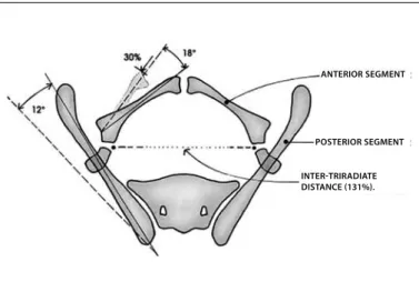
Figure 1 – Pelvic anatomy in cases of bladder exstrophy.
Figure 2 – Transverse and vertical osteotomies with fixation using one or two pins. The vertical osteotomy is in a hinged greenstick fracture.
of the cloacal membrane that prevents migration of the mesenchymal tissue and correct development of
the inferior abdominal wall(4).
The pelvic anatomy in these patients is abnormal, and it is extremely important to understand this, in order to understand the treatment and prognosis for
these patients. Studies by Sponseller et al(5) and Stec
et al(6) have demonstrated that, in addition to the
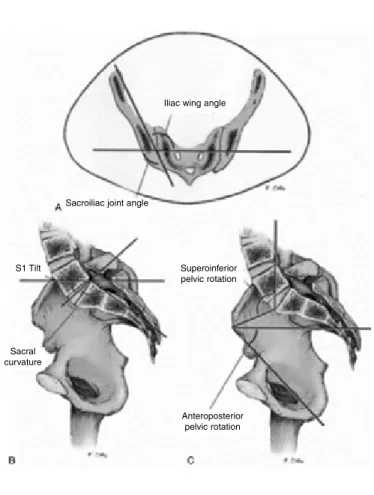
increased diastasis of the public symphysis, the pos-terior part of the pelvis presents external rotation of 12º, the acetabulum is retroverted, the anterior part has external rotation of 18º and the pubic branches are around 30% smaller than normal. In addition, the angle of the sacroiliac joint is 10º greater, there is internal rotation of the pelvis of around 15º, the volume is around 40% greater and the surface is 20% greater. These abnormalities consequently make the progression angle of the foot around 20 to 30º of external rotation greater than normal, but with improvement with increasing age, and the gait has a widened base, associated with dysplasia of hip
de-velopment(7,8). In patients who do not undergo
oste-otomy together with closure of the bladder, there is
higher incidence of premature hip arthrosis(9).
DESCRIPTION OF THE TECHNIQUE
The patient is positioned in dorsal decubitus with the pelvis raised on a pad. The bladder is isolated using a sterile drape. An incision of around 5 cm is made 1 to 2 cm distally to the anterosuperior iliac spine (an incision that is similar to what is used for Salter osteotomy). The lateral cutaneous nerve of the thigh is identified and protected. Both sides of the pel-vis are exposed subperiosteally along the iliac wings as far as the sciatic incisure, posteriorly as far as the medial ligaments of the sacroiliac joint and caudally
just above the triradiate cartilage (Figure 1)(6)
.
A small window is opened to the periosteum at the side of the iliac in order to control the osteotomy and for inserting the pins. After inserting a Hohmann spacer in the sciatic incisure, transverse osteotomy of the iliac is then performed using a Gigli saw or osteotome. The lower segment of the pelvis should then be capable of movement medially.
The vertical osteotomy is hinged and incomplete. It is performed parallel to and laterally to the sacroiliac
joint, thus creating a gutter with the posterior cortical bone intact. It is then tested by turning the iliac wings internally and closing the sulcus that was made in the
osteotomy, as a hinge (Figure 2)(9).
One or sometimes two fixator pins are placed in the lower segment of the ilium and one or two in the upper part. The incisions are closed, preferably using absorbable intradermal sutures, thereby allowing the pediatric urological surgeon to complete the genitou-rinary repair. After the urological procedure, the pu-blic diastasis can be closed in different ways. Someti-mes this is down by the urologist using a suture with nylon 2-0 thread while turning the pelvis internally, thus facilitating the suture. In older children or those with extreme diastasis, this may not be achieved and closure of the pelvis is stages may be needed, with gradual adjustment of the external fixator bar. After
Anterior segment
Posterior segment
inter-trirAdiAte distAnce (131%).
osteotomies
Post. Periosteum And cortex remAin
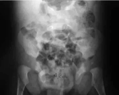
Figure 3 – Preoperative radiograph on patient with bladder exs-trophy, showing pubic diastasis of 4.2 cm.
closing the abdominal wall, the bars of the external fixator are inserted to keep the pelvis in the correct position.
We recommend that the external fixator should be used for four to six weeks. In the original technique, skin traction on each leg throughout the time of using the fixator was recommended in order to help to avoid early loosening of the pins. However, economic rea-lities and the need for beds in the hospitals where we perform osteotomy do not allow us to keep the child in hospital for all of this time. It our situation, we discharge the patient, who then remains resting, wi-thout loading the limb while the fixator is being used. Radiographic control is performed every seven to ten days. If the reduction of the diastasis of the symphysis is unsatisfactory, it can gradually be closed through using the bars of the external fixator. The pins are only removed after radiographic evidence of consolidation has been obtained.
The commonest complications that occur with this type of procedure include: infection of the pins, tran-sitory paralysis of the lateral cutaneous nerve of the thigh and delays in the consolidation process. There may also be complications consequent to the use of cutaneous traction, when this is used. Other, less com-mon complications include pseudarthrosis, arthrosis, lower-limb dysmetria and lesions of the sciatic nerve, femoral nerve and superior gluteal nerve. Deep
infec-tion is another, but rarer infecinfec-tion(10).
FIRST CASE REPORT
The first patient was five months old and was re-ferred to a pediatric surgery service with a diagnosis of bladder exstrophy, presenting preoperative pubic diastasis of 4.2 cm (Figure 3).
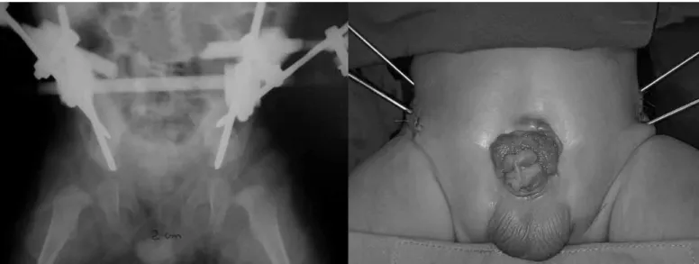
This patient underwent bilateral anterior pelvic osteotomy, performed by the present author in June 2005, using the technique subsequently described, concomitantly with bladder closure performed by the pediatric urological surgery team, at the age of five months. The procedure was made more difficult be-cause of a lack of image intensifier during the surgi-cal procedure, which made pin placement somewhat more laborious, with the need to produce intraoperati-ve radiographs in order to intraoperati-verify that their positioning was correct (Figure 4A). Reduction of the diastasis
to 2 cm was achieved in the immediate postoperative period (Figure 4B).
The patient remained in hospital for one week and no traction was used; only resting in bed. An external fixator was used for a total of eight weeks, and no type of complication was presented. Two years after the surgery, it was found that the patient had lost some of the reduction, but the result remained satisfactory, with diastasis of 3.8 cm and good urological function (Figure 5). In the late postoperative period, the patient was already walking, with gait on a normal base, and no external rotation of the legs was presented. There was no recurrence of or complication from the uro-logical abnormalities.
SECOND CASE REPORT
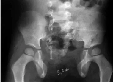
The second patient was referred at the age of five years, for correction of epispadia and for pelvic oste-otomy to be performed. Closure of the cloacal exstro-phy had already been performed at another hospital. Before the operation, the patient presented pubic di-astasis of 5.2 cm (Figure 6).
Figure 4 – A) Left: patient at the end of the orthopedic procedure, released to undergo bladder closure. B) Right: radiographic appe-arance during immediate postoperative period.
Figure 5 – A) Left: pelvic radiograph one year after the operation, showing diastasis of 3.8 cm. B) Right: appearance of closed bladder two years after the operation.
had the technical difficulty of lack of an image inten-sifier, which especially added difficulty to performing the vertical osteotomy, which was slightly oblique instead of vertical and parallel to the sacroiliac joint, as is recommended for the technique (Figure 8). The lack of intensifier also increased the duration of the operation because of the simple radiographs produced intraoperatively.
The patient remained in hospital for two weeks. No skin traction was used, but only resting in bed. An external fixator was used for eight weeks. No postop-erative complications were presented.
In January 2009, i.e. two years after the operation,
it was seen that the patient had lost some of the reduc-tion of the symphysis, to 3.6 cm (Figure 9). The gait projection angle before the operation was 35º, and this was reduced to 15º. Before the operation, the patient’s gait presented external rotation, which was found at the postoperative assessment to have been corrected. There was no recurrence or complications from the urological abnormalities.
DISCUSSION
pe-Figure 6 – Preoperative radiograph on patient with cloacal exs-trophy, showing public diastasis of 5.2 cm.
Figure 7 – Patient just after placement of the pins and before correction of the epispadia.
Figure 9 – Pelvic radiograph produced one year after the ope-ration, showing a diastasis of 3.6 cm.
Figure 8 – A) Left: radiograph produced during the immediate postoperative period, showing reduction of the diastasis to 1.0 cm and slightly oblique positioning of the osteotomies parallel to the sacroiliac joints. B) Right: patient during the postoperative period, showing the external fixator and the correction of the epispadia.
A B
diatric surgeons. In our institution, we have been following up these cases that were referred to us for almost ten years and, after this length of experience, we can conclude that performing pelvic osteotomy concomitantly with closure of the urological defect is highly beneficial for developing these patients’ gait and preventing arthrosis of the hip joint. It also gre-atly facilitates performing the urological procedure. This technique requires a shorter time to perform: it can be used for older patients and corrects the defor-mities better(7,9,11-15).
Stec et al(12,13) reported that there was an increase
the-Figure 10 – Increase of 10º in the angle of the sacroiliac joint and 15º in internal rotation of the pelvis.
se patients (Figure 10). This defect is currently not corrected by this type of technique and is one of the causes of the loss of reduction of the pubic diastasis that generally occurs in these patients later on. Other factors that lead to loss of diastasis include loosening of the pins, especially among younger children, and the fact that the pubic branches are 30% smaller than normal, thus causing lower growth. However, even with this loss of reduction of the symphysis, the result achieved is better than the condition of untreated pa-tients and fulfills the function of facilitating bladder closure and improving the patient’s functioning.
Early bladder closure, within the first days of life, has recently been advocated. This is done along with closure of the abdominal wall, with or without oste-otomy of the innominate bone. In patients under one month of age, closure of the pelvis can be achieved manually and the symphysis can be sutured, because the pelvis has high mobility, and this can be followed by the use of traction. Osteotomy of the pelvic bone not only facilitates closure through diminishing the pubic diastasis, because it decreases the tension and eliminates the need for flaps, but also is related to an increase in the continence rate among these patients, through better closure of the musculature of the pelvic floor and promoting placement of the urethra within the pelvic ring, thereby increasing the resistance of the bladder(11).
This osteotomy can also be applied to patients who have already undergone operations using different techniques from which satisfactory results were not achieved; to patients with cloacal exstrophy in whom the diastasis of the pelvic symphysis is greater; and to older children whose treatment was deferred.
There are also cases of failure of the exstrophy to close, even after osteotomy (performed using any technique), with consequent dehiscence or bladder prolapse that requires another urological intervention.
This new intervention is crucial for the new repair(16).
It is performed concomitantly with a new osteotomy, by means of the technique described here.
CONCLUSION
Other techniques for correcting all the abnorma-lities present in patients with bladder exstrophy will perhaps also be described in the future. However, pel-vic osteotomy is still the factor that has the greatest beneficial influence on the success rate for corrective
closure of exstrophy(13), and the bilateral anterior
te-chnique is among the most recommended ones.
1. Gearhart JP, Forschner DC, Jeffs RD, Ben-Chaim J, Sponseller PD. A combined vertical and horizontal pelvic osteotomy approach for primary and secondary repair of bladder exstrophy. J Urol. 1996;155(2):689-93.
2. Lancaster PA. Epidemiology of bladder exstrophy: a communication from the International Clearinghouse for Birth Defects monitoring systems. Teratology. 1987;36(2):221-7.
3. Shapiro E, Lepor H, Jeffs RD. The inheritance of the exstrophy-epispadias complex. J Urol. 1984;132(2):308-10.
4. Marshall VF, Muecke E. Congenital abnormalities of the bladder. Handbuch der Urologie. New York: Springer-Verlag; 1968. p. 165.
5. Sponseller PD, Bisson LJ, Gearhart JP, Jeffs RD, Magid D, Fishman E. The anatomy of the pelvis in the exstrophy complex. J Bone Joint Surg Am. 1995;77(2):177-89.
6. Stec AA, Pannu HK, Tadros YE, Sponseller PD, Fishman EK, Gearhart JP. Pelvic floor anatomy in classic bladder exstrophy using 3-dimensional computerized tomography: initial insights. J Urol. 2001;166(4):1444-9
REFERENCES
Sacroiliac joint angle
Superoinferior pelvic rotation
Sacral curvature
Anteroposterior pelvic rotation Iliac wing angle
7. Gökçora IH, Yazar T. Bilateral transverse iliac osteotomy in the correction of neonatal bladder extrophies. Int Surg. 1989;74(2):123-5.
8. Greene WB, Dias LS, Lindseth RE, Torch MA. Musculoskeletal problems in association with cloacal exstrophy. J Bone Joint Surg Am. 1991;73(4):551-60.
9. Sponseller PD, Jani MM, Jeffs RD, Gearhart JP. Anterior innominate osteotomy in repair of bladder exstrophy. J Bone Joint Surg Am. 2001;83(2):184-93.
10. Okubadejo GO, Sponseller PD, Gearhart JP. Complications in orthopedic ma-nagement of exstrophy. J Pediatr Orthop. 2003;23(4):522-8.
11. Gearhart JP, Jeffs RD. Management of the failed exstrophy closure. J Urol. 1991;146(2 Pt 2):610-2.
12. Stec AA, Wakim A, Barbet P, McCarthy EF, Lakshmanan Y, Sponseller PD, Ge-arhart JP. Fetal bony pelvis in the bladder exstrophy complex: normal potential
for growth? Urology. 2003;62(2):337-41.
13. Stec AA, Pannu HK, Tadros YE, Sponseller PD, Wakim A, Fishman EK, Gearhart JP. Evaluation of the bony pelvis in classic bladder exstrophy by using 3D-CT: further insights. Urology. 2001;58(6):1030-5.
14. Ben-Chaim J, Peppas DS, Sponseller PD, Jeffs RD, Gearhart JP. Applications of osteotomy in the cloacal exstrophy patient. J Urol. 1995;154(2 Pt 2):865-7.
15. Jones KB, Sponseller PD, Gearhart JP. Staged closure of the pelvis in secondary repair of cloacal exstrophy in the older patient. J Pediatr Orthop. 2002;22(1):67-72.
16. Nelson CP, King J, Sponseller PD, Gearhart JP. Repeat pelvic osteotomy in patients with failed closure of bladder exstrophy: applications and outcomes. J Pediatr Surg. 2006;41(6):1109-12.
NOTE FROM EDITOR
In the article “Prospective evaluation of patients undergoing total knee arthroplasty with and without placement of suction drains”, published in 2010, volume 45(6):539-53, the correct form of citation on page
552 is Carvalho Júnior et al(24).