Sialendoscopy Training: Presentation of a
Realistic Model
Gabriela Robaskewicz Pascoto
1Aldo Cassol Stamm
2Marcos Lyra
31Department of Otolaryngology, Avita Medicina, Florianópolis,
Santa Catarina, Brazil
2Department of Otorhinolaryngology, Complexo Hospitalar Edmundo
Vasconcelos, São Paulo, Brazil
3Department of Gynecology, Universidade Federal de Pernambuco,
Recife, Pernambuco, Brazil
Int Arch Otorhinolaryngol 2017;21:17–20.
Address for correspondence Gabriela Robaskewicz Pascoto, MD, Department of Otorhinolaryngology, Complexo Hospitalar Edmundo Vasconcelos, Rua Borges Lagoa 1450, São Paulo 04038-905, Brazil (e-mail: [email protected]).
Introduction
To improve the learning curve of residents, several surgical training simulators have been created, especially in neuro-surgery and plastic neuro-surgery areas.1,2Those surgical simulators have an important role in training, given the potentially dire consequences of technique errors.1
Among the several methods performed by otolaryngolo-gists is the sialendoscopy–the endoscopy of salivary glands.3 Despite sialendoscopy being a thirty-year-old technique, few surgeons know how to perform and deal with the delicate instruments. Laboratory training is fundamental for acquir-ing familiarity with the techniques of surgery and skill in handling instruments.1,2
Some courses have been organized around the world to teach sialendoscopy techniques. The current most commonly used model in such courses is a pig head, which provides the training in the papilla of salivary ducts very well. However, this model clearly has limitations.
First of all, the pig’s salivary glands do not present the pathologies, such as stones and strictures.
Second, the use of animal models can raise notable ethical objections and is even forbidden is some countries. Otherwise, the manipulation of animals is not allowed anywhere. Many sanitary laws may be respected to have such animals for training. Third, due to the post mortem rigor, is almost impossible to perform sialendoscopy in fresh human cadavers. In few hours the mandibular jaw cannot be easily opened.4,5
Keywords
►
salivary glands
►
learning
►
medical education
Abstract
Introduction
Several surgical training simulators have been created for residents and
young surgeons to gain experience with surgical procedures. Laboratory training is
fundamental for acquiring familiarity with the techniques of surgery and skill in handing
instruments.
Objective
The aim of this study is to present a novel simulator for training
sialendoscopy.
Method
This realistic simulator was built with a synthetic retractile,
thermo-sensible rubber which, when combined with different polymers, produces more than 30
different formulas. These formulas present textures, consistencies, and mechanical
resistance are similar to many human tissues.
Results
The authors present a training model to practice sialendoscopy. All aspects of
the procedure are simulated: month opening, dilatation of papillae, insert of the scope,
visualization of stones, extraction of these stones with grasping or baskets, and
fi
nally,
stone fragmentation with holmium laser.
Conclusion
This anatomical model for sialendoscopy training should be considerably
useful to abbreviate the learning curve during the quali
fi
cation of young surgeons while
minimizing the consequences of technical errors.
received
September 14, 2015 accepted
June 18, 2016 published online August 16, 2016
DOI http://dx.doi.org/ 10.1055/s-0036-1586250. ISSN 1809-9777.
Copyright © 2017 by Thieme-Revinter Publicações Ltda, Rio de Janeiro, Brazil THIEME
Objective
With the objective to solve these problems, the goal of this report is to present a realistic simulator for training sialendoscopy.
Method
Description of the Simulator
The authors, together with the Brazilian company ProDelphus (www.prodelphus.com), have worked on creating and devel-oping a new synthetic model that could provide a reliable model for sialendoscopy called MAX sialendoscopy.
This realistic simulator was built with a synthetic thermo-retractile, thermo-sensible rubber called Neoderma® (Pro-Delphus, Olinda, Brazil) which, when combined with differ-ent polymers, produces more than 30 differdiffer-ent formulas. These formulas present textures, consistencies, and mechan-ical resistance similar to many human tissues.
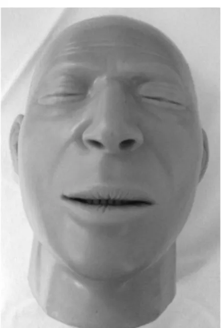
A special resin, in the shape of the anterior part of skull, constitute the basic structure of MAX sialendoscopy (►Fig. 1).
The model contains an articular joint of mandibular bone which allows the month opening (►Fig. 2). Inside the oral
cavity, it is possible to notice many realistic structures, such as tongue, teeth, uvula, and tonsils. The papillae of both parotid and submandibular glands are located in realistic sites.
Due to all this anatomical specificity, the surgeon can fully perform sialendoscopy procedure and techniques.
Results
The authors hereby present a training model to practice the sialendoscopy. It was possible to simulate all aspects of the procedure, from patient mouth opening to extracting stones from the salivary ducts.
The experienced surgeons could observe multiple opportunities for practice, such as dilatation of papillae (►Figs. 3and4), insertion of the scope (►Fig. 5), visualization Fig. 1 MAX simulator. Fig. 2 MAX with mouth openers.
Fig. 3 Dilatation of right parotid papilla. Fig. 4 Dilatation of left submandibular papilla.
International Archives of Otorhinolaryngology Vol. 21 No. 1/2017
Sialendoscopy Training: a Realistic Model Pascoto et al.
of stones (►Figs. 6 and 7), extraction of the stones with
grasping or baskets (►Fig. 8), andfinally, stone fragmentation
with holmium laser.
Discussion
The treatment of many salivary ducts’pathologies using the technique of sialendoscopy express a challenge for the sur-geon. All anatomical structures involved are delicate and once a rude movement is done, the entire procedure may be impaired. In any case, all instruments are delicate and the training to improve skills in handling them is the key for a successful sialendoscopy. In this context, the training based on surgical simulation is extremely helpful.
Many models have been created as the initial step in training residents and young surgeons to gain experience with surgical procedures.1,2
Previous investigators have reported class I data that supports the ability of simulators to improve performance in the operating room (OR).6–8
The use of realistic simulators can reduce the errors, maxi-mize the sensitivity in demonstrating skills transfer, and im-prove the learning curve prior to undertaking the real surgery.1
In this respect, our MAX sialendoscopy simulator is not only thefirst realistic simulator available for sialendoscopy, but also provides an alternative to the use of animal models and human cadaveric specimens.
Moreover, this simulator presents other advantages:
1. The material maintenance is convenient, and there is no need for any kind of special preparation. It is easier to maintain when compared with animal models.
2. The model expresses the appropriate size and the dimen-sions observed in salivary ducts. Thereby, the identifi ca-tion of anatomical landmarks and, more importantly, the depth perception is feasible.
3. This real simulator includes all necessary planning steps to perform sialendoscopy.
4. The ability to obtain realistic imaging studies provides knowledge of the pathological condition and its diagnosis. 5. There are not ethical restrictions to its use or the need for a
specific place to work with the simulator.
6. “Easy logistics”: possibility to organize courses and send models worldwide - no need for special care and/or legal authorizations.
Fig. 5 Main salivary duct vision. Fig. 6 Floating stone in the salivary duct.
Fig. 7 Stone trapped. Fig. 8 Using a basket to remove the stone.
On the other hand, it is important to emphasize some disadvantages of this simulator. Although the handmade and hand-tuned process of developing such a simulator has the most preferable characteristics (to represent the tissue properties adequately and to express the anatomical structures with precise localization), it can be difficult and extremely time consuming to create.
Another factor that must be considered is the estimated cost for each surgical unit. The single pig cranium, when compared with this realistic simulator, is far less expensive.
However, we stress that the benefits of such models, including their reproducibility of emergency situations, may be superior in their ability to augment the training of surgical residents. Prospective studies will be necessary to validate the effectiveness of this training model.
Conclusion
This report not only represents thefirst description of the anatomical model for sialendoscopy training but also rein-forces its promising prospects. It should be considerably useful to abbreviate the learning curve during the qualifi ca-tion of young surgeons while minimizing the consequences of technical errors.
Future studies will address the scientific validation using well-defined performance measures, possibly followed by integration of this new educational tool into the otorhinolar-yngology curriculum. We believe that this teaching method
will provide the ability to train out the learning curve for technical skills on a simulator rather than on a patient.
References
1 Coelho G, Warf B, Lyra M, Zanon N. Anatomical pediatric model for craniosynostosis surgical training. Childs Nerv Syst 2014;30(12): 2009–2014 10.1007/s00381-014-2537-x
2 Zymberg S, Vaz-Guimarães Filho F, Lyra M. Neuroendoscopic training: presentation of a new real simulator. Minim Invasive Neurosurg 2010;53(1):44–46 10.1055/s-0029-1246169 3 Ziegler CM, Steveling H, Seubert M, Mühling J. Endoscopy: a
mini-mally invasive procedure for diagnosis and treatment of diseases of the salivary glands. Six years of practical experience. Br J Oral Maxillofac Surg 2004;42(1):1–7 10.1016/s0266-4356(03)00188-8 4 Nakayama Y, Aoki Y, Niitsu H, Saigusa K. Forced oral opening for
cadavers with rigor mortis: two approaches for the myotomy on the temporal muscles. Forensic Sci Int 2001;118(1):37–42 5 Shapiro HA. Rigor mortis. BMJ 1950;2(4673):304
6 Lehmann KS, Ritz JP, Maass H, et al. A prospective randomized study to test the transfer of basic psychomotor skills from virtual reality to physical reality in a comparable training setting. Ann Surg 2005;241(3):442–449
7 Palter VN, Grantcharov T, Harvey A, Macrae HM. Ex vivo technical skills training transfers to the operating room and enhances cognitive learning: a randomized controlled trial. Ann Surg 2011;253(5):886–889
8 Seymour NE, Gallagher AG, Roman SA, et al. Virtual reality training improves operating room performance: results of a randomized, double-blinded study. Ann Surg 2002;236(4):458–463, discussion 463–464
International Archives of Otorhinolaryngology Vol. 21 No. 1/2017
Sialendoscopy Training: a Realistic Model Pascoto et al.