Clinicopathologic Analysis of Localized Nasal/Paranasal
Diffuse Large B-Cell Lymphoma
Hiroko Toda1, Yasuharu Sato1*, Katsuyoshi Takata1, Yorihisa Orita2, Naoko Asano3, Tadashi Yoshino1
1Department of Pathology, Okayama University Graduate School of Medicine, Dentistry, and Pharmaceutical Sciences, Okayama, Japan,2Department of Otolaryngology, Head and Neck Surgery, Okayama University Graduate School of Medicine, Dentistry, and Pharmaceutical Sciences, Okayama, Japan,3Department of Clinical Laboratory, Nagoya University Hospital, Nagoya, Japan
Abstract
Diffuse large B-cell lymphoma (DLBCL) comprises 2 molecularly distinct subgroups of non-germinal center B-cell-like (non-GCB) and germinal center B-cell-like ((non-GCB) DLBCLs, with the former showing relatively poor prognosis. In the present study, we analyzed the clinicopathological features of 39 patients with localized nasal/paranasal DLBCL. Immunohistochemistry-based subclassification revealed that 11 patients (28%) were of the GCB-type according to Hans’ algorithm and 11 (28%) were of the GCB-type according to Choi’s algorithm. According to both Hans’ and Choi’s algorithms, the non-GCB type was predominant. Nevertheless, prognosis was good. Overall survival did not differ significantly between the GCB and non-GCB subgroups (Hans’ algorithm:p= 0.57, Choi’s algorithm: p= 0.99). Furthermore, the prognosis of localized nasal/paranasal DLBCL was better than that of other localized extranodal DLBCLs. The prognosis of extranodal DLBCL is usually considered poorer than that of nodal DLBCL. However, in our study, no difference was noted between patients with localized nasal/ paranasal DLBCL and patients with localized nodal DLBCL. In conclusion, although the non-GCB subtype is thought to show poor prognosis, in our study, the prognosis for localized nasal/paranasal DLBCL patients was good irrespective of subclassification.
Citation:Toda H, Sato Y, Takata K, Orita Y, Asano N, et al. (2013) Clinicopathologic Analysis of Localized Nasal/Paranasal Diffuse Large B-Cell Lymphoma. PLoS ONE 8(2): e57677. doi:10.1371/journal.pone.0057677
Editor:Joseph S. Pagano, The University of North Carolina at Chapel Hill, United States of America ReceivedJanuary 2, 2013;AcceptedJanuary 24, 2013;PublishedFebruary 28, 2013
Copyright:ß2013 Toda et al. This is an open-access article distributed under the terms of the Creative Commons Attribution License, which permits unrestricted use, distribution, and reproduction in any medium, provided the original author and source are credited.
Funding:The authors have no support or funding to report.
Competing Interests:The authors have declared that no competing interests exist. * E-mail: [email protected]
Introduction
Although heterogeneous in nature, diffuse large B-cell lympho-ma (DLBCL) can be classified into 2 distinct subtypes on the basis of genetic profiling: the germinal center B-cell-like (GCB) phenotype and the non-germinal center B-cell-like (non-GCB) phenotype [1,2,3]. Notably, patients belonging to the former group have a better prognosis than those belonging to the latter group. Hans et al. reported that these DLBCL subtypes can be easily distinguished on the basis of immunohistological staining for CD10, BCL6, and MUM1 proteins [2]. Later, Choi et al. added 2 new antibodies, FoxP1 and GCET1 [4], and Choi’s algorithm is reported to achieve better prognostic classification than Hans’ algorithm [5]. Extranodal non-GCB-like DLBCL is generally characterized by poor prognosis regardless of its localized disease, but localized primary non-tonsillar oral DLBCL exhibits favorable prognosis even in cases of the non-GCB subtype [6]. Nasal/ paranasal DLBCL is uncommon, and the GCB and non-GCB subtypes of this disease have not yet been examined. In this study, we aimed to clarify the clinicopathological features of localized nasal/paranasal DLBCL.
Materials and Methods
Patients
We selected 39 Japanese patients diagnosed with localized nasal/paranasal DLBCL between 1995 and 2010 and reviewed our institution’s pathology department database to obtain the
medical records of these patients. We only evaluated localized lymphomas, because the primary sites of advanced lymphomas are difficult to determine. All 39 cases were diagnosed as primary extranodal DLBCLs. Patients were defined as having extranodal DLBCL when the disease was confined to one or more extranodal sites and showed no (or only minor) nodal involve-ment after the staging procedures [7,8]. This group of patients was then compared with 39 patients with localized nodal DLBCLs diagnosed at our institution [9]. The samples and the medical records (clinical history, treatment and survival data) used in our study was approved by the Institute Review Board (IRB) at Okayama University. Written informed consent was waived by our institutional review board, since our study was limited to the use of excess human tissue samples and medical records.
Histological Examination and Immunohistochemistry Surgically resected or biopsied specimens of localized nasal/ paranasal DLBCLs were fixed in 10% formaldehyde and
embedded in paraffin. Serial sections (4mm) were cut from each
1:200; Novocastra), BCL6 (D8, 1:100; SantaCruz), CD5 (4C7, 1:100; Novocastra), GCET1 (RAM341, 1:100; Abcam), CD10 (56C6, 1:50; Novocastra), MUM1 (MUM1p, 1:50; Dako), FoxP1 (JC12, 1:500; LifeSpan Biosciences, Seattle, USA), and Ki-67 (MIB-1, 1:5000; Novocastra). For each section, 10 high-power fields were recorded, quantitated, and averaged to calculate the estimated percentage of positively immunostained cells.
Negativ-ity for CD10, BCL6, and MUM1 was defined as ,30%
positively stained tumor cells, and positivity was defined as
.30% positively stained tumor cells. As an exception, for Choi’s
algorithm, negativity for MUM1 was defined as,80% positively
stained tumor cells, and positivity, as .80% positively stained
tumor cells. Negativity for GCET1 and FoxP1 staining was
defined as,80% positively stained tumor cells, and positivity, as
.80% positively stained tumor cells. Ki-67 immunoreactivity
was evaluated semi-quantitatively by using the average estimated percentage of positive cells in the 10 recorded high-power fields [4].
Statistical Analysis
All data were analyzed using STATA software (version 9.0; Stata Corp., College Station, TX, USA). Actuarial overall survival curves were calculated using the Kaplan-Meier method, and differences were examined using the log-rank test to determine significant prognostic factors [10]. Overall survival was defined as the time from diagnosis to death from any cause or to the last follow-up visit.
Results
Characteristics of the Nasal/Paranasal DLBCL Cases Table 1 and Table 2 summarize the characteristics of the localized nasal/paranasal DLBCL patients. The median age of the 39 patients was 76 years (range, 33–98 years). The patient population comprised 21 men and 18 women. According to Choi’s algorithm, 11 of the 39 patients (28%) were of the GCB-type and 28 (72%) were of the non-GCB- GCB-type. According to Hans’ algorithm, 11 (28%) were of the GCB- type and 28 of the 39 patients (72%) were of the non-GCB type (Table 3). According to the Ann Arbor classification, 33 patients were at clinical stage IE and 6 were at stage IIE. According to the
International Prognostic Index, 4 patients were at low-
interme-diate risk and 13 were at low risk. Histologically, all cases were
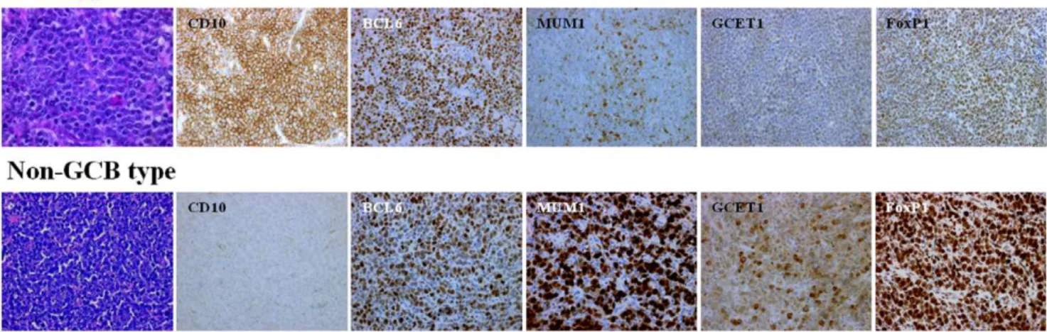
classified as DLBCL (Fig. 1). All patients were newly presenting with no prior treatment history.
Phenotypic Features of the Localized Nasal/Paranasal DLBCL Cases
Table 4 summarizes the phenotypic features of the localized nasal/paranasal DLBCL patients. The B-cell immunophenotype of the lymphomas was confirmed by immunoreactivity with antibodies to CD20 in 39 cases. Although no cases were positive for CD5, 8 (21%) were positive for CD10 and 25 (64%) were positive for BCL6. For MUM1 staining, 28 cases (72%) were positive according to Hans’ algorithm and 24 cases (62%) were positive according to Choi’s algorithm. Furthermore, 29 cases (74%) were positive for FoxP1 and 12 (31%) were positive for GCET1. Of the 11 cases (28%) classified as GCB- type according to Hans’ algorithm, 8 were CD10-positive cases (20%), and 3 were CD10-negative, BCL6-positive, MUM1-negative cases (8%). Of the 11 (28%) classified as GCB- type according to Choi’s algorithm, 3 were GCET1-positive, MUM1-negative cases (8%); 5 were GCET1-negative, CD10-positive cases (13%); and 3 GCET1-negative, CD10-negative, BCL6-positive, FoxP1-negative cases (8%). Of the 28 cases (72%) classified as the non-GCB- type according to Hans’ algorithm, 12 were CD10-negative, BCL6-negative cases (31%) and 16 were CD10-BCL6-negative, BCL6-positive, MUM1-positive cases (41%). Of the 28 cases (72%) classified as the non-GCB- type according to Choi’s algorithm, 9 were positive, MUM1-positive cases (23%); 8 were GCET1-negative, CD10-GCET1-negative, BCL6-positive, FoxP1-positive cases (20%); and 11 were GCET1-negative, CD10-negative, BCL6-negative cases (28%) (Fig. 2). The non-GCB- type was dominant according to both algorithms, but the prognosis for these cases was good. Overall survival did not differ significantly between the
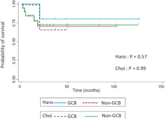
non-GCB type and non-GCB type groups (p =0.57, Hans’ algorithm,
p =0.99, Choi’s algorithm) (Fig. 3).
Therapeutic Response and Outcome
Follow-up clinical data were available for 28 patients. The duration of follow-up ranged from 1 to 125 months (mean, 29 months). Fifteen patients were initially treated with chemother-apy plus irradiation, 9 were treated with chemotherchemother-apy alone, and 2 were treated with irradiation alone. Twenty-two patients achieved complete remission. Although 7 patients relapsed, 3 of
Figure 1. Histological and immunohistochemical features.Diffuse infiltration and proliferation of large lymphoma cells (Hematoxylin–eosin staining).
doi:10.1371/journal.pone.0057677.g001
Localized Nasal/Paranasal DLBCL
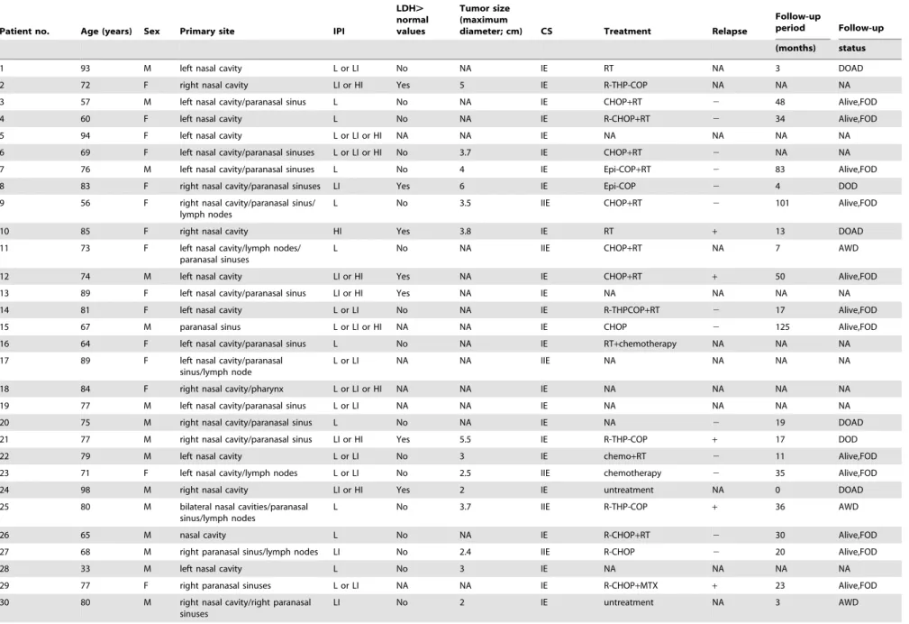
Table 1.Clinical data for 39 localized nasal/paranasal DLBCLs.
Patient no. Age (years) Sex Primary site IPI
LDH. normal values
Tumor size (maximum
diameter; cm) CS Treatment Relapse
Follow-up
period Follow-up
(months) status
1 93 M left nasal cavity L or LI No NA IE RT NA 3 DOAD
2 72 F right nasal cavity LI or HI Yes 5 IE R-THP-COP NA NA NA
3 57 M left nasal cavity/paranasal sinus L No NA IE CHOP+RT 2 48 Alive,FOD
4 60 F left nasal cavity L No NA IE R-CHOP+RT 2 34 Alive,FOD
5 94 F left nasal cavity L or LI or HI NA NA IE NA NA NA NA
6 69 F left nasal cavity/paranasal sinuses L or LI or HI No 3.7 IE CHOP+RT 2 NA NA
7 76 M left nasal cavity/paranasal sinuses L No 4 IE Epi-COP+RT 2 83 Alive,FOD
8 83 F right nasal cavity/paranasal sinuses LI Yes 6 IE Epi-COP 2 4 DOD
9 56 F right nasal cavity/paranasal sinus/
lymph nodes
L No 3.5 IIE CHOP+RT 2 101 Alive,FOD
10 85 F right nasal cavity HI Yes 3.8 IE RT + 13 DOAD
11 73 F left nasal cavity/lymph nodes/
paranasal sinuses
L No NA IIE CHOP+RT NA 7 AWD
12 74 M left nasal cavity LI or HI Yes NA IE CHOP+RT + 50 Alive,FOD
13 89 F left nasal cavity/paranasal sinus LI or HI Yes NA IE NA NA NA NA
14 81 F left nasal cavity L or LI No NA IE R-THPCOP+RT 2 17 Alive,FOD
15 67 M paranasal sinus L or LI or HI NA NA IE CHOP 2 125 Alive,FOD
16 64 F left nasal cavity/paranasal sinus L No NA IE RT+chemotherapy NA NA NA
17 89 F left nasal cavity/paranasal
sinus/lymph node
L or LI NA NA IIE NA NA NA NA
18 84 F right nasal cavity/pharynx L or LI or HI NA NA IE NA NA NA NA
19 77 M left nasal cavity/paranasal sinus L or LI NA NA IE NA NA NA NA
20 75 M right nasal cavity/paranasal sinus L No NA IE NA 2 19 DOAD
21 77 M right nasal cavity/paranasal sinus LI or HI Yes 5.5 IE R-THP-COP + 17 DOD
22 79 M left nasal cavity L or LI No 3 IE chemo+RT 2 11 Alive,FOD
23 71 F left nasal cavity/lymph nodes L or LI No 2.5 IIE chemotherapy 2 35 Alive,FOD
24 98 M right nasal cavity LI or HI Yes 2 IE untreatment NA 0 DOAD
25 80 M bilateral nasal cavities/paranasal
sinus/lymph nodes
L No 3.7 IIE R-THP-COP + 36 AWD
26 65 M nasal cavity L No NA IE R-CHOP+RT 2 30 Alive,FOD
27 68 M right paranasal sinus/lymph nodes LI No 2.4 IIE R-CHOP 2 20 Alive,FOD
28 33 M left nasal cavity L No 3 IE NA NA NA NA
29 77 F right paranasal sinuses L or LI NA NA IE R-CHOP+MTX + 23 Alive,FOD
Localized
Nasal/Pa
ranasal
DLBCL
ONE
|
www.ploson
e.org
3
February
2013
|
Volume
8
|
Issue
2
|
these patients achieved complete remission following alternative chemotherapy. Salvage treatments for the 7 relapsed patients were R-MFP (methotrexate, fluorouracil, low dose cisplatin, and rituximab), R-THP-COP (cyclophosphamide, vincristine, pred-nisolone, pirarubicin, and rituximab), R-MTX (methotrexate and rituximab), and CHASER (cyclophosphamide, high dose cytar-abine, dexamethasone, etoposide, and rituximab) plus radiation. At the time of reporting, 16 patients were disease- free and 3 patients had died of the disease.
Comparison of the Clinicopathological Characteristics of Localized Nasal/Paranasal DLBCL and Localized Nodal DLBCL
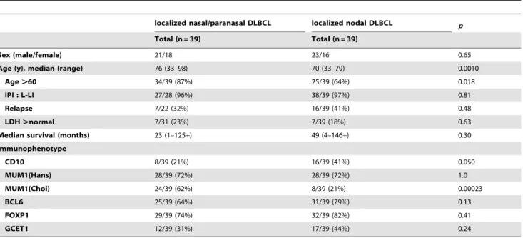
The clinicopathological characteristics of nasal/paranasal DLBCL and localized nodal DLBCL are summarized in Table 5. Nasal/paranasal DLBCL patients showed good prognosis. The slight difference in the overall survival between these patients and
patients with localized nodal DLBCL was not significant (p =0.30)
(Fig. 4). Moreover, analysis using thex2-test revealed a significant
difference between the 2 groups with regard to age distribution
and immunophenotype. Localized nasal/paranasal DLBCL
patients were more likely to be more than 60 years old than
localized nodal DLBCL patients (p =0.018). In addition, localized
nasal/paranasal DLBCL patients showed significantly higher positivity for MUM1 than localized nodal DLBCL patients
according to Choi’s algorithm (p =0.00023,x2-test).
Table 1. Cont. Patient no. Age (years) Sex Primary site IPI LDH . normal values Tumor size (maximum diameter; c m) CS Treatment Relapse Follow-up period Follow-up (months) status 31 78 F right nasal cavity L o r LI o r H I N A 2.5 IE NA NA NA NA 32 72 M left p aranasal sinuses L o r LI N o 5 IE NA NA NA NA 33 77 M right nasal cavity L o r LI o r H I N A N A IE R -THP-COP + RT + 23 AWD 34 76 M left n asal cavity/paranasal sinuses L N o 4 IE R-THP-COP + RT 2 9 Alive,FOD 35 78 F right nasal cavity L o r LI N o N A IE untreatment NA 2 AWD 36 68 M left n asal cavity/paranasal sinuses L N o 4 IE R-CHOP NA 1 DOD 37 74 F left n asal cavity L o r LI N o N A IE R -CHOP + RT 2 12 Alive,FOD 38 57 M right paranasal sinuses L N o 3 IE NA NA 27 Alive,FOD 39 83 M left n asal cavity/paranasal sinus LI No 3.6 IE R-THP-COP + RT + 48 Alive,FOD CHOP, cyclophospham ide, adriamycin, vincristine, prednisolone; CS, clinical stage; F, female; FOD, free of disease; AWD, alive with disease; DOD, dead of disease; DOAD, dead of another disease; IPI, International Prognostic Index; L, low; LDH, lactate dehydrogenase ; LI, low–intermediate; M, male; NA, not available; MTX, methotrexate; R-, w ith rituximab; R T, radiation; THP-COP, pirarubicin, cyclophosphamide, vincristine, predniso lone; Epi-COP, epirubicin, cyclophosphamide, vincristine, prednisolone. doi:10.1371/journal.pone. 0057677.t001
Table 2.Clinical characteristics of patients with localized nasal/paranasal DLBCL.
Characteristic No. of patients (%)
Sex
Male 21(54)
Female 18(46)
Age (y), median (range) 76 (33–98) Ann Arbor stage
I 33(85) II 6(15) LDH Normal Elevated 24(77) 7(23) PS 0–1 15(79)
2 or more 4(21)
IPI
L-LI 27(96)
HI-H 1(4)
Treatment
chemotherapy 8(32)
chemotherapy+RT 15(60)
RT alone 2(8)
Complete Remission
yes 22(88)
no 3(12)
doi:10.1371/journal.pone.0057677.t002
Localized Nasal/Paranasal DLBCL
Discussion
DLBCL is the most frequent and aggressive lymphoma,
representing a heterogeneous group that includesde novolarge
B-cell lymphomas, as well as transformed lymphomas from follicular or mucosa-associated lymphoid tissue lymphomas [11]. Recent studies have demonstrated that DLBCL can be further subclassified into 2 major prognostic categories according to Hans et al.: the GCB- type and the non-GCB- type [1,2].
However, Hans’ algorithm has been superseded by a new algorithm devised by Choi et al., and results obtained using Choi’s algorithm closely correlate with those of gene expression profiling for predicting prognosis [5]. In general, the non-GCB-type of DLBCL is associated with a significantly poorer prognosis than the GCB- type [1]; however, it has recently been established that this may not be true for extranodal DLBCL. Patients with localized primary non-tonsillar oral DLBCL presented with a
Figure 2. Distribution of GCB and non-GCB type according to Hans et al. and Choi et al. doi:10.1371/journal.pone.0057677.g002
Table 3.Clinical and phenotypic characteristics of patients with GCB-type and non-GCB-type DLBCL.
Total (n = 39) Hans’ algorithm P Choi’ algorithm p
GCB (n = 11) non-GCB (n = 28) GCB (n = 11) non-GCB (n = 28)
Sex (male/female) 21/18 6/5 15/13 0.96 6/5 14/14 0.80
Age (y), median (range) 76 (33–98) 75 (57–98) 77 (33–94) 0.61 77 (57–98) 76 (33–94) 0.98
Age.60 34/39 (87%) 10/11 (91%) 24/28 (86%) 0.66 10/11 (91%) 24/28 (86%) 0.66
PS.1 4/19 (21%) 0/5 (0%) 4/14 (29%) 0.18 0/5 (0%) 4/14 (29%) 0.18
B symptoms 1/29 (3%) 1/8 (13%) 0/21 (0%) 0.099 1/8 (13%) 0/21 (0%) 0.099
LDH.normal 7/31 (23%) 2/8 (25%) 5/23 (22%) 0.85 2/9 (22%) 5/22 (23%) 0.98 Median survival
(months)
23 (1–125+) 35 (11–125+) 20 (1–101+) 0.57 35 (11–48+) 23 (1–125+) 0.99
Immunophenotype
CD10 8/39 (21%) 8/11 (73%) 0/28 (0%) ,0.0001 6/11 (55%) 2/28 (7%) 0.00097
MUM1(Hans) 28/39 (72%) 5/11 (45%) 23/28 (82%) 0.022
MUM1(Choi) 24/39 (62%) 3/11 (27%) 21/28 (75%) 0.0058
BCL6 25/39 (64%) 9/11 (82%) 16/28 (57%) 0.15 10/11 (91%) 15/28 (54%) 0.029
FOXP1 29/39 (74%) 6/11 (55%) 23/28 (82%) 0.076
GCET1 12/39 (31%) 3/11 (27%) 9/28 (32%) 0.77
Abbreviations: GCB, germinal center B-cell; PS, performance status; BM, bone marrow; LDH, lactate dehydrogenase; FOXP1, forehead box protein 1; GCET1, germinal center B-cell expressed transcript 1.
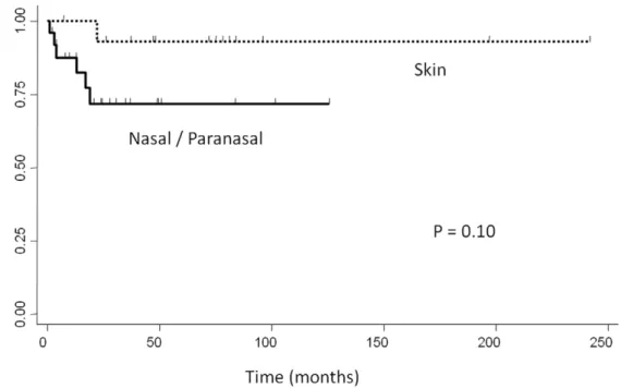
favorable clinical course despite having the non-GCB- type [6]. Similarly, in our study, the non-GCB- type of localized nasal/ paranasal DLBCL was the dominant type following subclassifi-cation according to both algorithms, but the prognosis of these patients was good. Moreover, the prognosis of localized nasal/ paranasal DLBCL was as good as that of primary cutaneous
DLBCL [12] (p =0.10) (Fig. 5) and was statistically better than
that of other localized extranodal DLBCLs (CNS [13], testis
[14], and adrenal gland [15]) (p =0.0002, p =0.0012, and
p =0.0044, respectively) (Fig. 6). Generally, extranodal
non-GCB-like DLBCLs are characterized by poor prognosis, and the incidence of non-GCB- type DLBCLs among extranodal DLBCLs is 83–100%, although this value differs according to the organ of manifestation [16,17,18,19]. According to previous reports, DLBCLs of the central nervous system [16], breast [17], stomach [20], leg type [21], testis [18], and intravascular type [19] are predominantly of the non-GCB- type, an observation consistent with the finding in our study of localized nasal/ paranasal DLBCL cases. However, patients with CNS, breast, and testicular DLBCL exhibit poor prognosis, regardless of the localized disease [16,17,18,22]. As shown for primary cutaneous B-cell lymphoma, findings of genes expression analysis suggest that primary non-leg-type cutaneous DLBCL and primary cutaneous DLBCL, leg type have expression profiles similar to those of GCB- type and non-GCB- type DLBCLs, respectively [23]. Therefore, primary non-leg-type cutaneous DLBCL is predominantly associated with an excellent prognosis [12,24]. According to the recent World Health Organization (WHO) classification, subsets of DLBCLs arising in peculiar extranodal sites have been categorized as distinct disease subgroups (primary
DLBCLs of the CNS, primary cutaneous DLBCLs, leg-type) or as distinct disease entities (primary mediastinal large B-cell lymphoma), on the basis of specific clinical and/or pathologic features [25,26]. When the cases in our study are included, extranodal disease is common among DLBCL patients [27]. It is thought that there are important clinical differences between nodal and extranodal DLBCL and that the most reliable distinction can be made in patients with stage I disease. For these patients, extranodal DLBCL is independently associated with poor survival [27]. Therefore, we also compared the clinicopathological profiles of localized nasal/paranasal DLBCLs with localized nodal DLBCLs. This analysis showed that localized nasal/paranasal DLBCL was associated with good prognosis and no difference was noted in the prognosis compared with localized nodal DLBCL. In recent years, the use of rituximab has improved the prognosis of DLBCL patients, and CHOP (cyclophosphamide, adriamycin, vincristine, and prednis-olone) therapy combined with rituximab (R-CHOP) is currently a standard chemotherapy for DLBCL [28]. In our study, no significant difference was noted in the number of patients treated with rituximab between localized nasal/paranasal DLBCL and
localized nodal DLBCL patients (p =0.24). Therefore, the
prognosis of localized nasal/paranasal DLBCLs was favorable regardless of treatment with rituximab. In conclusion, the prognosis of localized nasal/paranasal DLBCL patients was good irrespective of the disease subclassification, although the non-GCB- type of DLBCLs are usually thought to be associated with a poor prognosis.
Figure 3. Kaplan–Meier plot showing overall survival for patients with localized nasal/paranasal DLBCL. doi:10.1371/journal.pone.0057677.g003
Localized Nasal/Paranasal DLBCL
Table 4.Immunohistochemical findings of localized nasal/paranasal DLBCLs.
Patient
no. CD3 CD5 CD10 CD20
Ki-67 labeling (%)
MUM1 (Hans)
MUM1
(Choi) BCL6 EBER FOXP1 GCET1
subtype (Hans)
subtype (Choi)
1 2 2 2 + 21 U.D. 2 2 2 + 2 Non-GCB Non-GCB
2 2 2 p+ + 43 2 2 + 2 + 2 GCB GCB
3 2 2 + + 43 2 2 + 2 2 2 GCB GCB
4 2 2 2 + 80 + + 2 2 + 2 Non-GCB Non-GCB
5 2 2 2 + 59 + + 2 2 + 2 Non-GCB Non-GCB
6 2 2 2 + 54 + + + 2 + 2 Non-GCB Non-GCB
7 2 2 2 + 71 + + + 2 + 2 Non-GCB Non-GCB
8 2 2 2 + 82 + + 2 2 + 2 Non-GCB Non-GCB
9 2 2 2 + 71 2 2 2 2 + 2 Non-GCB Non-GCB
10 2 2 2 + 61 + + + 2 2 + Non-GCB Non-GCB
11 2 2 2 + 50 + + + 2 + 2 Non-GCB Non-GCB
12 2 2 2 + 34 + + + 2 2 + Non-GCB Non-GCB
13 2 2 2 + 55 + + + 2 + + Non-GCB Non-GCB
14 2 2 2 + 70 + + + 2 + + Non-GCB Non-GCB
15 2 2 + + 64 + + 2 2 2 + GCB Non-GCB
16 2 2 2 + 33 2 2 + 2 2 + GCB GCB
17 2 2 2 + 61 + + 2 2 + + Non-GCB Non-GCB
18 2 2 2 + 72 + + 2 2 2 2 Non-GCB Non-GCB
19 2 2 2 + 63 + + + 2 2 2 Non-GCB GCB
20 2 2 2 + 67 2 2 + 2 2 2 GCB GCB
21 2 2 2 + 80 + + 2 2 2 2 Non-GCB Non-GCB
22 2 2 + + 80 + + 2 2 + 2 GCB GCB
23 2 2 + + 72 2 2 + 2 + + GCB GCB
24 2 2 + + 90 + + + 2 + 2 GCB GCB
25 2 2 2 + 73 2 2 + 2 + + GCB GCB
26 2 2 2 + 81 + + + 2 + 2 Non-GCB Non-GCB
27 2 2 2 + 52 + + + 2 + 2 Non-GCB Non-GCB
28 2 2 2 + 72 + 2 2 2 + 2 Non-GCB Non-GCB
29 2 2 + + 97 + + + 2 + + GCB Non-GCB
30 2 2 2 + 90 2 2 2 2 + 2 Non-GCB Non-GCB
31 2 2 + + 77 + 2 + 2 + 2 GCB GCB
32 2 2 2 + 49 2 2 2 2 + 2 Non-GCB Non-GCB
33 2 2 2 + 88 + + + 2 + + Non-GCB Non-GCB
34 2 2 2 + 54 + + + 2 + 2 Non-GCB Non-GCB
35 2 2 2 + 58 + 2 + 2 2 2 Non-GCB GCB
36 2 2 2 + 92 + 2 + 2 + 2 Non-GCB Non-GCB
37 2 2 2 + 61 2 + + 2 + 2 Non-GCB Non-GCB
38 2 2 2 + 90 + 2 2 2 + 2 Non-GCB Non-GCB
39 2 2 2 + 83 + + + 2 + + Non-GCB Non-GCB
Figure 4. Comparison of overall survival between localized nasal/paranasal DLBCL and localized nodal DLBCL. doi:10.1371/journal.pone.0057677.g004
Table 5.Clinical and phenotypic characteristics of patients with localized nasal/paranasal DLBCL and localized nodal DLBCL.
localized nasal/paranasal DLBCL localized nodal DLBCL p
Total (n = 39) Total (n = 39)
Sex (male/female) 21/18 23/16 0.65
Age (y), median (range) 76 (33–98) 70 (33–79) 0.0010
Age.60 34/39 (87%) 25/39 (64%) 0.018
IPI : L-LI 27/28 (96%) 38/39 (97%) 0.81
Relapse 7/22 (32%) 16/39 (41%) 0.48
LDH.normal 7/31 (23%) 7/39 (18%) 0.63
Median survival (months) 23 (1–125+) 49 (4–146+) 0.30
Immunophenotype
CD10 8/39 (21%) 16/39 (41%) 0.050
MUM1(Hans) 28/39 (72%) 28/39 (72%) 1.0
MUM1(Choi) 24/39 (62%) 8/39 (21%) 0.00023
BCL6 25/39 (64%) 31/39 (79%) 0.13
FOXP1 29/39 (74%) 32/39 (82%) 0.41
GCET1 12/39 (31%) 17/39 (44%) 0.24
Abbreviations: GCB, germinal center B-cell; PS, performance status; LDH, lactate dehydrogenase. doi:10.1371/journal.pone.0057677.t005
Localized Nasal/Paranasal DLBCL
Figure 5. Comparison of overall survival between localized nasal/paranasal DLBCL and localized skin DLBCL. doi:10.1371/journal.pone.0057677.g005
Figure 6. Comparison of overall survival between localized nasal/paranasal DLBCL and localized adrenal, CNS, and testicular DLBCLs.
Author Contributions
Conceived and designed the experiments: YS. Performed the experiments: HT YS KT TY. Analyzed the data: HT NA YS YO. Wrote the paper: HT YS TY.
References
1. Alizadeh AA, Eisen MB, Davis RE, Ma C, Lossos IS, et al. (2000) Distinct types of diffuse large B-cell lymphoma identified by gene expression profiling. Nature 403: 503–511.
2. Hans CP, Weisenburger DD, Greiner TC, Gascoyne RD, Delabie J, et al. (2004) Confirmation of the molecular classification of diffuse large B-cell lymphoma by immunohistochemistry using a tissue microarray. Blood 103: 275–282. 3. Rosenwald A, Wright G, Chan WC, Connors JM, Campo E, et al. (2002) The
use of molecular profiling to predict survival after chemotherapy for diffuse large-B-cell lymphoma. N Engl J Med 346: 1937–1947.
4. Choi WW, Weisenburger DD, Greiner TC, Piris MA, Banham AH, et al. (2009) A new immunostain algorithm classifies diffuse large B-cell lymphoma into molecular subtypes with high accuracy. Clin Cancer Res 15: 5494–5502. 5. Meyer PN, Fu K, Greiner TC, Smith LM, Delabie J, et al. (2011)
Immunohistochemical methods for predicting cell of origin and survival in patients with diffuse large B-cell lymphoma treated with rituximab. J Clin Oncol 29: 200–207.
6. Sato Y, Onishi N, Morito T, Takata K, Mizobuchi K, et al. (2009) Patients with localized primary non-tonsillar oral diffuse large B-cell lymphoma exhibit favorable prognosis despite a non-germinal center B-cell-like phenotype. Cancer Sci 100: 42–46.
7. d’Amore F, Christensen BE, Brincker H, Pedersen NT, Thorling K, et al. (1991) Clinicopathological features and prognostic factors in extranodal non-Hodgkin lymphomas. Danish LYFO Study Group. Eur J Cancer 27: 1201–1208. 8. Krol AD, le Cessie S, Snijder S, Kluin-Nelemans JC, Kluin PM, et al. (2003)
Primary extranodal non-Hodgkin’s lymphoma (NHL): the impact of alternative definitions tested in the Comprehensive Cancer Centre West population-based NHL registry. Ann Oncol 14: 131–139.
9. Habara T, Sato Y, Takata K, Iwaki N, Okumura H, et al. (2012) Germinal Center B-Cell-Like versus Non-Germinal Center B-Cell-Like as Important Prognostic Factor for Localized Nodal DLBCL. J Clin Exp Hematop 52: 91–99. 10. Peto R, Pike MC, Armitage P, Breslow NE, Cox DR, et al. (1977) Design and analysis of randomized clinical trials requiring prolonged observation of each patient. II. analysis and examples. Br J Cancer 35: 1–39.
11. Pileri SA, Dirnhofer S, Went P, Ascani S, Sabattini E, et al. (2002) Diffuse large B-cell lymphoma: one or more entities? Present controversies and possible tools for its subclassification. Histopathology 41: 482–509.
12. Hembury TA, Lee B, Gascoyne RD, Macpherson N, Yang B, et al. (2002) Primary cutaneous diffuse large B-cell lymphoma: a clinicopathologic study of 15 cases. Am J Clin Pathol 117: 574–580.
13. Hattab EM, Martin SE, Al-Khatib SM, Kupsky WJ, Vance GH, et al. (2010) Most primary central nervous system diffuse large B-cell lymphomas occurring in immunocompetent individuals belong to the nongerminal center subtype: a retrospective analysis of 31 cases. Mod Pathol 23: 235–243.
14. Li D, Xie P, Mi C (2010) Primary testicular diffuse large B-cell lymphoma shows an activated B-cell-like phenotype. Pathol Res Pract 206: 611–615.
15. Mozos A, Ye H, Chuang WY, Chu JS, Huang WT, et al. (2009) Most primary adrenal lymphomas are diffuse large B-cell lymphomas with non-germinal center B-cell phenotype, BCL6 gene rearrangement and poor prognosis. Mod Pathol 22: 1210–1217.
16. Camilleri -Broe¨t S, Crinie`re E, Broe¨t P, Delwail V, Mokhtari K, et al. (2006) A uniform activated B-cell-like immunophenotype might explain the poor prognosis of primary central nervous system lymphomas: analysis of 83 cases. Blood 107: 190–196.
17. Yoshida S, Nakamura N, Sasaki Y, Yoshida S, Yasuda M, et al. (2005) Primary breast diffuse large B-cell lymphoma shows a non-germinal center B-cell phenotype. Mod Pathol 18: 398–405.
18. Al-Abbadi MA, Hattab EM, Tarawneh MS, Amr SS, Orazi A, et al. (2006) Primary testicular diffuse large B-cell lymphoma belongs to the nongerminal center B-cell-like subgroup: A study of 18 cases. Mod Pathol 19: 1521–1527. 19. Murase T, Yamaguchi M, Suzuki R, Okamoto M, Sato Y, et al. (2007)
Intravascular large B-cell lymphoma (IVLBCL): a clinicopathologic study of 96 cases with special reference to the immunophenotypic heterogeneity of CD5. Blood 109: 478–485.
20. Connor J, Ashton-Key M (2007) Gastric and intestinal diffuse large B-cell lymphomas are clinically and immunophenotypically different. An immunohis-tochemical and clinical study. Histopathology 51: 697–703.
21. Campo E, Chott A, Kinney MC, Leoncini L, Meijer CJ, et al. (2006) Update on extranodal lymphomas. Conclusions of the Workshop held by the EAHP and the SH in Thessaloniki, Greece. Histopathology 48: 481–504.
22. Zucca E, Conconi A, Mughal TI, Sarris AH, Seymour JF, et al. (2003) Patterns of outcome and prognostic factors in primary large-cell lymphoma of the testis in a survey by the International Extranodal Lymphoma Study Group. J Clin Oncol 21: 20–27.
23. Hoefnagel JJ, Dijkman R, Basso K, Jansen PM, Hallermann C, et al. (2005) Distinct types of primary cutaneous large B-cell lymphoma identified by gene expression profiling. Blood 105: 3671–3678.
24. Vural F, Saydam G, Cagirgan S, Ertekin B, Hekimgil M, et al. (2008) Primary cutaneous B-cell lymphoma: report of eight cases and review of the literature. Int J Dermatol 47: 675–680.
25. Deckert M, Engert A, Bru¨ck W, Ferreri AJ, Finke J, et al. (2011) Modern concepts in the biology, diagnosis, differential diagnosis and treatment of primary central nervous system lymphoma. Leukemia 25: 1797–1807. 26. Steidl C, Gascoyne RD (2011) The molecular pathogenesis of primary
mediastinal large B-cell lymphoma. Blood 118: 2659–2669.
27. Møller MB, Pedersen NT, Christensen BE (2004) Diffuse large B-cell lymphoma: clinical implications of extranodal versus nodal presentation–a population-based study of 1575 cases. Br J Haematol 124: 151–159. 28. Yamaguchi H, Hirakawa T, Inokuchi K (2011) Importance of relative dose
intensity in chemotherapy for diffuse large B-cell lymphoma. J Clin Exp Hematop 51: 1–5.
Localized Nasal/Paranasal DLBCL