Universidade Nova de Lisboa
Instituto de Higiene e Medicina Tropical
Regulation of the apoptosis pathway in
Rhipicephalus annulatus
ticks by the protozoan
Babesia bigemina
Catarina Sofia Bento Monteiro
DISSERTAÇÃO PARA A OBTENÇÃO DO GRAU DE MESTRE EM PARASITOLOGIA MÉDICA
Universidade Nova de Lisboa
Instituto de Higiene e Medicina Tropical
Regulation of the apoptosis pathway in
Rhipicephalus
annulatus
ticks by the protozoan
Babesia bigemina
Autor: Catarina Sofia Bento Monteiro
Orientador: Doutora Ana Isabel Amaro Gonçalves Domingos (IHMT, UNL)
Coorientador: Doutora Sandra Isabel da Conceição Antunes (IHMT, UNL)
i The results obtained during the development of this master project were reported at two international conferences:
Monteiro, C, Domingos, A, and Antunes, S 2017, ´Regulation of the apoptosis pathway in Rhipicephalus annulatus ticks by the protozoan Babesia bigemina´, COST action:
EuroNegVec, 11-13 September, Crete, Greece.
Monteiro, C, Domingos, A, and Antunes, S 2017 ´Is the apoptosis pathway in
ii
Acknowledgments
Quero, desde já, agradecer à Doutora Ana Domingos por se ter disponibilizado a orientar-me e por não ter desistido de mim.
À Doutora Sandra Antunes, pelo apoio constante e compreensão durante toda esta longa jornada. Admiro a simplicidade com que encaras a vida.
À Joana Couto pela boa disposição e prontidão em ajudar. És das pessoas mais genuínas que já conheci.
À Joana Ferrolho com quem partilho o mesmo gosto pela medicina veterinária. A vida até nos pode derrubar, mas nós temos a opção de nos voltar a erguer.
À Ana Rita pelos desabafos e conselhos.
À Samira, Gustavo, Martim, Catarina e Filipa com quem me cruzei durante este percurso.
À Cláudia, Filipa e Inês com quem partilhei o primeiro ano de mestrado e continuo a partilhar bons momentos.
Ao Samuel e Carrusca, com quem continuei a partilhar a vida académica no IHMT.
À Bianca, David, Pedro e Rodrigo pela partilha da vida académica e amizade.
À Estrela, por poder contar sempre contigo, tanto nos bons como maus momentos.
À team ZFortes, sem a qual estes últimos tempos teriam sido bem mais difíceis.
Aos restantes amigos pela partilha de bons momentos, sempre com boa disposição.
iii
Resumo
Regulação da via de apoptose em carraças Rhipicephalus annulatus pelo protozoário Babesia bigemina
Palavras-chave: Apoptose, Babesia, Carraça, Vacina.
As carraças são os vetores de agentes patogénicos com maior importância na área veterinária enquanto que em saúde humana aparecem em segundo, atrás dos mosquitos. Estes ectoparasitas hematófagos obrigatórios de vertebrados terrestres são capazes de transmitir um grande número de agentes patogénicos. Diferentes ixodídeos são responsáveis pela transmissão do protozoário Babesia, agente causal de babesiose numa
ampla variedade de animais incluindo humanos. A carraça Rhipicephalus annulatus,
considerado um dos mais importantes ectoparasitas do gado com grande impacto económico na produção animal, é o principal vetor de Babesia bigemina. O controlo de
carraças e agentes patogénicos transmitidos por carraças baseia-se sobretudo no uso de acaricidas. Contudo, a acumulação de resíduos químicos nos animais e produtos derivados de animais, bem como o aparecimento de carraças resistentes aos acaricidas e contaminação ambiental evidenciam a necessidade de desenvolver alternativas económica e ambientalmente seguras como as vacinas. A identificação e caracterização de antigénios com uma função essencial no desenvolvimento da carraça e/ou no “fitness” do parasita ainda estão a limitar o desenvolvimento de vacinas contra carraças e agentes patogénicos transmitidos por carraças. Focar as interações vetor-agente patogénico é uma abordagem que permite a identificação de antigénios de carraça com um possível efeito tanto na carraça como no agente patogénico. Estas interações têm sido ajustadas ao longo de uma coevolução duradoura: os agentes patogénicos invadem o hospedeiro e, as células hospedeiras respondem, forçando os agentes patogénicos a desenvolver novas estratégias moleculares para ultrapassar os seus mecanismos de defesa. Alguns agentes patogénicos transmitidos por carraças parecem ter desenvolvido estratégias para manipular diferentes processos metabólicos, como a apoptose, mecanismo de defesa celular baseado no sacrifício individual em beneficio do tecido ou organismo. Com estas premissas, o principal objetivo deste estudo foi a caracterização da via de apoptose durante a infeção por B. bigemina nas glândulas salivares das carraças, uma barreira que os agentes
patogénicos têm de ultrapassar e explorar para serem transmitidos com sucesso ao hospedeiro vertebrado. A informação sobre a regulação da apoptose por agentes patogénicos em carraças é escassa e nula no caso de Babesia spp. Assim, o sistema R. annulatus–B. bigemina foi usado para investigar se o parasita Babesia é capaz de afetar
o processo celular de apoptose. Com base num catálogo de sialotranscriptómica previamente obtido, seis genes apoptóticos de carraças foram selecionados para estudos quantitativos de expressão genética. Três genes pró-apoptóticos, DAP3, DAPK1 e VDAC
demonstraram estar significativamente diferenciadamente expressos na infeção por B. bigemina. Os três genes anti-apoptóticos AATF, BI-1 e API5 não mostraram expressão
diferencial significativa. No geral, os resultados sugerem que a Babesia pode ser capaz
iv
Abstract
Regulation of the apoptosis pathway in Rhipicephalus annulatus ticks by the
protozoan Babesia bigemina
Keywords: Apoptosis, Babesia, Tick, Vaccine.
Ticks are the vectors of pathogens of major importance in the veterinary area whereas in human health they appear in second, behind the mosquitoes. These obligate hematophagous ectoparasites of terrestrial vertebrates are capable of transmitting a large number of pathogens. Different ixodid ticks are responsible for the transmission of the protozoan Babesia, causal agent of babesiosis in a wide variety of animals including
humans. The tick Rhipicephalus annulatus, considered one of the most important
ectoparasites of cattle with great economic impact on animal production, is the main vector of Babesia bigemina. The control of ticks and tick-borne pathogens (TTBP) is
mainly based on the use of acaricides. However, the accumulation of chemical residues in animals and animal products, as well as the appearance of acaricide resistant ticks and environmental contamination evidence the need for the development of cost-effective and environmentally safe alternatives such as vaccines. Identification and characterization of antigens with a key role in the development of tick and/or parasite "fitness" are still limiting the development of anti-TTBP vaccines. Focusing the intricate vector-pathogen interactions is one approach that enables the identification of tick antigens with a potential effect on both tick and pathogen. These interactions have been adjusted through long-lasting coevolution: pathogens invade the host and host cells respond by forcing pathogens to develop new molecular strategies to bypass their defense mechanisms. Some tick-borne pathogens (TBP) appear to have developed strategies for manipulating different metabolic processes, such as apoptosis, a cellular defense mechanism based on individual sacrifice for the benefit of the tissue or organism. With these premises, the main objective of this study was the characterization of the apoptosis pathway during B. bigemina infection in the salivary glands (SG) of ticks, a barrier that pathogens have to
overcome and exploit to be successfully transmitted to the vertebrate host. Information regarding apoptosis regulation in ticks by pathogens is scarce and nothing is known in the case of Babesia spp. Thus, herein the system R. annulatus – B. bigemina was used to
investigate if Babesia parasite is able to affect the cellular process of apoptosis. Based on
a previously obtained sialotranscriptomic catalogue, six tick apoptotic genes were selected for quantitative gene expression studies. Three pro-apoptotic genes DAP3, DAPK1 and VDAC demonstrated to be significantly differentially expressed upon B. bigemina infection. The three anti-apoptotic genes AATF, BI-1 and API5 did not-show a
significant differential expression. Overall, the results suggest that Babesia may be able
v
Table of Contents
Introduction ... 1
1.1. Ticks ... 2
1.2. Rhipicephalus annulatus Life Cycle ... 4
1.3. Babesiosis ... 5
1.3.1.Babesia spp. Life Cycle ... 6
1.3.2.Babesia spp. - Vector Interactions: The Importance of Tick Salivary Glands in Infection ... 7
1.4. Apoptosis ... 9
1.5. Aims ... 10
Material and Methods ... 12
2.1. Rhipicephalus annulatus Ticks ... 13
2.2. Salivary Glands Extraction ... 13
2.3. RNA Extraction and cDNA Synthesis ... 14
2.4. Detection of Babesia bigemina in Ticks by Taq-man based qPCR ... 15
2.5. Selection of Apoptosis Pathway Target Genes ... 15
2.6. Gene Expression Analysis ... 16
2.6.1.qPCR Assay Optimization ... 16
2.6.2.qPCR Assay Design ... 16
2.6.3.Gene Expression Normalization... 17
2.7. Amplicon Identity Confirmation ... 17
Results ... 19
3.1. Salivary Glands Dissection and RNA Extraction ... 20
3.2. Babesia bigemina Infection Confirmation in Tick Salivary Glands ... 20
3.3. qPCR Optimization ... 21
3.4. Apoptotic Pathway Genes Differential Expression ... 22
3.5. Amplicon Identity Confirmation ... 25
Discussion ... 27
4.1. General Conclusions ... 34
vi
Index of Figures
Figure 1 Cornupalpatum burmanicum hard tick in a feather, conserved in an amber .... 2
Figure 2 One-host ixodid life cycle. Dorsal view of an engorged Rhipicehalus annulatus female ... 4
Figure 3 Babesia life cycle ... 7
Figure 4 Midgut and tick salivary glands importance during pathogen infection ... 8
Figure 5 Intrinsic and Extrinsic Cell Death Pathways ... 10
Figure 6 Tick salivary glands dissection steps ... 13
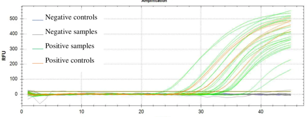
Figure 7 Detection of Babesia bigemina in Rhipicephalus annulatus salivary glands by TaqMan assay ... 21
Figure 8 Relative normalized expression of the apoptotic DAP3 gene in Rhipicephalus annulatus salivary glands ... 23
Figure 9 Relative normalized expression of the apoptotic DAPK1 gene in Rhipicephalus annulatus salivary glands ... 23
Figure 10 Relative normalized expression of the apoptotic VDAC gene in Rhipicephalus annulatus salivary glands ... 24
Figure 11 Relative normalized expression of the apoptotic AATF gene in Rhipicephalus annulatus salivary glands ... 24
Figure 12 Relative normalized expression of the apoptotic BI-1 gene in Rhipicephalus annulatus salivary glands ... 25
Figure 13 Relative normalized expression of the apoptotic API5 gene in Rhipicephalus annulatus salivary glands ... 25
Figure 14 Detailed apoptosis signalling pathway ... 46
Figure 15 Qualitative analysis of the RNA samples from the SG of the non-infected and Babesia bigemina-infected Rhipicephalus annulatus ticks ... 47
Figure 16 Confirmation of the DAPK1 and BI-1 amplicon size ... 48
Figure 17 Confirmation of the DAP3, AATF and VDAC amplicon size ... 48
vii
Index of Tables
Table 1 Primer sequences used for qPCR and respective optimal conditions ... 18
Table 2 Concentration of RNA extracted from R. annulatus salivary glands measure in Nanodrop ... 20
Table 3 Final amplification efficiencies of the references and target genes ... 22
Table 4 Study genes melting temperature ... 26
Table 5 Alignment of obtained amplicons with target genes ... 26
viii
Abbreviations
µl Microliter
µm Micrometre
µM Micromolar
AATF Apoptosis antagonizing transcription factor
AGE Agarose gel electrophoresis
AMPs Antimicrobial peptides
API5 Apoptosis inhibitor 5
BI-1 Bax inhibitor 1-related
BID BH3-interacting domain death agonist
bp Base pair
cDNA Complementary DNA
DAP3 Putative mitochondrial ribosome small subunit component mediator of apoptosis dap3
DAPK1 Death-associated protein kinase 1
DELE Death ligand signal enhancer
DISC Death-inducing signaling complex
DNA Deoxyribonucleic acid
DR Death receptors
dsDNA Double-stranded DNA
EDTA Ethylene diamine tetra acetic acid
ELF Elongation factor
ER Endoplasmic reticulum
g Gram
g G-force
i.v. Intravenous
IFA Indirect fluorescent antibodies
IFN Interferon
ix LIV Louping ill virus
MD midgut
min Minute
ml Millilitre
mM Millimolar
mRNA Messenger RNA
NGS Next-generation sequencing
nm Nanometer
ºC Celsius
PBS Phosphate-buffered saline
PCD Programed cell death
PCR Polymerase chain reaction
qPCR Real time PCR
RFU Relative fluorescence units
RNA Ribonucleic acid
RNAseq RNA sequencing
rRNA Ribosomal RNA
RT Reverse transcription
s Second s.s. sensu stricto
SG Salivary glands
spp. Species (plural)
TBD Tick-borne diseases
TBE Tris/Borate/EDTA
TBEV Tick-borne encephalitis virus
TBP Tick-borne pathogens
TNF Tumor necrosis factor
UV Ultraviolet
VDAC Voltage-dependent anion channel
2 1.1. Ticks
Ticks (Acari: Ixodida) are arthropod ectoparasites of a huge variety of terrestrial vertebrates comprising reptiles, birds, amphibians and mammals (including humans) (Anderson and Magnarelli, 2008; Hajdušek et al., 2013). Fossil records suggest that ticks
originated 65–146 million years ago in the Cretaceous period from the Mesozoic Era (Figure 1) (Klompen and Grimaldi, 2001; Nava et al., 2009). Currently, there are
approximately 900 tick species divided into three families – Argasidae, Ixodidae and Nutalliellidae, this last one with only one species (Estrada-Peña, 2015). These arthropods exert a direct physical injury as a result of their biting and blood feeding activities and may cause allergic reactions and paralysis. Besides that, ticks act as vectors being capable to transmit the widest spectrum of pathogens such as bacteria, viruses and protozoa. This versatility is related with the fact of the ticks are strict obligate hematophagous arachnids, which allows them to acquire, and therefore, multiply, maintain and transmit disease-causing pathogens to their hosts (Anderson and Magnarelli, 2008; Hajdušek et al., 2013).
Ticks are the most important arthropod vectors of animal diseases (Arthur, 1962) and second to mosquitos as vector of human diseases (de la Fuente et al., 2008; de la Fuente
et al., 2016c), whereby 10% of the known tick species represent high medical and
veterinary concern all around the world (de la Fuente et al.,2017), although their diversity
is greatest in tropical and subtropical regions (Anderson and Magnarelli, 2008; Hajdušek et al., 2013).
Tick-borne diseases (TBD) have been noticeable by the last few decades regarding their emergence, resurgence and expansion in Europe, Asia and North America (Mansfield et al., 2017), possibly due to the climate change, drug resistance, exploitation
of land resources, global movement of people and animals, among others (Colwell et al.,
3
2011). Tick-borne encephalitis, Lyme disease, human granulocytic anaplasmosis, for example, are transmittable to humans making TBD a global threat not only to livestock but also to human health. Babesiosis, anaplasmosis and theileriosis undermine cattle health, welfare and fitness thereby causing significant economic losses to the livestock
production (Hajdušek et al., 2013). In Portugal, five endemic TBD are described, as many
as in the other southern Mediterranean countries. These are the Lyme borreliosis, Q fever, Mediterranean spotted fever, tick-borne encephalitis and tick-borne relapsing fever (CEVDI Annual Report 2017).
Tick and TBD control in animals is mainly based on the use of chemical acaricides (Valle and Guerrero, 2018). Several disadvantages as the toxicity to animals and humans, resistance of ticks to acaricides and environmental contamination led to the development of vaccines, cost-effective and environmentally safe alternatives (Domingos et al., 2013).
The only commercially available anti-tick vaccine is based on the recombinant antigen Bm86. Developed in the 1990s, this vaccine results in the reduction of the number, weight and reproductive capacity of engorging female Rhipicephalus (Boophilus) microplus
(Canestrini, 1888) and Rhipicephalus (Boophilus) annulatus (Say, 1821), and
consequently in the reduction of cattle tick infestations and tick-borne pathogens (TBP) (de la Fuente et al., 2016b; Domingos et al., 2013). However, more efficient vaccines
need to be developed for the control of ticks and TBP since this vaccine has limited efficacy against all tick stages, different tick species and geographical R. microplus ticks.
Next-generation sequencing (NGS) represents a fundamental tool to the in-depth study of tick molecular dynamics and TBP interface. Transcriptomics, proteomics and functional genomics (García-García et al., 1999; Rodriguez-Valle et al., 2012; Valle and Guerrero,
2018) have brought new insight regarding this subject characterizing tick antigens that could become therapeutic targets.The study of the molecular interactions between ticks and pathogens may evidence key molecules that could be tested as potential vaccines that would target not only ticks but also the pathogens they harbour.
Babesia sp.-tick relationship is one of the most important in the veterinary field.
These intraerythrocytic apicomplexan organisms can be vectored by different ixodid tick and typically, different species are associated whit specific vertebrate and arthropod hosts. B.
bigemina and B. bovis are mainly vectored by R. microplus and R.annulatus ticks, specially
4
1.2. Rhipicephalus annulatus Life Cycle
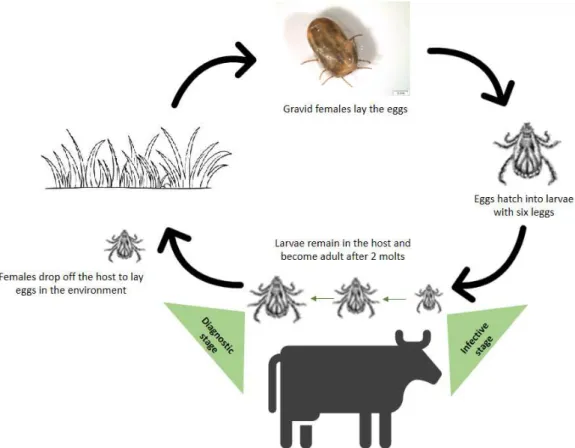
Ixodid ticks life cycle is a complex process of morphological and physiological modifications in the same individual encompassing four development stages - egg, larvae, nymph and adult (male and female). After egg hatching, the six-legged larvae emerge and a blood meal is required in order to moult to the next immature stage, the nymph. In the same way as larvae, eight-legged nymph need a blood meal to develop into adults. Ixodid ticks feed once in each active stage, being that the immature stages feed during 2.5 to 8 days while the adults feed 5 to 12 days. Mating also occurs in the host, followed by female drop-off. Fertilized females may ingest 100 to 120 times their body mass in blood, essential for the proper maturation of the eggs (Anderson and Magnarelli, 2008; Estrada-Peña, 2015).
Rhipicephalus annulatus are one-host ticks (Figure 2), i.e., all the stages develop
on only one host, except the eggs that are deposited in suitable microenvironments (Figure 2). These ticks are part of the Boophilus complex, a Rhipicephalus subgenus, known as
cattle ticks (Jongejan and Uilenberg, 2004).
5
These ixodid ticks, so-called pasture or field-dwelling ticks, can be found in the forest, brush and grassland habitats (Anderson and Magnarelli, 2008; Sonenshine and Roe, 2014). They take about three weeks to complete their life cycle (Jongejan and Uilenberg, 2004), depending on the conditions to which they are subject. Between the last are the abiotic environmental conditions, such as temperature, relative humidity and photoperiod (Anderson and Magnarelli, 2008) that “have a direct effect on tick development, questing activity and longevity” (de la Fuente et al., 2017). Cattle are the
preferred hosts for R. annulatus,occasionally occurring on other large animals, such as
horses and deer. However, this tick species was already found in humans and dogs (CFSPH, 2007). This species is associated with the transmission of pathogens that cause babesiosis (Babesia bovis and Babesia bigemina) and anaplasmosis (Anaplasma
marginale) (Peter et al., 2009) which are regarded as very important diseases in cattle
leading to large economic losses (Suarez and Noh, 2011; Bock et al.,2004; Shkap et al.,
2007).
1.3. Babesiosis
Babesiosis is a worldwide TBD caused by apicomplexan hemoparasites of the protozoan genus Babesia (Piroplasmida: Babesiidae) (Chauvin et al., 2009; Vannier et
al.,2015). It is the most common blood-borne disease of parasitic origin, following the
trypanosomiasis, which affects many mammalian and some avian species (Gohil et al.,
2013; Hunfeld et al., 2008). More than 100 Babesia spp. are reported and biologically
distinct by their exclusive invasion of erythrocytes and transovarial transmission (Babesia
spp. s.s.) in ixodid ticks, their primary vector (Chauvin et al.,2009; Hunfeld et al.,2008;
Antunes et al.,2017). Babesia spp. can be grouped according to their size. Example of
the small babesiae (1.0– 2.5µm), in which the merozoites are smaller than the erythrocyte radius, are B. ovis, B. microti, B. divergens and B. gibsoni. Large babesiae (2.5–5.0µm),
in which the merozoites are longer than the erythrocyte radius, are B. bigemina, B. canis,
B. major and B. caballi, for example (Chauvin et al.,2009; Hunfeld et al.,2008).
It was Babes (1888) that first identified the commonly called Babesia as the
6
Texas Cattle Fever. Human babesiosis was only confirmed in 1956 (Skrabalo and Deanovic, 1957) and, ever since, babesiosis is considered as a potential life-threatening emerging zoonosis of humans (Hunfeld et al., 2008; Vannier et al., 2015). Human
babesiosis is sporadically reported in Europe, Africa, Asia, Australia and South America (Vannier et al., 2015) contrarily to the high endemicity area of Eastern United States,
where B. microti has the major number of human cases attributed (Hunfeld et al.,2008). The other known species also liable as causing disease in humans are “Babesia microti
and B. microti-like, Babesia duncani and B. duncani-type organisms, Babesia divergens
and B. divergens–like organisms, Babesia venatorum, and KO1” (Vannier et al., 2015).
The increasing outdoor activities are a proposed explanation for a higher exposure to TBP. However, more than 170 blood transfusion-transmitted cases have been reported, a fifth of which led to death. One only case of transplacental transmission is known (Vannier et al.,2015).
The cattle industry is particularly impaired by Babesia parasites leading to
enormous economic impact due to death, abortion, sterility, reduced meat, milk and leather production and the cost of treatments and prevention. B. bigemina and B. bovis
are the foremost responsible species and are transmitted by the R. annulatus and R.
microplus cattle ticks in tropical and subtropical regions (Antunes et al.,2012; Gohil et al., 2013) against to the northern Europe where B. divergens can be transmitted by the Ixodes ricinus tick (Gohil et al.,2013). B. ovata and B. major are also known to cause
disease in cattle (Suarez and Noh, 2011).
1.3.1. Babesia spp. Life Cycle
During a blood meal, Babesia-infected tick introduces sporozoites into the
vertebrate host, which directly penetrate the erythrocytes, the only cells infected by
Babesia species (Chauvin et al.,2009; Uilenberg, 2006). Sporozoites differentiate into
trophozoites that undergo asexual replication, resulting in two or four merozoites (Uilenberg, 2006; Vannier et al., 2015), depending on whether it is a large or small Babesia spp., respectively (Bennett et al.,2015). Upon rupture of the infected red blood
cells, merozoites invade new host cells to repeat the replicative cycle (merogony) (Chauvin et al.,2009; Gohil et al.,2013; Vannier et al.,2015), until death of the host or
7
when some trophozoites develop into gametocytes, ingested by ticks during feeding. Gamete differentiation and zygote formation occur in the tick gut. Zygote then transforms into kinete, which replicates by asexual division (Chauvin et al., 2009; Hunfeld et al.,
2008; Uilenberg, 2006) and further invade tick ovaries and salivary glands through the hemolymph (Liu and Bonnet, 2014). Infection acquired during one life stage can be passed on to the next (transstadial transmission) although Babesia spp. s.s., commonly
called large Babesia spp.,can also be transovarially transmitted via the eggs, whereby all
the tick stages are potentially infective, persisting over several tick generations, even without new infections (Chauvin et al.,2009; Hunfeld et al., 2008; Uilenberg, 2006).
Transovarial transmission is an absolute necessity for TBP infecting one-host tick species (Liu and Bonnet, 2014; Šimo et al., 2017), such as the R. annulatus - transmitted B. bigemina. Once in the larvae, kinetes become sporozoites in the salivary glands (SG),
which are only form transmitted to the vertebrate host during tick nymph stages for B.
bigemina (Suarez and Noh, 2011). Figure 4 illustrates the life cycle of B. bigemina.
1.3.2. Babesia spp. - Vector Interactions: The Importance of Tick Salivary
Glands in Infection
Midgut (MD) and tick salivary glands (SG) are very important tissues during infection showing different roles during pathogen infection, multiplication, and transmission (Antunes et al.,2017). In ticks, as hemoparasites, the MD is the first barrier
8
that pathogens encounter where initial uptake and replication occurs before migration into hemolymph and other tissues. SG are the vehicle for pathogen transmission during feeding and are the final place for replication, constituting another barrier that pathogens need to surpass (Alberdi et al.,2016a,b) (Figure 4). Infection pressures cells to respond
with the activation of several metabolic pathways including apoptosis (Bedner et al.,
1998; Mansfield et al.,2017). It was described that during infection with Louping ill virus
(LIV) and tick-borne encephalitis virus (TBEV), and the bacterium Anaplasma
phagocytophilum, tick cell lines present a comparable transcriptional response, whereas
an upregulation of key regulators of apoptosis was observed (Alberdi et al., 2016b;
Mansfield et al.,2017). The protozoan parasites Plasmodium spp., Giardia lamblia and Trichomonas vaginalis also showed to inhibit apoptosis in host cells as a parasite survival
strategy (Bruchhaus et al., 2007; Shemarova, 2010). Yet, little is known about the
influence of Babesia infection in the host tick. Apoptosis reflects the organism survival
strategy, sacrificing some for the benefit of the whole. Thus, the ability to restrain this mechanism will be for sure an important advantage for pathogen survival.
1.4. Apoptosis
Apoptosis, also called type I programmed cell death (PCD), is an evolutionarily conserved process (Ichim and Tait, 2016) consisting in a homeostatic mechanism to maintain cell populations in tissues (Elmore, 2007), with essential roles that range from proper development during ontogenesis to protection of viral, bacterial and parasitic
9
infections (Bruchhaus et al., 2007). Apoptosis is associated with characteristic
morphologic and biochemical signs, such as cell shrinkage, chromatin condensation, DNA fragmentation, membrane blebbing, exposure of phosphatidylserine and protein cleavage (Bruchhaus et al., 2007; Elmore, 2007; Shemarova, 2010; Zandbergen et al.,
2010).
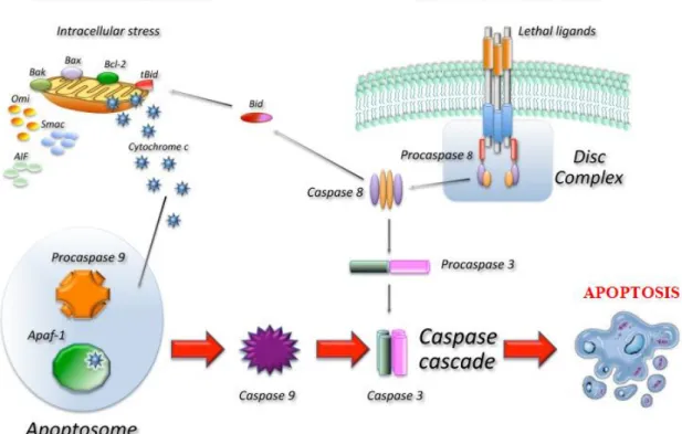
There are two major apoptotic signalling pathways - the intrinsic, so-called mitochondrial, and extrinsic, so-called death receptor, pathways (Figure 5), that are linked whereby molecules of one pathway can influence the other (Elmore, 2007). Various protein families promote or inhibit caspase activation, which is “essential for the morphological and biochemical hallmarks of apoptosis” (Ichim and Tait, 2016), upon binding to protein complexes such as the apoptosome or the death-inducing signaling complex (DISC) (Apoptosis Handbook, Novus Biologicals). Intrinsic apoptosis pathway is triggered by various stimulus, such like DNA damage and endoplasmic reticulum (ER) stress, which activate BH3-only protein family, resulting in the loss of mitochondrial integrity, release of cytochrome c, and the subsequent activation of caspase-9. In turn, caspase-9 activates caspase-3 and caspase-7, leading to apoptosis (Ichim and Tait, 2016; Portt et al., 2011). In contrast, extrinsic apoptosis pathway is triggered by external
stimulation and mediated by cell surface death receptors, such as Fas, tumor necrosis factor (TNF), or TRAIL receptors able to activate the initiator caspases-8 and -10 that cleave and activate the effector caspase-3 and caspase-7, leading to apoptosis (Ichim and Tait, 2016). Crosstalk between the intrinsic and extrinsic pathways can occur through caspase-8 cleavage and activation of the BH3-only protein BH3-interacting domain death agonist (BID), whose product is required in some cell types for death receptor-induced apoptosis (Ichim and Tait, 2016; Taylor et al.,2008). A more detailed apoptosis signalling
pathway is shown in Appendix 1.
The impact of the bacterial infection by A. phagocytophilum in the apoptotic
pathway have been studied in the both tick and mammalian cells showing that this pathogen can manipulate cell machinery to counteract apoptosis (Carlyon and Fikrig, 2003; Lee and Goodman, 2006; Galindo et al.,2012; Ayllon et al.,2013, 2015; Alberdi et al.,2016a,b). However, nothing is known about apoptosis related to the Babesia genus.
10
order to provide information on new strategies for the prevention and control of babesiosis, such as the identification of new targets for drug and vaccine development.
1.5. Aims
Infection facilitation by regulation of specific defense mechanism such as cell death confers some pathogens an important parasitic adaptive trait. Some studies have reported this pathogen ability focusing on the vertebrate host but information regarding the influence in ticks is scarce. The only tick-transmitted pathogen studied so far is A.
phagocytophylum showing that as in vertebrate host cells also in the vector the bacteria is
able to avoid destruction of the cells.
11
molecular interactions between the tick Rhipicephalus annulatus and Babesia bigemina
in order to suggest a novel approach to antigen selection for vaccine development against ticks and tick-transmitted pathogens. Focusing on the apoptosis pathway, the transcriptional response of R. annulatus salivary glands to B. bigemina infection was
13
2.1. Rhipicephalus annulatus Ticks
The non-infected and Babesia bigemina-infected Rhipicephalus annulatus female
ticks used in this study were provided by the Kimron Veterinary Institute, Israel. Ticks were maintained at the tick rearing facilities of the Kimron Veterinary Institute, in accordance with standards detailed in the Guide for Care and Use of Laboratory Animals. Two 3-4 months old male Friesian calves free of babesiosis were used to obtain the ticks after being tested for antibodies to Babesia spp. infection do with an indirect fluorescent
antibody (IFA) assay (Shkap et al.,2005) and kept under strict tick-free conditions. One
calf was inoculated i.v. with cryopreserved 2X 10⁸B. bigemina (Moledet strain), to obtain the infected ticks. Engorged adult female ticks were collected from both the infected and non-infected calves after feeding and maintained at 28ºC and 80% humidity. Ticks were
promptly stored in 500μl of RNA later (Ambion, CA, USA) at –20ºC, partially opened to exposed internal organs to the solution. Ten of each control and test groups were randomly selected to proceed with tissue dissection.
2.2. Salivary Glands Extraction
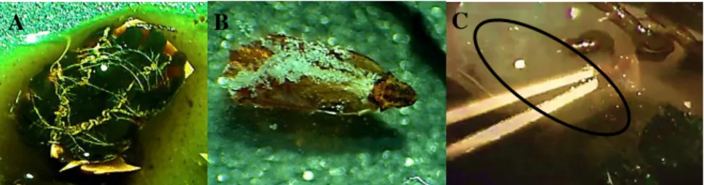
Each tick was submerged in a drop of 1X phosphate-buffered saline (PBS) (VWR, Pennsylvania, USA), to prevent dehydration. With fine-tipped forceps, ticks were stabilized by holding the basis capitulum and the scutum removed to expose the organs (Fig. 6 – A). Salivary glands (SG) (Fig. 6 – B), located bilaterally along the legs of the tick (Fig. 6 – C), were removed with fine-tipped forceps and place in a new drop of PBS. This wash step was gently repeated until the SG were cleaned of blood. SG were dissected under a stereoscopic microscope at 3.5X magnification (Motic SMZ-171B, China).
Figure 6 Representation of tick salivary glands dissection steps performed in Rhipicephalus bursa (originalfrom Catarina Monteiro).
14 2.3. RNA, DNA Extraction and cDNA Synthesis
SG total RNA was promptly extracted and purified with the GRS FullSample Purification Kit GK26.0050 (GRISP Research Solutions, Porto, Portugal), according to the manufacturer´s instruction. Briefly, SG were disrupted in 2ml centrifuge tubes with
400μl of DRP lysis buffer (GRS kit) and 4μl of β-mercaptoethanol (Bio-Rad, CA, USA), using a VWRTM pellet mixer (VWR). Five minutes later, sample lysates were centrifuged at 15.000g (Micro Star 17, VWR) for 90s and 0.8 volumes of 100% ethanol added to supernatants. Mixtures were transferred to RNA mini spin columns and centrifuged at
15.000g for 60s. One hundred μl of DNAse I mixed in reaction buffer was added to the spin columns placed in new collection tubes and incubate at room temperature for 10min. Afterwards, 400μl of wash buffer RNA 1 were added and columns centrifuged at 15.000g
for 60s followed by the addition of 600μl of wash buffer RNA 2 and centrifugation at 15.000g for 60s. Flow-through was discarded in each step. To dry the matrix, RNA columns were centrifuged at 15.000g for 3min and transferred into 1.5ml microcentrifuge tubes. After, 25μl of nuclease-free water was pipetted to the center of the spin columns, incubated at room temperature for 3min and then centrifuged at 15.000g for 60s. The eluted RNA was promptly used or stored at -80°C. RNA concentration and purity were determined by measuring the ratio of the UV absorbance at 260nm and 280nm, using the Nanodrop-1000 V3.7.1 spectrophotometer (Thermo Fisher Scientific, Waltham, MA, USA) and with QIAxcel (Qiagen, Hilden, Germany). The QIAxcel system uses capillary gel electrophoresis to enable fast separation of nucleic acids based on size, enabling higher detection sensitivity, less sample wastage and automated loading and analysis. Briefly, 1μl of each sample was pipetted into a 12-tube strip, one of them with 1μl of the QX RNA size marker. An equal volume of QX RNA denaturation buffer was added and the solution heated at 70ºC for 2min, then placed on ice for 1 min. After centrifuged, the 10μl total volume was completed with QX RNA dilution buffer and immediately analysed.
The cDNA was synthesized from 190ng/μl of RNA using the iScript cDNA synthesis kit (Bio-Rad) according to the manufacturer's directions, using a 100TM Thermal Cycler (Bio-Rad). In summary, 4μl of 5X iScript reaction mix, 1μl of iScript reverse
15
(RT) at 46ºC, 1min of RT inactivation and a hold optional step at 4ºC. cDNA was then stored at -20°C for downstream application.
2.4. Detection of Babesia bigemina in Ticks by Taq-man based qPCR
A TaqMan-based qPCR was performed to detect the presence of B. bigemina in
the sampled ticks using the pair of forward and reverse primers BiF-BiR (BiF: 5´-
AATAACAATACAGGGCTTTCGTCT-3´; BiR:
5´-AACGCGAGGCTGAAATACAACT-3´) and TaqMan fluorescence-labelled probe BiP (5´-TTGGAATGATGGTGATGTACAACCTCA-3´) that specifically amplify a fragment of the 18S rRNA gene, previously described by Kim et al., (2007). PCR
amplifications were carried out on a CFX Connect™ Real-Time PCR Detection System (Bio-Rad) with the Xpert Fast Probe kit (Grisp). Triplicate 20μl reactions were performed
with 10μl of Xpert Fast Probe 2X Mastermix, 400 nM of reverse and forward primers, 100nM of probe, 1μl of cDNA template and nuclease-free water up to the final volume. The conditions of the PCR were: initial denaturation 10min at 95ºC followed by 45 cycles: 20s at 95ºC and 1min at 55ºC. Negative controls were prepared with no template. PCR efficiency was determined generating sequence-specific standard curves with fivefold serial dilutions of DNA from the positive controls. The data was analysed using the Bio-Rad CFX Manager Software version 3.1. Samples with quantification cycle (Cq) values above 39 were considered negative for the presence of the pathogen.
2.5. Selection of Apoptosis Pathway Target Genes
Genes coding for putative mitochondrial ribosome small subunit component mediator of apoptosis dap3 (DAP3), death-associated protein kinase 1 (DAPK1),
apoptosis antagonizing transcription factor (AATF), apoptosis inhibitor 5 (API5), bax
inhibitor 1-related (BI-1) and voltage-dependent anion channel (VDAC) were chosen from
a R. annulatus RNAseq based sialotranscriptomic catalogue previously obtained by the
16 2.6. Gene Expression Analysis
Gene expression analysis were assessed in the SG by qPCR using the minimum information for publication of qPCR experiments (Bustin et al.,2010).
2.6.1. qPCR Assay Optimization
In order to identify an annealing temperature that provides efficient and specific amplification of the targets, annealing temperatures for the primer sets were optimized by running a temperature gradient on an CFX Connect™ Real-Time PCR Detection System (Bio-Rad) with a thermal cycling comprising 95°C for 3min, 40 cycles of 95°C for 10s and 48ºC – 62ºC for 30s, and a melting curve step of 55°C - 95°C, 0.5°C/s. Primers concentrations were then optimized between 0.3μM and 1μM. Final annealing temperatures and primers concentrations are in Table 1.
2.6.2. qPCR Assay Design
qPCR assay using SYBR® Green-based detection was designed and carried out
on an CFX Connect™ Real-Time PCR Detection System (Bio-Rad) with a thermal cycling protocol comprising a 3min denaturation and polymerase activation step at 95°C, 45 cycles of 95°C for 10s and 30s of specific annealing temperature for each primer sets in study. A melting curve step at the end of every amplification reaction, to ensure the production of single specific products and no primer-dimers as well as nonspecific products, is formed by easing the temperature in small increments (55°C - 95°C; 0.5°C/s) and monitoring the fluorescence signal at each step. When the double-stranded DNA (dsDNA) is denatured, SYBR® Green I Die is released and the fluorescence significantly reduced. Melting temperature (Tm) corresponds to the temperature at which 50% DNA denaturation occurs.
17
3.1. Efficiency is established to be between 90% and 115%. The differential gene expression was considered significant when p-value < 0.05.
2.6.3. Gene Expression Normalization
For accurate and reliable gene expression analysis, normalization of cDNA concentration relative differences between samples of the genes of interest against reference genes is essential, in order to infer steady-state mRNA levels. Data were normalised using the genes elongation factor (ELF), Glyceraldehyde-3-Phosphate
Dehydrogenase (GAPDH), TATA box binding protein (TATA),β-actin, β-tubulin and 16S
as internal controls, once these genes are described as constitutively expressed (Nijhof et
al.,2009). Reference gene validation was based on the geNorm algorithm (Vandesompele et al., 2002) and their expression stability measured with the coefficient variation
(CV<0.5) and M-value (M<1) (Hellemans et al.,2007), all included in the CFX Manager
Software version 3.1 (Bio-Rad).
2.7. Amplicon Identity Confirmation
Table 1 Primer sequences used for qPCR and respective optimal conditions.
Gene Accession Number Forward/reverse primer sequences (5´-3´) Fragment Length (bp) Temp anneal (ºC) Primer (μM)
β-actin AY255624 GACATCAAGGAGAAGCTYTGC
CGTTGCCGATGGTGAT 127 58 0.6
GAPDH CK180824 AGTCCACCGGCGTCTTCCTCA
GTGTGGTTCACACCCATCACAA 123 53.5
0.7
TATA CV453818 CTTGTCCTCACACACAGCCAGTT
GTGAGCACGACTTTTCCAGATAC 122
59.8 ELF EW679365 CGTCTACAAGATTGGTGGCATT
CTCAGTGGTCAGGTTGGCAG 109
0.5
β-tubulin CK179480 AACATGGTGCCCTTCCCACG
GCAGCCATCATGTTCTTTGC 140
56 16S MF946466 TTAACTGGGGCGGTTAAAAA
AACATCGAGGTCGCAAACTT 147
DAPK1 GACK01000273 AGGGTCACACGGACGTTATC
GTCTCCATGCTCGTCAGTCA 173 61.6
API5 GACK01008908 AACGTAACCCCCAGTTCCTC
GCCACCTTCAGCTTGTTCTC 175 50
BI-1 JO843858 GATGCCCAACACTGACACAG
CTCCTGCCAGAACACCTTTC 159 54.5
VDAC GU994210 CGCGACCTGTTCAACAAGAA
CGTGTTGTCGGTGTTCCATT 198 59.6
DAP3 GACK01006243 CAACCTGACCACTACGCAGA
GCAGGAGAGCTCCCATACAG 134 60 0.7
AATF GACK01004895 AGGCAGTGCAGGTCTCCTTA
GCTTCTGCAACTGGATCCTC 179 54 0.9
20 3.1. Salivary Glands Dissection and RNA Extraction
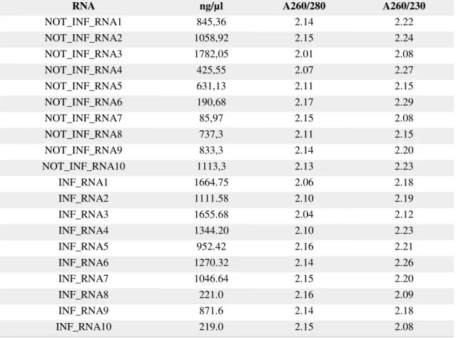
In this study, 20 R. annulatus female ticks were used to extract total RNA. Ten
ticks were fed in a B. bigemina infected calf and ten ticks were fed in a healthy calf, free
of babesiosis. Before cDNA synthesis, RNA concentration was determined as described and the results are present in the following table; RNA quality was evaluated with QIAxcel and the result presented in Appendix 2.
Table 2 Concentration of RNA extracted from Rhipicephalus annulatus salivary glands measure in Nanodrop. A260/280: ratio of the sample absorvance at 260 and 280 nm; A260/230: ratio of the sample absorvance at 260 and 230 nm.
RNA ng/µl A260/280 A260/230
NOT_INF_RNA1 845,36 2.14 2.22
NOT_INF_RNA2 1058,92 2.15 2.24 NOT_INF_RNA3 1782,05 2.01 2.08
NOT_INF_RNA4 425,55 2.07 2.27
NOT_INF_RNA5 631,13 2.11 2.15
NOT_INF_RNA6 190,68 2.17 2.29
NOT_INF_RNA7 85,97 2.15 2.08
NOT_INF_RNA8 737,3 2.11 2.15
NOT_INF_RNA9 833,3 2.14 2.20
NOT_INF_RNA10 1113,3 2.13 2.23
INF_RNA1 1664.75 2.06 2.18
INF_RNA2 1111.58 2.10 2.19
INF_RNA3 1655.68 2.04 2.12
INF_RNA4 1344.20 2.10 2.23
INF_RNA5 952.42 2.16 2.21
INF_RNA6 1270.32 2.14 2.26
INF_RNA7 1046.64 2.15 2.20
INF_RNA8 221.0 2.16 2.09
INF_RNA9 871.6 2.14 2.18
INF_RNA10 219.0 2.15 2.08
3.2. Babesia bigemina Infection Confirmation in Tick Salivary Glands
To confirm the infection by B. bigemina in the ticks, it was used a qPCR assay.
The TaqMan fluorescence-labelled probe used increases the specificity of the reaction. This assay was developed to amplify a fragment of the 18S rRNA gene. The absence of
21
The ten SG from ticks fed on a B. bigemina infected calve were found to be positive while
the ten SG from ticks that fed on the naïve calve were found to be negative (Figure 7).
3.3. qPCR Optimization
Gene expression of the six-targeted apoptosis-related genes (DAP3, DAPK1,
AATF, API5, BI-1, VDAC), as well as those of the reference genes (β-actin, GAPDH, TATA, ELF, β-tubulin, 16S) was accessed through a qPCR assay using SYBR®
Green-based detection, as described above. After testing different annealing temperatures and the best primer concentration (the final conditions of each gene are described in table 1), the assays were validated by the absence of amplification in the negative controls and by the efficiency of each reaction plate (Table 2). Reaction efficiency, which describes how much the target is being produced in each cycle, was automatically calculated by the CFX manager software that displays it under the standard curve using serial dilutions of a representative SGs pool, whose slope of the derived line are created with, at least, two points but as many points as possible were used.
In order to normalize the expression of the genes in study, their relative quantity was normalized to the relative quantities of the reference genes. Reference genes stability between the different conditions (infected/uninfected) was calculated with CFX manager software. It is established that a correct and accurate normalization is achieved by using two to five reference genes (Vandesompele et al.,2002). Herein, only the combinations
___ Negative controls
___ Negative samples ___ Positive samples
___ Positive controls
22
of reference genes showing a CV below 0.5 and M below 1 were used assuring the most stable reference genes.
Table 3 Final amplification efficiencies of the references and target genes.
Gene Amplification efficiency (%)
β-actin 91.4
GAPDH 100.1
TATA 105.7
ELF 99.3
β-tubulin 103.1
16S 88.3
DAPK1 87.0
API5 109.1
BI-1 101.5
VDAC 98.6
DAP3 88.0
AATF 100.3
3.4. Apoptotic Pathway Genes Differential Expression
For the evaluation of differential expression of the selected genes, the relative expression of the genes was measure in both the conditions studied, infected and uninfected and after compared. If the relative expression in the control condition is higher than in the test condition then the gene is assumed to be down-regulated while if the relative expression in the control is lower than in the test condition, the gene is up-regulated. This regulation is represented by the fold change that can be either positive or negative. The statistical significance of such result is given by the p-value. Overall our results show that from the six studied genes DAP3 and API5 are more expressed in the
infected samples while DAPK1, VDAC, BI-1 and AATF are more expressed in the control
samples (uninfected). Significant differential expression was demonstrated in DAP3,
DAPK1 and VDAC (p-value < 0.05). Detailed information regarding each gene is below.
DAP3gene expression showed to be significantly up-regulated in the R. annulatus
ticks SG when infected with the B. bigemina parasite (fold-change = 4.204; p = 0.005)
(Figure 8). Normalization was made using the 16S and β-tubulin reference genes (CV =
23
DAPK1 gene expression showed to be significantly down-regulated in the R. annulatus ticks SG when infected with the B. bigemina (fold-change = -3.546; p = 0.011)
(Figure 9). Normalized expression was calculated using the β-tubulin, β-actin, ELF and
TATA as reference genes (CV=0.359; M-value=0.766).
Similarly, the expression of the gene VDAC showed to be significantly down-regulated in the R. annulatus ticks SG when infected with B. bigemina (foldchange =
-1.512; p = 0.034) (Figure 10). The expression normalization was made using the β -tubulin, β-actin, ELF and TATA reference genes (CV=0.310; M-value=0.664).
Figure 9 Relative normalized expression of the apoptotic DAPk1 gene in Rhipicephalus annulatus salivary glands. The grey column corresponds to the uninfected SG and the green to the B. bigemina infected SG. Infection results in a downregulation (fold-change = -3.546; p = 0.011).
24
In the case of the AATF gene expression, under the conditions undertaken in the present study, it was demonstrated that in infected SG there is a down-regulation of the gene without statistical significance (fold-change = -1.016; p = 0.086) (Figure 11). Normalization was assured against the β-tubulin, β-actin and ELF reference genes (CV =
0.245; M-value =0.617).
After normalization with the β-tubulin, β-actin and ELF reference genes (CV =
0.224; M-value=0.541), it was demonstrated that the BI-1 gene is more expressed in the control samples (non-infected) in comparison to the infected samples (Figure 12). However, there is no a statistical significance (fold-change = -1.724; p = 0.441).
Figure 10 Relative normalized expression of the apoptotic VDAC gene in Rhipicephalus annulatus salivary glands. The grey column corresponds to the uninfected SG and the green to the B. bigemina infected SG. Infection results in a downregulation (fold-change = -1.512; p = 0.034).
25
Finally, API5 gene relative expression was evaluated in tick SG in B. bigemina
infected and non-infected SG. Results show that in infected SG there is an overexpression of this gene but without being statistically significant (fold-change = 2.514; p = 0.295) (Figure 13). For expression normalization the 16S and β-tubulin reference genes were
used (CV =0.151; M-value =0.438).
3.5. Amplicon Identity Confirmation
For confirmation of the identity of the amplified cDNA fragments three different strategies were used. The first approach was the analysis of the melt temperature (Table 4), followed by AGE (Appendix 3) and finally, Sanger sequencing was used to validate the identity of the genes studied. From the analysis of the melt curves, it was perceived that only one amplicon per pair of primers was being formed. All the samples and/or replicates that did not show the correct melt peak were discarded from the analysis.
Figure 12 Relative normalized expression of the apoptotic BI-1 gene in R. annulatus SG. The grey column corresponds to the uninfected SG and the blue to the B. bigemina infected SG. Infection results in a downregulation (fold-change = -1.724; p = 0.441).
26
The AGEs confirmed that the fragments amplified during the qPCRs presented the expected size (the fragment sizes are described in table 1 and AGE results are in appendix 3). Afterwards, randomly selected PCR products were purified using a commercial kit. Amplicons were sequenced and the obtained sequences compared with those that were deposited in the NCBI database and the transcripts previously obtained by the team. Results are shown in table 5 and appendix 4. With the exception of API5,
sequencing confirmed the identity of all target genes. The quality of the API5 obtained
sequence did not allow a correct alignment with the available sequence. Nevertheless, the previous analysis suggest that the fragment being amplified is the correct one. The BI-1
and AATF query cover values of 27 and 22%, respectively, are low because the obtained
sequences were compared with the available ones from Amblyomma maculatum and
Rhipicephalus pulchellus, respectively thus less homology between sequences can be
expected.
Table 4 Study genes melting temperature. Reference gene Melting temperature (ºC)
DAP3 81.0-81.5
DAPK1 83.5-84.0
AATF 80.0-80.5
API5 82.5-83.5
BI-1 81.0
VDAC 85.0-85.5
Table 5 Alignment of obtained amplicons with target genes.
Query cover E value Identity Accession number
DAPK1 90 % 3e-68 100 % GACK01000273
DAP3 84 % 7e-29 92 % JAA58791
VDAC 97% 1e-81 99% GU994210
API5 - - - -
BI-1 27% 7e-08 100% JO843858
28
The protozoan parasites B. bigemina mainly vectored by the R. annulatus ticks are
responsible for bovine babesiosis commonly named tick fever or red water fever. Due to the great impact on cattle health and economy is urgent the search for more effective strategies for the control of tick and TBP. The molecular interactions at the vector-pathogen interface may be targeted in order to impair pathogen infection and/or multiplication within the tick or block transmission to the vertebrate host. Research has shown that during the long-lasting tick-pathogen co-evolution, microorganisms have developed important strategies to manipulate or modulate tick response to infection, without impairing tick survival, enhancing their capacity of infection, replication and transmission guaranteeing the survival of both (de la Fuente et al.,2016a; Šimo et al.,2017). Moreover, it was demonstrated that tick
gene expression is modified in response to pathogen infection in different tick stages and various tick organs, including SG (Antunes et al., 2012; Heekin et al., 2012, 2013;
Sunyakumthorn et al.,2012; Cotté et al.,2014; Alberdi et al.,2016b; Cabezas-Cruz et al.,2016, 2017; Mansfield et al.,2017; Martins et al.,2017; Kalil et al.,2017; Šimo et al., 2017; Thangamani et al.,2017). SG are an important barrier that pathogen need to
overcome and exploit in order to successfully be transmitted to the vertebrate host (Alberdi et al., 2016b; Šimo et al., 2017), making this tissue a primary target in the
discovery of potential protective tick antigens. Within this tissue, pathogens need to interact with SG proteins in order to invade cells and multiply inside them before being released to the vertebrate bloodstream taking advantage of the vasodilator, anticoagulant, anti-inflammatory, and immunosuppressive properties of saliva (Šimo et al., 2017).
During SG infection different metabolic processes are affected, including the important apoptosis pathway (Ayllón et al., 2015). As previously mentioned, several pathogens,
especially those capable of invading and multiplying within host cells (Ashida et al.,
2011), as B. bigemina, have developed mechanisms to inhibit the cellular process of
apoptosis. Information regarding pathogen influence in tick apoptotic response is scarce and few systems, as the A. phagocytophylum – Ixodes sp., are partially studied so far.
Infection of tick SGwith A. phagocytophilum inhibit the intrinsic pathway of apoptosis
by down-regulating Porin expression, favoring bacterial infection, and tick cells respond to infection, promoting the apoptosis through induction of the extrinsic apoptotic pathway (Ayllón et al.,2015). However, nothing is known about apoptosis in the Babesia genus.
It is important to study the PCD manipulation in host cells by Babesia in order to provide
29
identification of new targets for drug and vaccine development. Base on this, six apoptotic genes of a R. annulatus RNAseq based sialotranscriptomic catalogue of genes
up-regulated in an infected tick population were selected and evaluated in B. bigemina
infected and uninfected R. annulatus. The results obtained are discussed below.
Mitochondrial Ribosome Small Subunit Component Mediator of Apoptosis dap3 (DAP3)
The commonly called death associated protein 3 (DAP3), highly conserved from yeast to human (Mukamel and Kimchi, 2004), is a 46-kDa protein containing a GTP binding domain and a P-loop, essentials for the induction of cell death (Mukamel and Kimchi, 2004). It is believed to be involved in both the intrinsic and extrinsic apoptotic pathways (Wazir et al.,2015b). Kissil and collaborators (1995) firstly described DAP3
interaction with the extrinsic pathway through the antisense RNA-mediated inactivation of the DAP3 in HeLa cells that shown to be protected from IFN-γ-induced cell death.
Nowadays, it is known that DAP3 interfere with the extrinsic pathway by Fas, TNF-α or TRAIL death receptors (DR) stimulation (Wazir et al.,2015a; Xiao et al.,2015) although
recent studies suggest that DAP3 association is strongest with Fas receptor-related DISC and its actions are enabled by binding with death ligand signal enhancer (DELE) (Wazir
et al.,2015a). Other studies have shown DAP3 interaction with the intrinsic pathway of
apoptosis intimately related to the mitochondrial biogenesis (Berger et al.,2000). It was
demonstrated that a loss of DAP3 expression leads to lethal mice embryonic mutations
(Kim et al.,2007) and overexpression of human, mouse and nematode DAP3 is sufficient
to cause cell death as a result of mitochondrial fragmentation (Mukamel and Kimchi, 2004; Xiao et al.,2015). In the present study, DAP3 gene was found to be overexpressed
under B. bigemina infection of R. annulatus female ticks (fold-change = 4.204; p = 0.005).
This result suggests that infection is leading to the natural cell response with the activation of apoptosis. Due to the role of DAP3 in both the extrinsic and intrinsic pathways, it is not clear which is via that is being induced.
Death-Associated Protein Kinase I (DAPK1)
30
conserved gene from invertebrates to chordates and mammals (Singh et al.,2016) and in
Caenorhabditis elegans and Drosophila, the only encoded DAPK family member
(Chuang and Chisholm, 2014). In mammals, DAPK1 gene is transcribed into a single
mRNA of 6.3kb (Singh et al., 2016) encoded for a 160 kDa protein with a conserved
Ca2+/CaM autoregulatory domain, 10 ankyrin repeats, 2 putative P-loop consensus sites and a ROC-COR domains, which overlap with a cytoskeletal-binding region, a death domain and a serine-rich C-terminal tail (Chuang and Chisholm, 2014; Shiloh et al.,2014;
Singh et al.,2016). DAPK1 is a critical component in the ER stress-induced cell death
pathway (belonging to the intrinsic apoptotic pathway) that transmits upstream signaling events into two distinct directions, caspase activation and autophagy, leading to cell death. Stimuli include Fas, INF-γ, and TNF-α (Singh et al.,2016). DAPK1 can activate
the tumor suppressor activity of p53 and “p53 activation molecularly links DAPK to the classical death pathway involving mitochondrial-based activation of the caspase cascade” (Bialik and Kimchi, 2006). This process requires the presence of p19ARF that promotes the ubiquitin-dependent degradation of p53 (Bialik and Kimchi, 2006). Currently identified as a putative tumor suppressor gene, nothing is known about their role in parasitic infections. In the present study, DAPK1 gene shown to be significantly
downregulated (fold-change = -3.546; p = 0.011). A deficiency in DAPK1 mRNA levels affects the intrinsic apoptotic pathway since it could no longer interact with p53. This potential DAPK1 modulation by B. bigemina benefitsthe parasitic dissemination within
cells.
Voltage-Dependent Anion Channel (VDAC)
The voltage-dependent anion channel (VDAC), commonly called mitochondrial porin, is a protein of the outer mitochondrial membrane that plays a central role in the intrinsic apoptotic pathway, leading to cell life or death, mediated by its association with various ligands and proteins (Shoshan-Barmatz et al.,2010). With a molecular weight of
30–35kDa, VDAC “consists of a polypeptide having unfolded alternating hydrophobic and hydrophilic amino acids, forming from 13 to 19 transmembrane β-chains composed
of a single α-helix at the amino terminus”. This conformation separates the apolar environment from the polar one, forming a wide barrel-shaped channel through the membrane (Rodríguez-Hernández et al.,2015). It is known that some pathogens have
31
namely by interfering with the mitochondrial membrane permeability. VDAC allows the flux of small molecules into the mitochondrial intermembrane space and the release of pro-apoptotic molecules, such as the cytochrome c, into the cytosol (Rodríguez-Hernández et al., 2015). VDAC upregulation is strongly correlated with apoptosis
induction and several mechanisms have been proposed. Among them, mitochondrial outer membrane permeabilization (MOMP) increase, VDAC oligomerization, increase of VDAC-ANT complexes and ROS production (Shoshan-Barmatz et al., 2010). A
BmVDAC-like protein that participates during B. bigemina invasion of R. microplus
midgut cells was recently identified by Rodríguez-Hernández and collaborators (2012) through proteomic analysis. Based on PCR results, BmVDAC gene expression shown to be significantly increased in infected ticks compared to uninfected ones at 24h post-repletion (Rodríguez-Hernández et al., 2015). VDAC also showed to be critical in T. gondii infection with a modified expression in infected cells (Nelson et al., 2008). “In
mosquitoes, VDAC plays a role during Plasmodium sp. invasion of the midgut; likewise,
the dissemination of B. burgdorferi through the tick midgut might be associated with the
ability of VDAC to bind a tissue-type plasminogen activator” (Antunes et al., 2017).
Contrary to the results obtained by Rodríguez-Hernández et al.,(2015), in the present
study, VDAC gene was significantly down-regulated under B. bigemina infection of R.
annulatus ticks (fold-change = -1.512; p = 0.034). Once VDAC is required for apoptosis
induction, the results obtained suggest that the parasite influences the levels of VDAC
mRNA. The different tick species used in the studies may explain the difference or even different infection status or levels. Nevertheless, more studies focusing this molecule are necessary to clarify the Babesia spp. potential manipulation of VDAC expression.
Apoptosis Antagonizing Transcription Factor (AATF)
Apoptosis antagonizing transcription factor (AATF), also called Che-I, Ded (Passananti and Fanciulli, 2007) and Traube (the murine ortholog) (Desantis et al.,2015),
is a RNA polymerase II binding protein of 558 aminoacids, highly conserved among eukaryotic species (Lezzi and Fanciulli, 2015; Passananti and Fanciulli, 2007). This
protein consists of an “N-terminal acidic domain, a canonical leucine zipper, and three LXXLL motifs for nuclear receptor binding. It also contains two nuclear and two putative
32
by binding specific transcription factors to the general transcription machinery (Passananti and Fanciulli, 2007). For example, after DNA damage AATF are released from the E2F target genes and recruits to p53 promoter (Floridi and Fanciulli 2007; Hopker et al.,2012; Passananti and Fanciulli, 2007) where “AATF binds to the PUMA, BAX and BAK promoter regions to repress p53-driven expression of these pro-apoptotic genes” (Höpker et al.,2012). So, AATF belongs to the intrinsic apoptotic pathway. AATF
anti-apoptotic activity was originally identified by Page and collaborators (1999) in rats for its ability to antagonize the Dlk-induced apoptosis (Lezzi and Fanciulli, 2015). Up to now, information about the anti-apoptotic activity of this protein derives from studies performed in neural tissue but nothing is known about AATF possible involvement in the
control of parasitic infection. In the present study, AATF expression under B. bigemina
infection of R. annulatus ticks did not show significant regulation (fold-change = -1.016;
p = 0.086). Interestingly, it was noticed that a mouse embryo mutant for Traube shows a
decrease in the number of ribosomes and Drosophila AATF mutant restrain the
development of the egg chamber at the same stage as the mutants affecting the synthesis of ribosomes (Lezzi and Fanciulli, 2015). In this context, more studies need to be performed to understand if B. bigemina is able to restrict AATF transcription, for example,
through the reduction of ribosomal activity as a way of escape from AATF anti-apoptotic
activity.
Bax Inhibitor-1 (BI-1)
Bax inhibitor-1 (BI-1) is a multi-spanning membrane protein of the endoplasmic reticulum (ER) with anti-apoptotic activity (Bultynck et al.,2012; Urra et al.,2013). It
was originally identified by Xu and Reed (1998) in cDNA library screens for human proteins capable of suppressing BAX-induced apoptosis in yeast. BAX, a Bcl-2 family member, promotes the intrinsic apoptotic pathway when induced by ROS (Rolland et al.,
2014). During ER stress, calcium and protein disulfide isomerases (PDI) are released from the ER to the cytosol, inducing apoptosis by MOMP deregulation. In addition, “BiP at the cytosol translocates to the plasma membrane, where it serves as a cell surface receptor for the pro-apoptotic protein Par-4” (Urra et al.,2013). BI-1 acts like an
anti-apoptotic protein by increasing ER calcium content (Urra et al.,2013), as well as reducing
ROS production (Bultynck et al., 2012; Rolland et al., 2014). Furthermore, BI-1 was