Arq Neuropsiquiatr 2005;63(2-A):321-323
Division of Neurology Hospital das Clínicas/ São Paulo University, São Paulo SP, Brasil (HC/FMUSP): 1Staff Neurologist; 2Fellow, Behavioral and Cognitive Neurology; 3PhD, Assistant Professor; 4Chief Resident; 5Patrícia Paula Santoro: MD, PhD, Staff Otorrinolaryngologist, Otorrinolaryngology Department; 6MD, Chief, MRI Section, Radiology Division; 7Division of Physiotherapy, Speech Pathology and Occupational Therapy; 8Chief, Neurology Division.
Received 6 September 2004. Accepted 10 December 2004.
Dra. Suely Kazue Nagahashi Marie - Centro de Investigações em Neurologia FMUSP - Avenida Dr. Arnaldo, 455/4110 - 01246-903 São Paulo SP - Brasil. E-mail: [email protected]
BILATERAL OLIVARY HYPERTROPHY AFTER
UNILATERAL CEREBELLAR INFARCTION
Case report
Adriana Bastos Conforto
1, Jerusa Smid
2, Suely Kazue Nagahashi Marie
3,
Jovana Gobbi Marchesi Ciríaco
4, Patrícia Paula Santoro
5,
Claudia da Costa Leite
6, Letícia Lessa Mansur
7, Milberto Scaff
8ABSTRACT - We describe a case of bilateral olivary hypertrophy and palatal tremor after unilateral cere-bellar infarction. Hypertrophic olivary degeneration (HOD) is associated with hypersignal in the inferior olivary nucleus (ION), on T2-weighted images. HOD has been more often observed ipsilaterally to a cen-tral tegmentum tract lesion or concen-tralaterally to a dentate nucleus or a superior cerebellar peduncle lesion. Double innervation of each ION from either dentate nucleus may have underlied the imaging and clini-cal findings in this 63 year-old male patient.
KEY WORDS: hypertrophic olivary degeneration, palatal tremor, cerebellar infarction, denervation super-sensitivity.
Hipertrofia olivar bilateral após infarto cerebelar unilateral: relato de caso
RESUMO - Relatamos um caso de hipertrofia olivar bilateral e tremor cerebelar após infarto cerebelar uni-lateral. A degeneração olivar hipertrófica (DOH) é associada ao aumento de sinal no núcleo olivar inferior (NOI), em sequências pesadas em T2. A DOH tem sido mais frequentemente observada ipsilateralmente a lesões do trato tegmentar central ou contralateralmente a lesões do núcleo denteado ou do pedúnculo cerebelar superior. A inervação bilateral de cada NOI por ambos os núcleos denteados pode ter sido responsável pelas características clínicas e radiológicas neste paciente de 63 anos.
PALAVRAS-CHAVE: degeneração olivar hipertrófica, tremor de palato, infarto cerebelar, hipersensibilidade por denervação.
Hypertrophic olivary degeneration (HOD) is a parti-cular kind of transsynaptic degeneration that occurs after lesions of the dentato-olivary pathway. On T2-weighted images, the affected olive is typically in-creased in size and shows a hyperintense signal. Usu-ally, unilateral HOD is observed ipsilaterally to a cen-tral tegmentum tract (CTT) lesion or concen-tralateral- contralateral-ly to a dentate nucleus (DN) or a superior cerebel-lar peduncle (SCP) lesion1. We report a case of
bila-teral HOD after unilabila-teral cerebellar infarction.
CASE
A 63-year-old man with a history of smoking had sud-den onset of dysarthria, ataxia, and diplopia. Months
322 Arq Neuropsiquiatr 2005;63(2-A)
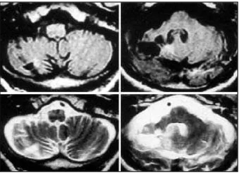
left. Saccadic eye movements were hypometric. The gag reflex was normal bilaterally. Laringoscopy revealed bi-lateral cyclical laryngeal spasms, with rhythmic bilater-al vocbilater-al cord adduction. No other cranibilater-al nerve abnormbilater-al- abnormal-ities were present. Brain MRI at 1.5 T revealed a right cerebellar hemisphere infarction and bilateral olivary hy-pertrophy (Fig 1). In addition, a left occipital and a left frontal infarction were observed, as well as multiple small deep white matter lesions, hyperintense on T2-weight-ed and FLAIR images, in both cerebral hemispheres. Di-gital subtraction angiography (DSA) revealed diffuse mural irregularities and moderate stenosis in the basilar artery, and right vertebral artery (VA) occlusion at its origin. A transesophageal echocardiogram was normal. Laboratory results revealed hypertrigliciridemia, hypercholesterole-mia, increased erythrocyte sedimentation rate (55 mm/ hour), eosinophilia, positive anti-RNP antibodies (1/640) and increased policlonal gammaglobulin content (2.35 mg/dl). Cerebrospinal fluid analysis also showed increased gammaglobulin content (17.5%). Electroneuromyogra-phy demonstrated a cronic sensorimotor demyelinating polyneuropathy affecting the lower limbs. Microangiopa-thical changes without vasculitic features were present in sural nerve biopsy. The right cerebellar infarction was attributed to right VA occlusion and consequent involve-ment of the right posterior inferior cerebellar artery ter-ritory in the cerebellum, in a patient with diffuse vasculo-pathy caused by atherosclerosis or vasculitis. The patient gave informed consent for the publication of this report.
DISCUSSION
The Guillain-Mollaret triangle (Fig 2) is compo-sed of the ipsilateral inferior olivary nucleus (ION) in the medulla, the contralateral DN in the cerebel-lum, and the red nucleus (RN)1-8. Fibers from the
ION project to the contralateral cerebellar cortex through the olivocerebellar tract via the inferior cerebellar peduncle (ICP) and then to the DN. Den-tate fibers enter the SCP and cross the midline in the decussation of the brachium conjuntivum, in the vicinity of the red nucleus (RN) before descending to the ipsilateral ION through the CTT. Lesions af-fecting the contralateral DN or SCP, or the
ipsilat-Fig 1. FLAIR (top; TR = 8402 ms; TE = 142 ms) and T2-weighted (bottom; TR = 4500 ms; TE = 99.9 ms) images performed two years after the cerebellar infarction disclose: involvement of the right cerebellar hemisphere including the dentate nucleus (right); ipsilateral volu-metric reduction of the right brachium pontis and pontine tegmentum (right); bilateral oli-vary hypertrophy with increased signal on T2-weighted image (left).
Arq Neuropsiquiatr 2005;63(2-A) 323
eral CTT are associated with deafferentation of the ipsilateral ION3,9. ION deafferentation may cause
supersensitivity denervation and HOD2. HOD is
his-tologically characterized by neuronal vacuolation and swelling, gliosis and demyelination in the ION1,6,7.
The process may follow not only vascular but also neoplastic, inflammatory, infectious and degener-ative lesions affecting the dentato-olivary path-way1,2,7,8. In autopsied patients with
cerebrovascu-lar lesions of the dentate-olivary tracts, neuronal hypertrophy was initially observed 20-30 days after the onset of the causative lesions and was maxi-mal 6-7 months later10,11. Three or four years later4,
HOD is substituted by olivary atrophy1,7.
MRI has been shown to correlate with patho-logic descriptions of HOD. Increased signal inten-sity on T2-weighted images in the ION has been re-ported one month after cerebellar and/or brain-stem infarctions, probably reflecting gliosis and increased water content1,3. ION enlargement may
resolve in 10-16 months but the increase in signal on T2-weighted images may still be seen years lat-er3. Differential diagnosis of the increase in signal
observed in the medulla include tumoral, demyeli-nating, vascular and inflammatory lesions. Increase in ION size, lack of involvement of medullary struc-tures other than the ION and lack of contrast enhance-ment favor a diagnosis of HOD3,8. The presence of
a contralateral cerebellar lesion or an ipsilateral pon-tine lesion further supports the diagnosis3.
Palatal tremor, previously known as palatal myo-clonus, consists of continuous rhythmic jerks of the soft palate and sometimes of other brainstem or spinal-innervated muscles2-4,7,12. Laryngeal
invol-vement, as in our patient, with associated dyspho-nia and dyspnea have been rarely described4.
Symp-tomatic palatal tremor (SPT) is due to a focal lesion within the dentato-olivary pathway while in essen-tial palatal tremor (EPT) no etiologies are identi-fied4. Unilateral or bilateral SPT have been
report-ed in association with contralateral HOD12, and
bi-lateral SPT in association with bibi-lateral HOD2-4,7.
Dif-ferent pathogenetic mechanisms have been pro-posed to explain how HOD leads to SPT4,5,10,12. In
se-ven of eight patients who had presented SPT after cerebrovascular lesions of the dentate-olivary path-way, SPT occurred 1-2 months after the causative injury, and was maximal 1-2 years later10.
Correla-tions between clinical presentation and histopatho-logical findings revealed that SPT appeared just after the onset of HOD changes, and was maximal soon after their peak. Spontaneous, rhythmic
firi-ng of the deafferented inferior olives due to disinhi-bition10might rhythmically stimulate the
contralat-eral dentate to lower motor neuron projections4,
therefore producing SPT. Alternatively, the denta-to-olivary pathway might become unable to inhib-it cranial nerve motor nuclei firing6,10. Maintenance
of SPT after substitution of HOD by atrophy has been reported6,10, suggesting long-term deficiency in
mo-dulation of motor nuclei firing due to olivary dys-function. This dysfunction may accompany olivary atrophy as well as hypertrophy.
After cerebellar lesions, HOD is usually contra-lateral to the affected DN2. Bilateral lesions
affect-ing both CTTs, or one CTT and the contralateral SCP have been associated with bilateral HOD3,9,10.
Re-ports of bilateral HOD and SPT after unilateral ce-rebellar lesions, as in the present case, have been rare in the literature1,7,10. It has been suggested that
supratentorial lesions causing interruption of oth-er pathways that might also modulate olivary func-tion could rarely lead to HOD7. Our patient had
mul-tiple asymptomatic white matter lesions in both ce-rebral hemispheres and we cannot completely ex-clude this hypothesis. However, double innervation of each ION from either DN may have more likely underlied bilateral HOD in this case since the clini-cal manifestation, SPT, was observed after a sympto-matic unilateral cerebellar infarction7.
REFERENCES
1. Goyal M, Versnick E, Tuite P, et al. Hypertrophic olivary degeneration: metaanalysis of the temporal evolution of MR findings. AJNR 2000; 21:1073-1077.
2. Matsuo F, Ajax ET. Palatal myoclonus and denervation supersensitivi-ty in the central nervous system. Ann Neurol 1979;5:72-78. 3. Salamon-Murayama N, Russell EJ, Rabin BM. Diagnosis please. Case
17: hypertrophic olivary degeneration secondary to pontine hemorrhage. Radiology 1999;213:814-817.
4. Deuschl G, Wilms H. Clinical spectrum and physiology of palatal tre-mor. Mov Disord 2002;17:(Suppl 2):S63-S66.
5. Deuschl G, Mischke G, Schenck E, Schulte-Mönting, Lücking CH. Symptomatic and essential rhythmic palatal myoclonus. Brain 1990; 113:1645-1672.
6. Lapresle J. Rhythmic palatal myoclonus and the dentato-olivary path-way. J Neurol 1979;220:223-230.
7. Visintini D, Trabattoni G, Tedeschi F, Lechi A, Granella F, Calzetti S. Pa-latal “myoclonus” and inferior olive hypertrophy with one-sided cere-bellar lesion. Clinico-pathological report of one patient. Funct Neurol 1986;1:63-70.
8. Krings T, Foltys H, Meister IG, Reul J. Hypertrophic olivary degener-ation following pontine haemorrhage: hypertensive crisis or cavernous haemangioma bleeding? J Neurol Neurosurg Psychiatry 2003;74:797-799.
9. Hommet CD, De Toffol B, Cottier JP, Autret A. Bilateral olivary hypertro-phy and palatal myoclonus. Surg Neurol 1998;49:215-216.
10. Nishie M, Yoshida Y, Hirata Y, Matsunaga M. Generation of symptoma-tic palatal tremor is not correlated with inferior olivary hypertrophy. Brain 2002;125:1348-1357.
11. Goto N, Kaneko M. Olivary enlargement: chronological and morphome-tric analyses. Acta Neuropathol (Berl) 54:275-282.