336
IMAGES IN NEUROLOGY
SMART syndrome: a late reversible complication
of radiotherapy
Síndrome SMART: uma complicação tardia reversível da radioterapia
Fabio de Vilhena Diniz
1, Laura Cardia Gomes Lopes
2, Luiz Henrique Martins Castro
2, Ricardo Nitrini
2,
Claudia da Costa Leite
1, Leandro Tavares Lucato
1A 43-year-ol d female had a cerebellar pilocytic
astrocy-toma operated 27 years ago. Reoperation and radiation
ther-apy were employed in a recurrence 13 years ago. One week
before admission, progressive drowsiness, disorientation in
time and space, slurred speech and worsening of a residual
right hemiparesis begun. Cerebrospinal fl uid (CSF) analysis
and electroencephalography (EEG) were unremarkable. She
improved without specifi c therapy.
Recently recognized stroke-like migraine attacks after
radiation therapy (SMART) is a syndrome which consists of
prolonged, unilateral neurological symptoms that are
spon-taneously reversible
1-4. Magnetic resonance (MR) fi ndings are
fundamental for diagnosis, characterized by transient,
dra-matic, but reversible cortical gadolinium enhancement of the
aff ected cerebral hemisphere
1-3(Figs 1 and 2). Appropriate
di-agnostic workup should exclude other didi-agnostic possibilities.
1Department of Radiology, Hospital das Clínicas da Faculdade de Medicina da Universidade de São Paulo (HCFMUSP), São Paulo SP, Brazil; 2Department of Neurology, HCFMUSP, São Paulo SP, Brazil.
Correspondence: Fabio de Vilhena Diniz; Avenida Dr. Enéas C. Aguiar 255/5084; 05403-000 São Paulo SP - Brasil; E-mail: [email protected] Confl ict of interest: There is no confl ict of interest to declare.
Received 05 October 2012; Received in fi nal form 10 December 2012; Accepted 17 December 2012.
Fig 1. Magnetic resonance (MR) exam obtained in the beginning of the clinical picture. Axial FLAIR (A) and coronal T2-weighted (B) images disclose slight cortical hyperintensity in the left insula, temporal and parietooccipital regions (arrows), which presents intense gyriform enhancement in the corresponding axial T1-weighted post gadolinium image (arrowhead in D). There is also slight cortical hyperintensity in diffusion-weighted image (C), but without clear reduced diffusion in apparent diffusion coeffi cients map (not shown). Notice also postoperative changes in the left cerebellar hemisphere, seen in coronal T2-weighted image (B), and nonspecifi c hyperintense periventricular lesions in axial FLAIR image (A).
A
B
C
D
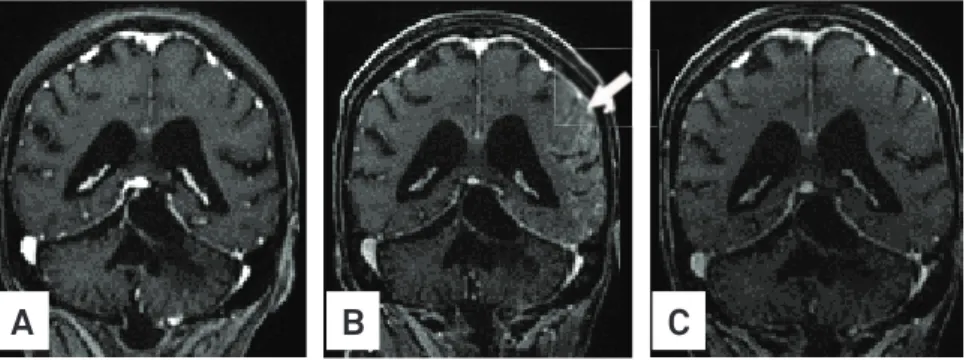
Fig 2. Coronal reformatted images from post contrast 3D-SPGR images obtained in three distinct time frames. (A) Image obtained 16 months before the beginning of the acute clinical fi ndings demonstrates only cerebellar postoperative changes, in a regular follow-up exam. (B) Image obtained from the same exam as Fig 1, showing the intense gyriform enhancement in the left temporal and parietooccipital regions (arrow). (C) After 14 days, a new magnetic resonance (MR) exam shows no abnormal enhancement.
A
B
C
337
Arq Neuropsiquiatr 2013;71(5):336-3371. Bartleson JD, Krecke KN, O’Neill BP, Brown PD. Reversible, strokelike migraine attacks in patients with previous radiation therapy. Neuro Oncolo 2003;2:121-127.
2. Black DF, Bartleson JD, Bell ML, Lachance DH. SMART: stroke-like migraine attacks after radiation therapy. Cephalagia 2006;26:1137-1142.
3. Lachance DH, Black DF, Bartleson JD. SMART: stroke-like migraine attacks after radiation therapy. Neurology 2005;64:A220.
4. Murthy SN, Cohen ME. Pseudomigraine with prolonged aphasia in a child with cranial irradiation for medulloblastoma. J Child Neurol 2002;17:134-138.