Arq Neuropsiquiatr 2006;64(2-B):398-401
Sarah Network of Rehabilitation Hospitals - Fortaleza CE, Brazil: 1,2,3,5Radiologist; 4Pediatrician. Received 6 October 2005, received in final form 8 December 2005. Accepted 7 February 2006.
Dr. Mauro Nakayama - Av. Juscelino Kubitschek 4500 - 60861-630 Fortaleza CE - Brasil. E-mail: [email protected]
MRI AND
1
H-MRS FINDINGS OF THREE PATIENTS
WITH SJÖGREN-LARSSON SYNDROME
Mauro Nakayama
1, Daniel G.F. Távora
2, Thereza C.L. Alvim
3,
Alexandre C.B. Araújo
4, Rômulo L. Gama
5ABSTRACT - Sjögren-Larsson syndrome (SLS) is a rare autosomal recessive neurocutaneous disorder caused by deficiency of the microsomal enzyme fatty aldehyde dehydrogenase. Patients present the classical tri-ad of congenital ichthyosis, mental re t a rdation and spastic di- or tetraplegia. Magnetic resonance imag-ing (MRI) of the brain usually shows hypomyelination involvimag-ing the periventricular white matter. Cere b r a l p roton MR spectroscopy (1H-MRS) reveals a characteristic abnormal lipid peak. We re p o rt three cases of
SLS from different families with the typical clinical triad. The MRI and 1H-MRS findings are discussed.
KEY WORDS: Sjögren-Larsson syndrome, magnetic resonance imaging, proton spectroscopy.
S í n d rome de Sjögren-Larsson: achados à ressonância magnética e espectroscopia de prótons em três pacientes
RESUMO - A síndrome de Sjögren-Larsson (SJL) é distúrbio raro, autossômico recessivo, caracterizado pela tríade clássica de ictiose congênita, re t a rdo mental e tetraplegia ou diplegia espástica. Trata-se de um erro inato do metabolismo dos lipídios, causado pela deficiência da enzima microssômica aldeído graxo desidro-genase. Os achados de imagem do encéfalo na SJL demonstram atrofia cerebral e alteração da substância branca. A espectroscopia de prótons, com poucos casos relatados, caracteriza-se pelo elevado pico de lipí-dios e redução de N-acetil-aspartato. Apresentamos três casos de SJL, com ênfase nos achados da re s s o n â n-cia magnética e da espectroscopia de prótons.
PALAVRAS-CHAVE: síndrome de Sjögen-Larsson, ressonância magnética, espectroscopia de prótons.
S j ö g ren-Larsson syndrome (SLS) is a rare autoso-mal recessive neurocutaneous disorder characterized by the clinical triad of congenital ichthyosis, mental re t a rdation and spastic diplegia or tetraplegia. Addi-tional findings include retinal pigmentary degener-ation in the macular region, epilepsy, hypoplasia of teeth, kyphosis and metaphyseal dysplasia1 , 2. SLS is
an inborn error of lipid metabolism caused by a defici-ency of the microsomal enzyme fatty aldehyde dehy-d rogenase (FALDH), a component of the fatty alco-hol: NAD+o x i d o reductase enzyme complex. FA L D H
deficiency may lead to an accumulation of long-chain fatty alcohols with structural consequences for cell-membrane integrity, which disrupt the barrier func-tion of the skin and the white matter of the brain3.
R e p o rts on central nervous system imaging of SLS have described brain atrophy and white matter dis-ease characterized by hypomyelination4-6. However,
t h e re are only a few re p o rts on brain proton magne-tic resonance spectroscopy (1H-MRS), with typical
find-ings of abnormal high peak in spectral curve, bet-ween 0.8 and 1.6 ppm, corresponding to the range of lipids. It is also re p o rted a nonspecific diminished peak of N-Acetyl Aspartate (NAA) at the periventric-ular white matter1,2,7.
We present three cases of SLS from diff e rent fam-ilies with the typical clinical triad. The magnetic re s o-nance imaging (MRI) and 1H-MRS findings are
discus-sed.
CASES
Case 1 – A 16-year-old girl presented with ichthyosis
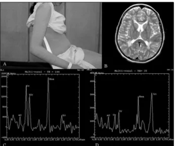
and spastic diplegia. She was the first child of consangui-neous Brazilian parents. She was born after an uneventful full term pre g n a n c y, and developmental delay was noticed at very early infancy, sitting at 2 years of age and crawling at 4 years. Generalized ichthyosis and pruritus were addi-tional features, with gradual worsening (Fig 1A). She had no past history of epilepsy.
Arq Neuropsiquiatr 2006;64(2-B) 399
examination disclosed mental re t a rdation, spasticity in both lower limbs, increased patellar reflexes and ankle clonus. Babinski`s sign was positive bilaterally. EEG and nerve con-duction studies were normal. Multislice computed tomog-raphy (CT) scan of the bra in showed diffuse periventricu-lar white matter low density and a mild cerebral atro p h y (not shown).
MRI at 1.5 T demonstrated diffuse and symmetric high signal areas in the periventricular white matter on T2-weighted images, most prominent around the trigones (Fig 1B). The area of abnormally high signal intensity on T2-weighted images had a decreased signal intensity on T1-weighted images. 1H-MRS (single and multivoxel, TE=35 ms and 144 ms) was performed with the voxel located at the peritrigonal area and showed a high lipid peak and a slight-ly low NAA peak (Figs 1C and 1D).
Case 2 – A 11-year-old boy presented with cutaneous
symptoms since birth, delayed milestones and speech devel-opment. He was the only child of consanguineous Brazilian p a rents, born at about 32 weeks of gestational age thro u g h a cesarean delivery. He was examined by a neurologist dur-ing early infancy and a diagnosis of cerebral palsy was ma-de. The ichthyosis was confirmed by skin biopsy. The par-ents noted a cognitive developmental delay and diff i c u l t walking. He also had dysarthria.
On examination, his height was 143 cm (percentile 50) and his weight was 36 kg (percentile 50). Neurological exa-mination revealed spastic diplegia, increased deep tendon reflexes, positive Babinski`s sign and ankle clonus. He was able to walk a short distance with aids, and had no diff i-culties in crawling. Conduction nerve studies were norm a l . Brain MRI showed bilateral and symmetric slightly
ab-n o rmal sigab-nal iab-nteab-nsity at the peritrigoab-nal white matter oab-n T2-weighted images with normal signal intensity on T1-weighted images (Figs 2A and 2B). 1H-MRS (single and mul-tivoxel, TE=35 ms and 144 ms) demonstrated a significant-ly high and sharp lipid peak and a low NAA peak (Fig 2C).
Case 3 – A 4-year-old boy was born after an
unevent-ful unevent-full term pregnancy and norm al delivery. The pare n t s a re 3rdd e g ree cousins. Developmental delay was noted sin-ce very early infancy. He began sitting after his fourth anni-versary with assistance and could speak only a few words. Examination revealed spastic tetraplegia, mental re t a r-dation, scoliotic deformity and ichthyosis on trunk and limbs. His weight was 15 kg (percentile 25) and his height 100 cm (percentile 25). Neurological examination demon-Fig 1. Patient 1. (A) Photographs of the skin abnormalities of
the trunk and limbs (ichthyosis). (B) Axial T2-weighted FSE (Fast Spin Echo) images show prominent hyperintensity in the periventricular white matter. (C) and (D) Multivoxel 1H - M R S .
Peritrigonal spectral analysis shows a high lipid peak at TE=144 ms (C) and a less prominent lipid peak at TE=35 ms (D).
Fig 2. Patient 2. (A) Axial FLAIR (Fluid Attenuated Inversion R e c o v e ry) images and (B) axial T2-weighted FSE (Fast Spin Echo) images shows minimal hyperintensity in the peritrigonal white m a t t e r. (C) Single voxel 1H-MRS (TE=35 ms) shows a sharp and
abnormally high peak at the spectral range of lipids.
Fig 3. Patient 3- Single voxel 1H-MRS (TE=35 ms). The voxel is
400 Arq Neuropsiquiatr 2006;64(2-B)
strated increased deep tendon reflexes and clonus. Nerv e conduction studies were normal.
Brain MRI study show ed periventricular w hite matter high signal on T2-weighted images and increased lipid peak on 1H-MRS (single and m ultivoxel, TE=35 ms and 144 ms) (Fig 3). The area of abnormally high signal intensity on T2-weighted images had a decreased signal intensity on T1-weighted images.
MRI and 1H-MRS of the brain were obtained in a single session for each patient using the same 1.5 T MR unit (Signa Horizon LX Echo Speed; GE Medical System, Milwaukee, WI, USA), and a standard head coil. Imaging was carr i e d out with axial (5 mm and 3 mm), sagittal (5 mm) and coro-nal (5 mm) slices, with the following sequences: spin-echo (SE) T1-weighted: echo time (TE), 10 ms; repetition time (TR), 500 msec; and fast spin-echo (FSE) T2-weighted: (TE/TR) 100/2000 msec.
The conventional T2-weighted MR images were used to position a spectroscopic volume of interest (VOI), for sin-gle and multivoxel spectro s c o p y, in areas of abnormal sig-nal in the periventricular white matter. Spectroscopy vol-ume selection was perf o rmed by using a PROBE-P sequence ( P roton Brain Examination) a version of the PRESS sequence ( P o i n t - resolved Proton Spectroscopy Sequence) (TR=2000, TE=35 ms or TE=144 ms). Each voxel measured 20 mm x 20 mm x 20 mm. MR spectroscopy data were accumulated after the optimal water signal was suppressed by the chemical shift-selective technique. Institutional review board appro v a l was obtained. A verbal consent was obtained from all par-ents.
DISCUSSION
SLS is a rare autosomal recessive disorder with a clinical triad of ichthyosis, mental re t a rdation and spastic diplegia or tetraplegia. It was first described in 1957, with an incidence of 1:200000 births8.
SLS is due to deficient activity of fatty aldehyde d e h y d rogenase. This enzyme catalyses the oxidation of long chain aldehyde to fatty acids2,3. Due to
defi-ciency of this enzyme, there is an accumulation of al-dehyde-modified lipids or fatty alcohol in the skin and in the myelin. FALDH plays an essential role in leucotriene B (LTB4) metabolism and a defective de-gradation of LTB4 may be responsible for consider-able pruritus in patients with SLS. FALDH gene has been mapped to the SLS locus on band 17p11.22,9.
Ichthyosis is a generalized hyperkeratosis of the t runk, joints and the dorsal aspects of the hands and the feet. Most patients have erythema at birth, with worsening of cutaneous symptoms during the first year of life. Pruritus is a prominent feature that is n o t found in other types of ichthyotic skin disorders, and has been recognized as an important symptom that strongly suggests the diagnosis of SLS8.
N e u rological features are nonspecific; however,
mental re t a rdation and developmental delay are usu-ally obvious at 1-2 years of age. Spasticity may be ap-parent before age 3 years and is more severe in the lower limbs1,2.
Photophobia, macular dystrophy and decre a s e d visual acuity are the most prominent ophthalmolog-ic abnormalities, and may be caused by accumula-tion of long-chain fatty alcohols or fatty aldehydes2.
T h e re is a paucity of case re p o rts on SLS imaging findings. Gomori et al.5studied six siblings by CT and
found low confluent density areas in the cere b r a l white matter, and stated that the severity of the CT findings correlated with the severity of the neuro-logical symptoms. Altinok et al.6reported MRI
find-ings of three siblfind-ings with diagnosis of SLS confirm e d by enzyme analysis. Brain MRI showed diffuse white matter abnormalities mostly in re t rotrigonal and peri-ventricular areas, with no contrast enhancement.
Ve ry limited data are available about 1H-MRS in
this condition. 1H-MRS directed to periventricular
le-sions revealed an abnormally high peak at the lipid range and decreased peak of NAA. A decreased NAA is an indicator of neuronal and axonal damage or dysfunction. It has been re p o rted that the incre a s e d relative concentration of lipids correspond pre c i s e l y to the high signal areas observed on T2-weighted MRI1,2,7,10.
Van Domburg et al.1evaluated 11 SLS patients,
and serial MRI findings showed evidence of delayed myelination and a variable degree of dysmyelination in young patients, particularly at the subcortical asso-ciation areas of the frontal, parietal and temporal lobes. At the early infancy stage there were no MRI evidence of demyelination or dysmyelination. Six pa-tients underwent 1H-MRS investigation directed to
the white matter which showed an abnormal lipid peak, both at a TE=30 and 135 ms. The most intense lipid peaks were localized on periventricular and per-itrigonal areas.
Willemsen et al.2re p o rted a clinical, biochemical
and molecular characteristics of 19 patients with SLS who presented periventricular white matter abnor-malities on MRI and abnormal lipid peak on 1H - M R S
studies. Miyanomae et al.7described a SLS case whose
MRI showed high signal on T2-weighted and low sig-nal on T1-weighted images at the peritrigosig-nal are a s .
1H-MRS of those lesions revealed increased lipid peak.
They speculated that such lipids in the periventricu-lar regions with high T2 signal might be pathogno-monic of SLS.
Arq Neuropsiquiatr 2006;64(2-B) 401
been observed in other degenerative diseases, includ-ing peroxisomal disorders and multiple sclerosis, the spectral peaks were usually broad and disappeare d on long TE sequences11,12.
In a recent paper, Willemsen et al. reported that abnormalities on MRI and proton spectroscopy dur-ing the first years of life and that the lipid peak at 1.3 ppm in the proton MRI s p e c t rum of the cere b r a l white matter may offer a quantitative parameter for monitoring the effects of therapeutic interv e n t i o n s1 3.
The MRI and 1H-MRS findings of the three SLS
pa-tients studied by us correlate well with those des-cribed by the reviewed literature, showing periven-tricular white matter changes on MRI and a signifi-cantly high and sharp lipid peak on 1H-MRS. In a series
of 18 patients, Willemsen et al. found normal levels of NAA at 1H-MRS, by comparison with healthy
sub-j e c t s1 1. In our cases, we found slightly decreased NAA/
Cr ratio. However, it must be stressed that we did not compare these results with a control group.
1H-MRS findings of the patient 2 revealed a high
lipid peak, although only minimal signal abnormal-ity was found in the periventricular white matter. We speculate that 1H-MRS may demonstrate an
accumu-lation of free lipids in the white matter even before an abnormality become detectable by morphologi-cal MRI.
The diagnosis of SLS should be considered in any neonate or infant with a congenital ichthyosis asso-ciated to neurological features. Biochemical confir-mation can be obtained by demonstration of enzyme deficiency of FALDH measured in cultured skin fibro
b-lasts or leukocytes, or by the presence of abnorm a l metabolites of LTB4 in the urine.
We conclude that 1H-MRS may be a useful tool
for confirmation of SLS diagnosis, suggesting local accumulation of unusual free lipids or lipophilic sub-stances at periventricular white matter.
REFERENCES
1. Van Domburg PHMF, Willemsen MAAP, Rotteveel JJ, et al. Sjögre n -Larsson syndrome: clinical and MRI/MRS findings in FA L D H - d e f i-cient patients. Neurology 1999;52:1345-1352.
2. Willemsen MAAP, Ijlst L, Steijlen PM, et al. Clinical, biochemical and molecular genetic characteristics of 19 patients with the Sjögre n - L a r s s o n syndrome. Brain 2001;124:1426-1437
3. Rizzo WB, Craft DA. Sjögren-Larsson syndrome: deficient activity of the fatty aldehyde dehydrogenase component of fatty alcohol: NAD+ oxireductase in cultured fibroblasts. J Clin Invest 1991;88:1643-1648. 4. Hussain MZ, Aihara M, Oba H, et al. MRI of white matter changes in
the Sjögren-Larsson syndrome. Neuroradiology 1995;37:576-577. 5. Gomori JM, Leibovici V, Zlotogorski A, Wi rguin I, Haham-Zadeh S.
Computed tomography in Sjögren-Larsson syndrome. Neuro r a d i o l o g y 1987;29:557-559.
6. Altinok D, Yildiz Y T, Seçkin G, Altinok G, Tacal T, Eryilmaz M. MRI of t h ree siblings with Sjögren-Larsson syndrome. Pediatr Radiol 1999; 29:766-769.
7. Miyanomae Y, Ochi M, Yoshioka H, et al. Cerebral MRI and spectros-copy in Sjögren-Larsson syndrome: case report. Neuro r a d i o l o g y 1995;37:225-228.
8. Te rcedor J, Garcia A. Síndromes neurocutáneos queratósicos. Rev Neurol 1997;25 (Supl 3):S238-S242.
9. Pigg M, Jagell S, Sillen A, Weissenbach J, Gustavson KH, Wadelius C. The Sjögren-Larsson syndrome gene is close to D17S805 as determined by linkage analysis and allelic association. Nat Genet 1994;8:361-364. 10. Mano T, Ono J, Kaminaga T, et al. Proton MR spectroscopy of Sjögre n
-Larsson syndrome. AJNR 1999;20:1671-1673.
11. Wolinsky JS, Narayana PA, Fenstermacher MJ. Proton magnetic reso-nance spectroscopy in multiple sclerosis. Neurology 1990;40:1764-1769. 12. B ruhn H, Kruse B, Korenke GC, Hanefeld F, Hanicke W, Merboldt KD. P roton NMR spectroscopy of cerebral alterations in infantile pero x i s o-mal disorders. J Comput Assist Tomogr 1992;16:335-344.