Functional magnetic resonance imaging and swallowing:
systematic review
Ressonância magnética funcional e deglutição: revisão
sistemática
Luciana Grolli Ardenghi1, Alana Verza Signorini2, Ana Carolina Battezini3, Silvia Dornelles4, Carlos Roberto de Mello Rieder5,6
ABSTRACT
Purpose: Evaluate the feasibility and safety of functional magnetic re-sonance imaging (fMRI) for the evaluation of cortical areas involved in swallowing. Research strategy: The search was conducted from April 2003 to April 2013, using the keywords“functional magnetic resonance imaging” or “fMRI” and “dysphagia” and “functional magnetic reso-nance imaging” or “fMRI” and “swallowing” in “PubMed” database.
Selection criteria: Studies were reviewed by analyzing their titles and abstracts with the following inclusion criteria: research involving human subjects, use of neurofunctional tests, reference to swallowing function, adult and/or elderly population analysis and association with neurolo-gical disorders. Results: the search strategy resulted in 1167 citations, from which only 35 met the eligibility criteria. Conclusion: the func-tional magnetic resonance imaging was considered safe and feasible for evaluating cortical areas involved in swallowing. However, the reports of functional magnetic resonance usage differed between the reviewed studies and the variability in the methodology used, made meaningful comparisons difficult.
Keywords: Disorders of swallowing; Neuroimaging; Swallowing; Neu-rology; Neurological diagnostic techniques
RESUMO
Objetivo: Avaliar a viabilidade e segurança da ressonância magnética funcional para a avaliação de áreas corticais envolvidas na deglutição.
Estratégia de pesquisa: Conduziu-se uma busca entre abril de 2003 e abril de 2013, usando as palavras chave “functional magnetic resonance imaging” or “fMRI” and “dysphagia” e “functional magnetic resonan-ce imaging” or “fMRI” and “swallowing” na base de dados PubMed.
Critérios de seleção: Os estudos foram revisados por análise de seus títulos e abstracts e os critérios de inclusão utilizados foram: pesquisas envolvendo seres humanos, utilização de exames neurofuncionais, refe-rência à função de deglutição, análise de população adulta e/ou idosa, relação com patologias neurológicas. Resultados: A estratégia de busca resultou em 1167 citações, das quais apenas 35 preencheram os critérios de elegibilidade. Conclusão: A ressonância magnética funcional foi con-siderada segura e viável para a avaliação de áreas corticais envolvidas na deglutição. Entretanto, os relatos de utilização de ressonâcia magnética funcional diferiram entre os estudos revisados e houve variabilidade na metodologia utilizada, dificultando as comparações.
Descritores: Transtornos da deglutição; Neuroimagem; Deglutição; Neurologia; Técnicas de diagnóstico neurológico
Work developed at the Universidade de Passo Fundo – UPF – Passo Fundo (RS), Brazil.
(1) Program of Graduate Studies in Medical Sciences (Doctorate), School of Medicine, Universidade Federal do Rio Grande do Sul – UFRGS – Porto Alegre (RS), Brazil.
(2) Program of Graduate Studies in Medical Sciences (Master degree), School of Medicine, Universidade Federal do Rio Grande do Sul – UFRGS – Porto Alegre (RS), Brazil.
(3) Speech-Language Pathology Course, Medical School, Universidade de Passo Fundo – UPF – Passo Fundo (RS), Brazil.
(4) Health and Human Communication Department, Speech-Language Pathology Course, Universidade Federal do Rio Grande do Sul – UFRGS – Porto Alegre (RS), Brazil.
(5) Neurology Service, Clinics Hospital of Porto Alegre, Universidade Federal do Rio Grande do Sul – UFRGS – Porto Alegre (RS), Brazil. (6) Universidade Federal Ciências da Saúde de Porto Alegre – UFCSPA – Porto Alegre (RS), Brazil.
Conflict of interests: No
Authors’ contribution: LGA main researcher, research preparation, schedule development, literature collection, review and data analysis, article writing, article submission and procedures; AVS research development, literature review, data collection and analysis, article writing; ACB research preparation, literature review, data collection and analysis, article writing; SD guiding, research preparation, development schedule, data analysis, article writing correction, final version ap-proval; CRMR advisor, research preparation, development schedule, data analysis, article writing correction, final version approval.
Correspondence address: Luciana Grolli Ardenghi. R. General Firmino, 476/701, Centro, Palmeira das Missões (RS), Brazil, CEP: 98300-000. E-mail: [email protected]
INTRODUCTION
Swallowing is one of the processes that ensure the human being survival and its disorders require global health care. Among its consequences are dehydration, malnutrition and pulmonary complications involving aspiration, pneumonia and deficits in the quality of life(1). Initially, research in
neu-rophysiology swallowing restricted the process to functional mechanisms of the brain stem. Subsequently, however, thanks to advances in the use of functional neuroimaging techniques, the cerebral cortex contribution in the swallowing control has been better understood(2).
The complexity involving swallowing is the result of a sequence of coordinated movements, including pressure balance, sensory processing of ingested food and synergistic movement of the muscles involved in the oral, pharyngeal and laryngeal regions. It is synchronized with other stomatognathic functions such as breathing and chewing and, when voluntarily performed, requires a cognitive processing and attention ac-tivation(3). From the first swallowing component until the last
stage of the solid bolus movement, multiple oral and perioral muscles are used(4).
The knowledge of the swallowing neurophysiology, in-cluding understanding the role of different brain structures in the movement synergy, can bring new perspectives of dysphagia management and treatment. Studies in the fMRI brain field (functional MRI) in recent decades have helped to understand the brain spatial and temporal activities of swallo-wing by means of a noninvasive procedure. Although fMRI is an important tool for understanding the brain mechanisms involved in swallowing, the methodologies and paradigms used are varied.
PURPOSE
The purpose of this systematic review was to identify and compare brain activation during swallowing, analyzing the tasks used and stimuli provided to patients in healthy adults, elderly and with dysphagia.
Data collected for the functional MRI studies were analyzed in order to verify the safety and reliability of the findings, in a comparative way.
RESEARCH STRATEGY
To identify the swallowing and brain imaging studies, a search of publications available in PubMed and MEDLINE databases was conducted. In order to achieve a broad base of literature review, the research including studies published since 2003. The survey was conducted using the following keywords: “functional magnetic resonance imaging” or “fMRI” and “dysphagia” and “functional magnetic resonance imaging” or “fMRI” and “swallowing”.
SELECTION CRITERIA
The studies analyzed by two examiners independently, were selected according to the following inclusion criteria:
- Published between 2003 and 2013; - Original studies involving human subjects;
- tudies, whose objective was the evaluation of swallowing through the brain neuroimaging;
- Analysis on adult or elderly population. Exclusion criteria used for this work were: - Samples with animals;
- Samples with fetuses, children and adolescents;
- Evaluation of related functions such as speech, voice, among others;
- Peripheral structures evaluation: larynx, pharynx, tongue, vocal folds;
- No use of fMRI.
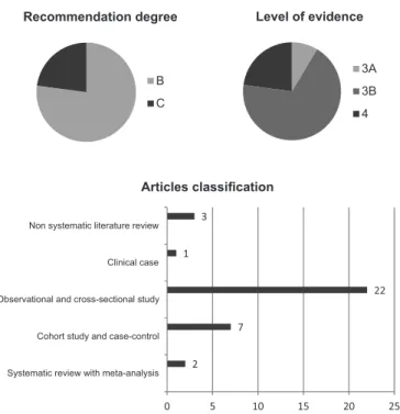
Full texts were classified according to the type of study (case study, meta-analysis, systematic review or experimen-tal); type of protocol used to collect the cortical functioning of images; the sample size; the population characteristics and results found. The tests results were compared between two evaluators and the criteria classification was revalued in a con-sensus meeting, analyzing differences. Differences occurred in the articles classification exclusion. Therefore, the hierarchy among the topics for deletion was established and unselected items were reclassified.
The search for studies published between April 2003 and April 2013 resulted in 1167 articles, of which 397 were repea-ted. 728 abstracts were excluded for not using neurophysiology assessment techniques, studies not related to swallowing func-tions, anatomical functioning and/or functional studies sensory oral motor system, among others. 35 complete articles that met the proposed criteria for this systematic review were included.
Figure 1 shows the breakdown of the articles analysis.
DATA ANALYSES
Determining the recommendation degree and level of evidence
Articles framed in defined criteria were assessed by two researchers on the type of study, level of evidence and recom-mendation grade, according to the Oxford Centre for Evidence-Based Medicine Classification. In a consensus meeting, studies were organized (Figure 2).
RESULTS
Population studied
specific features to the swallowing cortical representation(3,7-25).
Research on elderly and patients with dysphagia underwent compared to control groups. There was a tendency to use he-althy adults for comparison with elderly people(1,26,27), except in
two researches(5,28). Studies involving dysphagia patients groups
or patients with specific diseases were compared with those who evaluated healthy individuals, matched by chronological age and elderly patients(6,29-32).
Tasks used
A chronological analysis of the presented studies, obser-ved that initially the concern was the confirmation of cortical activation mechanisms for swallowing
Therefore, procedures such as saliva(1,3,5,7,16-20,22,23,26,29-32),
wa-ter(5,11,12,17,24-26,28,29,31), barium(5,26,29,31) swallowing were performed,
comparing swallow and not swallow(1,7,14,27). Later on, studies
began to analyze differences in the cortical areas, responsible for each motor phase, differentiating the role of the structures involved. To this end, electrical stimulation(24), swallowing by
means of air pulses in the oral cavity(13,31), gustatory
stimula-tion(12,17,24), olfactory(9) and stimulation of the esophagus(1,6,15 )
were employed.
In an attempt to assess the representativeness of sensory--motor organs, the authors proposed an examination of cortical responses to movements used in rehabilitation procedures, such as orofacial exercises(1,14,21,27) and maneuvers used in
rehabilitation(18).
Even on population with dysphagia, they used swallowing of different types of food, such as water(5,29-31), barium(5,29,31)
and saliva(5,29-32). In the case of food introduction, they were
generally administered by positioning a catheter in different portions of the oral treatment, varying in time and food volume administration. Agar-agar seaweed(10) was used as solid food,
for being devoid of flavor, and chewing gum(8) to evaluate the
chewing and swallowing.
Stimulation type and swallowing monitoring
The motivation to study the stimuli used to trigger the swallowing process emerged as a result of competitive activa-tion of stimuli on brain areas. The overlap of the swallowing task with the trigger for its elicitation makes it difficult to determine the areas truly activated during the swallowing act.
Researches show that tasks requiring attention activate a group of cortical regions related to the nature of the task (sen-sory, motor, cognitive, visual or auditory).
Guidance provided to different groups of surveyed subjects included visual(3,7,8,10,14,16-19,23-28,30-32), hearing(6,16,21,29) and sensorial
clues(5,9,11,13,15,17,19,24,29). In studies examining the food
swallo-wing, its ingestion was the track for the patient to swallow, i.e., the absence of formal statement was a major factor, differen-tiating it from studies with other types of tasks.
Figure 1. Selection of abstracts and articles complete texts
Figure 2. Distribution of items according to the recommendation de-gree, level of evidence (Oxford Centre for Evidence-Based Medicine) and its classification
Recommendation degree
B C
Level of evidence
3A 3B 4
2 7
22 1
3
0 5 10 15 20 25
Systematic review with meta-analysis Cohort study and case-control Observational and cross-sectional study Clinical case Non systematic literature review
Articles classification
and classified into clinical cases, meta-analyzis, systematic reviews and experimental studies.
The use of fMRI techniques provides high resolution ana-tomical images, which can detect human tissue variations (5).
The biggest advantage of this technique is related to security, for it is a non-invasive method of investigation of the human brain operating processes, capable of detecting relatively small signal changes, with high resolution reliability and location in areas used in the neuronal activity. Furthermore, it allows many brain areas to be analyzed with relative ease(5).
The fMRI disadvantages making the swallowing operation depend on the examined patient positioning, who should be in a supine position during mapping, which means an unfamiliar position, making it difficult to search different food types, beyond saliva. It is assumed that swallowing without food intake, i.e., saliva only, when lying down, occurs in people’s various everyday situations, either asleep, or awake. However, the authors agree that it is doubtful that the neural swallowing activation would vary according to body positioning(3,14,33).
Other factors that may influence the signal accuracy include the use of medication and its effects on the hemodynamic response of neural excitability, the motion artifacts required for certain motor tasks in assessment and the patient’s degree of relaxation(5). It is evident that the swallowing cortical
representation involves various related fields, as a network of sequential or simultaneous activation, found in the pre--motor cortex, the sensorypre--motor, cingulate gyrus and some limbic regions(14). Therefore, the clinical findings suggest
that there may be the existence of a functioning network for swallowing(3).
In an attempt to elucidate the activated locations contribu-tions during swallowing, an experiment using the “swallow, not swallow” paradigm was designed aiming to compare these conditions. The tasks have been previously trained with patients and, for the swallowing task, saliva was used. The experiment was considered satisfactory and the suggested tasks suitable to achieve the proposed objectives(7).
The orofacial exercises and tongue, lips and jaw movements have been used in order to monitor the activated cortical areas, serving as a control task for the swallowing analysis(3,14,21,27).
Movements involving the tongue mobility can be used to monitor the activated cortical areas, serving as an important comparative feature between the cortex regions.
The use of short air pulses directed to the posterior oral cavity and the oropharynx, monitored during fMRI, aimed to analyze that region processing during swallowing. The authors argue that the use of this type of stimulation useful for research is effective and well tolerated by the subjects, allowing explo-ring the oral and oropharyngeal sensory systems, increasing the swallowing frequency(13).
Cortical regions activated
Current evidence on the cortical regions involved in swallowing have been obtained thanks to fMRI studies results
in humans(34). Multiple regions of the brain are seen as
res-ponsible for this task, however, the functional contributions of each region remain uncertain(3). Studies suggest that the
neuronal activity is located in different cortical regions, in-cluding pre-central, post-central gyrus and insula for saliva automatic swallowing, saliva voluntary swallowing and food bolus swallowing(13). Meta-analysis studies show that networks
involved in swallowing water and saliva are distributed and partially overlapping in the cerebral cortex(35).
The back of the oral cavity and oropharynx have a decisive role in the swallowing process. The experience of monitoring them concluded that their stimulation can activate a cortical network bilaterally distributed, which overlaps the regions previously involved in the pharynx sensory motor functions, as well as the tongue movement, chewing and swallowing. The direction of the food bolus through the oral cavity is preceded by the oral preparatory stage, which is highly dependent on the sensory motor cortical integration(13).
In the meta-analysis articles, it is concluded that when swallowing water, there is greater activation in the sensory motor cortex, left parietal lobe and right anterior insula. For saliva swallowing, a higher activation in the left sensory motor cortex, right motor cortex and bilateral cingulate gyrus was found. The comparison between the two tasks revealed evidence of increased participation of cortical areas for water than for saliva swallow, in the inferior parietal lobe, right post-central gyrus and right anterior insula. The higher activation for saliva was found in the supplementary motor area, bilateral anterior cingulate gyrus and bilateral pre-central gyrus, which are cru-cial to the initiation and control of movement(34).
Combined techniques such as fluoroscopy video and fMRI were used to observe the oropharyngeal motor behaviors, asso-ciated with brain regions. Data were obtained through healthy adults’ response analyses, who were passively watching to a movie. The fluoroscopy video use determined the beginning of swallowing and controlled the cortical activation analysis. The results found with fMRI were the activation of regions commonly identified as swallowing network areas. However, differences included the Brodmann activation areas 8 and 41 and precuneus, which may be related to the nature of the addi-tional proposed task, requiring a visual demand(20).
and programming were triggered by visual, auditory and au-diovisual stimuli provided(16).
Studies showed no correlation concerning the lateraliza-tion aspects of the swallowing funclateraliza-tion in the cerebral cortex. The preparatory and oral stages have been linked by varying degrees of the bilateral cortical activation(3,4). It was found that
for the tongue lifting there is a preferred activation in the left cortex(4). It is noted that this lateralization may be related to
the dominance of the left hemisphere for language and lingual functions, including those associated to swallowing(4).
The swallow and not swallow skills, tested in an experiment, suggest that pre-central, post-central gyrus and the anterior cingulate gyrus primarily contribute to the act of swallowing. However, contrary to what was expected, there was no diffe-rence in the cuneus and precuneus activation in both tasks, suggesting that these regions mediate the input signal of the swallowing process(7).
Studies results suggest the occurrence of a bilateral activa-tion in the neuronal extensive network, including pre-central gyrus (primary motor cortex) and multiple activations in the primary sensory motor cortex, supplementary motor cortex, prefrontal cortex, Helsch gyrus, cingulate gyrus, insula, Broca’s area and superior temporal gyrus. The increased activation leads some researchers to suggest that these areas are not specific for swallowing, but indicate the location for related functions with the tongue, larynx, pharynx and face also(5).
Hemispheric representation
The models of cerebral lateralization were analyzed using fMRI, by comparing preparing tasks for swallowing, swallo-wing, tongue pressure and clearing of oropharyngeal tract. The study included 10 young and 9 elderly people. The results showed that the hemispheric lateralization was more frequent in the young group for swallowing and planning and that the elderly people tended to combine laterality during swallowing and clearing the oropharynx(27).
Depending on each investigation’s context, the swallowing lateralization may show different fMRI results. Comparing the cortical activation results, measuring water and saliva, it was concluded that there was initiation of the cortical activity, primarily in the right hemisphere for water and bilateral for saliva(4).
It is noteworthy that the cortex primary sensory and motor areas are constantly driven in healthy adult subjects. Other activated areas also include the cingulate cortex and the insu-lar cortex. When comparing saliva swallowing and the tongue lifting movements, it was found that approximately 60% of the subjects showed strong lateralization in post-central gyros for the left hemisphere, during the swallowing task. In 40% of the subjects, there was an activation similar to the language elevation, showing that there is no equivalence between the sensory motor oral cortex in different hemispheres, representing
a non equivalent functionality in the above mentioned tasks. Regarding the type of task requested, the study found that the activated regions for swallowing and the language movements are the same, being consistent with the pericentral lateral cortex, the front-parietal operculum and the anterior cingulate cortex, showing higher activation in the tongue mobility than for the swallowing task(3).
Oral exercises have been widely used in the rehabilitation of patients with dysphagia, as it is believed that they affect nerve centers in various ways. Therefore, in a study analyzing the brain activity of eight healthy adults, performing labial contraction exercises, stretching lip, tongue protrusion, lateral movements of the tongue and roll a ball in the oral cavity, it was found that many regions increased their brain activity. The regions commonly activated during the lips and tongue exercises include the pre-central gyrus and cerebellum. The rolling the ball in the oral cavity activation activity was longer, compared with the other three exercises(21).
The current trend in neuroimaging studies is to analyze the relationship between the cerebral cortex and sub-cortical regions, responsible for the swallowing task, in its different phases. The normal subjects fMRI during saliva swallowing and the tongue elevation showed that motor responses activate the pericentral lateral cortex, the anterior parietal cortex and the supplemental motor area adjacent, suggesting that these re-gions may serve both functions. However, the tongue elevation activates a larger volume of cortex than saliva(33).
To view the cortical activation of all swallowing stages, it was proposed to 10 healthy volunteers, responding to a visual stimulus, to perform the following movements: swallow, pre-pare to swallow, raise the tongue and throat clearing randomly. These movements were helpful to identify the neural location of various swallowing components. The more activated areas during the throat clearing, when compared to other tasks, were the posterior insula and small portions of the pre-central gyrus and post-central, bilaterally. The tongue elevation showed high activation in the pre-motor cortex portions, the primary sensory motor cortex and parietal lobes. Planning swallowing did not show greater activation of any particular region. When swallo-wing was compared to other tasks, there were more significant activation in the cerebellum, the thalamus, the cingulate gyrus and in all areas of the sensory motor cortex, bilaterally(14).
DISCUSSION
Study of cortical representation in subjects with dysphagia
Studies on the neurophysiology of swallowing after neuro-logical injuries can provide information on the brain operating implications, identifying the swallowing cortical representa-tion(2). For speech therapy, these studies are important for the
implications for rehabilitation. The motor aspects and sensory implications compose a set of monitored skills, due to its im-portance on the rehabilitation procedures(2).
One study analyzing a eight weeks pre-treatment and post-treatment of a dysphagic patient with fMRI swallowing barium, water and saliva showed increased activation in the contralateral lesion, indicating plasticity related to treatment. To confirm these findings, muscle and dietary conditions were also considered, highlighting the patient food benefits(5).
There is evidence of changes in swallowing cortical activa-tion networks in healthy adults and patients with Alzheimer’s disease (AD). In a study aiming to analyze swallowing through saliva, water and barium use, the control group subjects - heal-thy adults - showed higher cortical activation in the swallowing network than the group with AD, for water and saliva swallo-wing. There was great activation of the insula and operculum for the experimental group, in agreement with previous studies showing that these areas are activated with the swallowing onset. Patients need to make additional efforts to “bind” the swallowing centers, putting the function in a conscious alert-ness(31). The study highlights that there is a preclinical condition
of dysphagia in patients with this pathology, and therefore patients should be carefully evaluated(31).
The comparison between subjects diagnosed with neuro-genic dysphagia post stroke was performed using the saliva swallowing task during the fMRI procedure. The results showed activation opposite to the lesion, in areas related to swallowing, i.e., patients with lesions on the right hemisphere showed great activation on the left hemisphere and vice versa for patients with injury on the left side. These findings enable the analysis of cortical adaptation phenomenon of the neurological damage process(32).
Similarly to the previous study, patients with Amyotrophic Lateral Sclerosis (ALS), with and without dysphagia, were compared to healthy individuals. Results concluded that the non dysphagia group had neurophysiologic responses similar to the control group. However, dysphagic patients showed reduced signal, specifically in the primary motor sensory cortex(30).
The implications on neuroplasticity are particularly impor-tant in speech rehabilitation processes. In a case study with a dysphagic patient, the neural activation was compared for barium, saliva and water swallowing. The authors state that results should be analyzed with caution, but point out that, in neurofunctional post-treatment evaluations, more areas in both hemispheres, ipsilaterally and opposite to the lesion were activated(5).
Studies with fMRI in populations of dysphagic patients have limited use in determining results due to difficulties rela-ted to patient’s positioning, postural changes during the exam and its duration. However, these studies may have important information on adaptations of the brain in neurological damage in acute or chronic disease’s phase(5).
In studies with dysphagic patients, it is still necessary to
analyze the characteristics of neurological damages, such as location, size, etc. In neurodegenerative diseases, implications as the pathology diagnosis time, the intensity of symptoms and treatments provided to the patient may also produce variants to be studied.
The rehabilitation processes variables, such as time, intensi-ty and treatment characteristics need to be carefully controlled, so that data can be generalized.
Study of aging and the swallowing cortical representation
It is reported that extra cortical activation is found in elderly, when compared to young adults, in swallowing the same type of bolus and it is believed that this difference may be related to the first group increased effort needed(5). This activation
increase was found in somatosensorial areas in both cerebral hemispheres and may be due to inaccuracy in swallowing, according to age. The effects on the swallowing physiology have been shown in all phases, but specifically the increase of the oral phase and the reduced pharyngeal sensitivity lead to difficulties in the swallowing reflex trigger(1,5). Moreover, it is
noted that the areas involved in the sensory processing, sen-sory integration and motor coordination, show limited cortical activity in elderly patients(3).
Physiological changes in the functional behavior of the elderly swallowing are described in literature. Through water and saliva swallowing, the activation trigger of multiple cortical regions was found, including anterior cingulate cortex, perisyl-vian and pericentral lateral. The activation of the post-central gyrus was lateralized to the left hemisphere in swallowing of the two comparative situations. The activation relationship between saliva and water swallowing showed a fourfold in-creased volume for water related to saliva, particularly in the right hemisphere of the pre-motor and prefrontal cortex. This finding may suggest a compensatory response in water demand, compared to saliva, due to the sensory motor functional abilities decline related to age(28).
Comparing healthy adults and elderly in saliva, water and barium swallowing, through video-fluoroscopic and fMRI analyses, we found increased brain activity in elderly cortex regions, indicating they required greater effort to swallow. This factor may be related to the increased need of the sphincter response to swallow different bolus types and also an additio-nal effort in memorizing tasks inherent to these procedures. The barium swallowing recruited more regions and saliva swallowing an increased number of activated areas, with non associated specific functions for this function, bilaterally(26).
Therefore, authors point out that investigation with functional neuroimaging means showed that swallowing includes regularly cortical cognitive areas(29).
The effects and results found in the analysis of the selected studies are related to the ability to objectively measure the chan-ges undergone in the cortical tissue and therefore, generalize this information to the rehabilitation process.
Different paradigms have been used by researchers and ap-plied in different population groups and, just recently, research models have been applied in dysphagic patients. It is difficult to generalize the results obtained in researches, due to the varia-bility of tasks and stimuli used that generate activation of non specific areas to swallowing, however related to the received stimulus. The establishment of guidelines for fMRI studies is suggested, in order to standardize and classify applied proto-cols, facilitating the comparison with similar studies.
CONCLUSION
The study of swallowing neurophysiology is a valuable method for understanding the normal and pathological phy-siology, which can contribute to the rehabilitation techniques innovation. Despite differences in paradigms used in the studies, it appears that the activated areas during swallowing are not specific, i.e., the swallowing cortical representation seems to be multifocal and overlapped to the motor areas responsible for controlling the oral motor gestures.
Therefore, an extensive field of research opens up in the swallowing neurophysiology and its rehabilitation, when the typical physiological mechanisms are understood, in aging and disease.
REFERENCES
1. Malandraki GA, Perlman AL, Karampinos DC, Sutton BP. Reduced somatonsensory activations in swallowing with age. Hum Brain Mapp. 2009;32(5):730-43. http://dx.doi.org/10.1002/hbm.21062 2. Michou E, Hamdy S. Cortical input in control of swallowing. Curr
Opin Otolaryngol Head Neck Surg. 2009;17(3):166-71. http://dx.doi. org/10.1097/MOO.0b013e32832b255e
3. Martin RE, MacIntosh BJ, Smith RC, Barr AM, Stevens TK, Gati JS et al. Cerebral areas processing swallowing and tongue movement are overlapping but distinct: a funcional magnetic resonance imaging study. J Neurophysiol. 2004;92(4):2428-43. http://dx.doi. org/10.1152/jn.01144.2003
4. Leopold N, Daniels SK. Supranuclear control of swallowing. Dysphagia. 2010;25(3):250-7. http://dx.doi.org/10.1007/s00455-009-9249-5
5. Malandraki GA, Johnson S, Robbins J. Functional MRI of swallowing: from neurophysiology to neuroplasticity. Head Neck. 2011;33 Suppl 1:S14-20. http://dx.doi.org/10.1002/hed.21903 6. Wang K, Duan LP, Zeng XZ, Liu JY, Xu-Chu W. Differences in
cerebral response to esophageal acid stimuli and psychological
antecipation in GERD subtypes: a fMRI study. BMC Gastroenterol. 2011;11:28. http://dx.doi.org/10.1186/1471-230X-11-28
7. Toogood JA, Barr AM, Stevens TK, Gati JS, Menon RS, Martin RE. Discrete functional contribuitions of cerebral cortical foci in voluntary swallowing: a functional magnetic resonance imaging (fMRI) “Go, No Go” study. Exp Brain Res. 2005;161(1):81-90. http://dx.doi.org/10.1007/s00221-004-2048-1
8. Solstysïk DA, Hyde JS. Strategies for block-design fMRI experiments during task-related motion of structures of the oral cavity. Neuroimage. 2006;29(4):1260-71. http://dx.doi.org/10.1016/j. neuroimage.2005.08.063
9. Marciani L, Pfeiffer J, Hort J, Head K, Bush D, Taylor AJ et al. Improved methods for fMRI studies of combined taste and aroma stimuli. J Neurosci Methods. 2006;158(2):186-94. http://dx.doi. org/10.1016/j.jneumeth.2006.05.035
10. Paine PA, Hamdy S, Chitnis X, Gregory LJ, Giampietro V, Brammer M et al. Modulation of activity in swallowing motor cortex following esophagel acidification: a functional magnetic resonance imaging study. Dysphagia. 2008;23(2):146-54. http://dx.doi.org/10.1007/ s00455-007-9114-3
11. Shibamoto I, Tanaka T, Fujishima I, Katagiri, Uematsu H. Cortical activation during solid bolus swallowing. J Med Dent Sci. 2007;54(1):25-30.
12. Kami NY, Goto TK, Tokumori K, Yoshiura T, Kobayashi K, Nakamura Y et al. The development of a novel automated taste stimulus delivery system for fMRI studies on the human cortical segregation of taste. J Neurosci Methods. 2008;172(1):48-53. http:// dx.doi.org/10.1016/j.jneumeth.2008.04.009
13. Sörös P, Lalone E, Smith R, Stevens T, Theurer J, Menon RS et al. Funcional MRI of oropharygeal air-pulse stimulation. Neuroscience. 2008;153(4):1300-8. http://dx.doi.org/10.1016/j. neuroscience.2008.02.079
14. Malandraki GA, Sutton BP, Perlman AL, Karampinos DC, Conway C. Neural activation of swallowing and swallowing-related tasks in healty young adults: an attempt to separate the components of deglutition. Hum Brain Mapp. 2009;30(10):3209-26. http://dx.doi. org/10.1002/hbm.20743
15. Kern M, Chai K, Lawal A, Shaker R. Effect of esophageal acid exposure on the cortical swallowing network in healthy human subjects. Am J Physiol Gastrointest Liver Physiol. 2009;297(1):G1520-8. http://dx.doi.org/10.1152/ajpgi.00062.2009 16. Kawai T, Watanabe Y, Tonogi M, Yamane SA, Yoshiaki Y, Callan A.
Visual and auditory stimuli associated with swallowing: an fMRI study. Bull Tokyo Dent Coll. 2009;50(4):169-81. http://dx.doi. org/10.2209/tdcpublication.50.169
17. Babaei A, Kern M, Antonik S, Mepani R, Ward BD, Li SJ et al. Enhancing effects of flavored nutritive stimuli on cortical swallowing network activity. Am J Physiol Gastrointest Liver Physiol. 2010;299(2):G422-9. http://dx.doi.org/10.1152/ajpgi.00161.2010 18. Peck KK, Branski RC, Lazarus C, Cody V, Kraus D, Haupage S et
19. Lowell SY, Poletto CH, Knorr-Chung BR, Reynolds RC, Simonyan K, Ludlow CL. Sensory stimulation activates both motor and sensory componentes of the swallowing system. Neuroimage. 2008;42(1):285-95. http://dx.doi.org/10.1016/j. neuroimage.2008.04.234
20. Paine TL, Conway CA, Malandraki GA, Sutton BP. Simultaneous dynamic and functional MRI scanning (simulscan) of natural swallows. Magn Reson Med. 2011;65(5):1247-52. http://dx.doi. org/10.1002/mrm.22824
21. Ogura E, Matsuyama M, Goto TK, Nakamura Y, Koyano K. Brain activation during oral exercises used for dysphagia rehabilitation in healthy human subjects: a functional magnetic resonance imaging study. Dysphagia. 2011;27(3):353-60. http://dx.doi.org/10.1007/ s00455-011-9374-9
22. Lowell SY, Reynolds RC, Chen G, Horwitz B, Ludlow CL. Functional connectivity and laterality of the motor and sensory componentes in the volitional swallowing network. Exp Brain Res. 2012;219(1):85-96. http://dx.doi.org/10.1007/s00221-012-3069-9 23. Babaei A, Ward D, Ahamad S, Patel A, Nencka A, Li SJ et al.
Reproducibility of swallow-induced cortical BOLD positive and negativa fMRI activity. Am J Physiol Gastrointest Liver Physiol. 2012;303(5):G600-9. http://dx.doi.org/10.1152/ajpgi.00167.2012 24. H u m b e r t I A , J o e l S . Ta c t i l e , g u s t a t o r y, a n d v i s u a l
biofeedback stimuli modulate neural substrates of deglutition. Neuroimage. 2012;59(2):1485-90. http://dx.doi.org/10.1016/j. neuroimage.2011.08.022
25. Mihai PG, Halbach OVBU, Lotze M. Differentiation of cerebral representation of occlusion and swallowin with fMRI. Am J Physiol Gastrointest Liver Physiol. 2013;304(10):G847-54. http://dx.doi. org/10.1152/ajpgi.00456.2012
26. Humbert IA, Fitzgeral ME, McLaren DG, Johnson S, Porcaro E, Kosmatka K et al. Neurophysiology of swallowing: effects of age and bolus type. Neuroimage. 2011;44(3):982-91. http://dx.doi. org/10.1016/j.neuroimage.2008.10.012
27. Malandraki GA, Sutton BP, Perlman AL, Karampinos DC. Age-related differences in laterality of cortical activations in swallowing. Dysphagia. 2010;25(3):238-49. http://dx.doi.org/10.1007/s00455-009-9250-z
28. Martin R, Barr A, MacIntosh B, Smith R, Stevens T, Taves D et al. Cerebral cortical processing of swallowing in older adults. Exp Brain Res. 2007;176(1):12-22. http://dx.doi.org/10.1007/s00221-006-0592-6
29. Humbert IA, McLaren DG, Kosmatka K, Fitzgerald M, Johnson S, Porcaro E et al. Early deficits in cortical control of swallowing in Alzheimer’s disease. J Alzheimers Dis. 2010;19(4):1185-97. http:// dx.doi.org/10.3233/JAD-2010-1316
30. Li S, Chen Q, Yu B, Xue K, Luo C, Xu Y et al. Structural and functional changes mapped in the brains of amyotrophic lateral sclerosis patients with/without dysphagia: a pilot study. Amyothroph Lateral Scler. 2009;10(5-6):280-7. http://dx.doi. org/10.3109/17482960902893342
31. Humbert IA, Mclaren DG, Malandraki GM, Johnson SC, Robbins J. Swallowing intentional off-state in aging and Alzheimer’s disease: preliminar study. J Alzheimers Dis. 2011;26(2):347-54. http://dx.doi. org/10.3233/JAD-2011-110380
32. Li S, Luo C, Yu B, Yan B, Gong Q, He Q et al. Functional magnetic resonance imaging study on dysphagia after unilateral hemispheric stroke: a preliminar study. J Neurol Neurosurg Psychiatry. 2013;80(12):1320-9. http://dx.doi.org/10.1136/jnnp.2009.176214 33. Humbert IA, Robbins J. Normal swallowing and functional magnetic
resonance imaging: a systematic review. Dysphagia. 2007;22(3):266-75. http://dx.doi.org/10.1007/s00455-007-9080-9
34. Miller AJ. The neurobiology of swallowing and dysphagia. Dev Disabil Res Rev. 2008;14(2):77-86. http://dx.doi.org/10.1002/ddrr.12 35. Sörös P, Inamoto Y, Martin R. Functional brain imaging of swallowing: an activation likelihood estimation meta-analysis. Hum Brain Mapp. 2009;30(8):2426-39. http://dx.doi.org/10.1002/ hbm.20680
ERRATUM
On page 167:
Which read:
“Ana Carolina Batezzini”
Read:
“Ana Carolina Battezini”