The Effect of Mesenchymal Stem Cells on
Fertility in Experimental Retrocervical
Endometriosis
O efeito das células-tronco mesenquimais na fertilidade
em endometriose retrocervical experimental
Jaqueline Pedroso de Abreu
1Carmen Lúcia Kuniyoshi Rebelatto
2Carolina Antunes Savari
1Luiz Guilherme Achcar Capriglione
3Lye Miyague
3Lucia de Noronha
1Vivian Ferreira do Amaral
41Center for Health and Biological Sciences, Pontifícia Universidade Católica do Paraná (PUC-PR), Escola de Medicina, Curitiba, Paraná, Brazil
2Experimental Laboratory of Cell Culture, PUC-PR, Curitiba, Paraná, Brazil
3Center for Health and Biological Sciences, Escola de Ciências Agrárias e Medicina Veterinária, PUC-PR, Curitiba, Paraná, Brazil 4Department of Obstetrics and Gynecology, Universidade Federal do
Paraná (UFPR), Curitiba, Paraná, Brazil Rev Bras Ginecol Obstet 2017;39:217–223.
Address for correspondnece Jaqueline Pedroso de Abreu, MSc, Departamento de Tocoginecologia–HCUFPR, Gen. Carneiro, 181 - Alto da Glória, 80060-900, Curitiba, PR, Brazil (e-mail: [email protected]).
Keywords
►
endometriosis
►
mesenchymal stem
cell
►
fertility
►
rabbits
Abstract
Purpose
To evaluate the effect of mesenchymal stem cells (MSCs) on fertility in
experimental retrocervical endometriosis.
Methods
A total of 27 New Zealand rabbits were divided into three groups:
endometriosis, in which endometrial implants were created; mesenchymal, in which
MSCs were applied in addition to the creation of endometrial implants; and control, the
group without endometriosis. Fisher
’
s exact test was performed to compare the
dichotomous qualitative variables among the groups. The quantitative variables were
compared by the nonparametric Whitney and Kruskal-Wallis tests. The
Mann-Whitney test was used for post-hoc multiple comparison with Boniferroni correction.
Results
Regarding the beginning of the fertile period, the three groups had medians
of 14
12.7, 40
5, and 33
8.9 days respectively (
p
¼
0.005). With regard to fertility
(number of pregnancies), the endometriosis and control groups showed a rate of
77.78%, whereas the mesenchymal group showed a rate of 11.20% (
p
¼
0.015). No
differences in Keenan
’
s histological classi
fi
cation were observed among the groups
(
p
¼
0.730). With regard to the macroscopic appearance of the lesions, the
mesen-chymal group showed the most pelvic adhesions.
Conclusion
The use of MSCs in endometriosis negatively contributed to fertility,
suggesting the role of these cells in the development of this disease.
received October 5, 2016 accepted after revision February 2, 2017 published online April 11, 2017
DOIhttps://doi.org/ 10.1055/s-0037-1601484. ISSN 0100-7203.
Introduction
Endometriosis is a disease characterized by the presence of glands and/or endometrial stroma outside the uterine cav-ity.1 Endometriosis can be macroscopically divided into three clinically distinct entities: superficial peritoneal en-dometriosis, ovarian endometriosis (endometrioma), and deep endometriosis.2,3Deep endometriosis is estimated to occur in20% of women with the disease.4
The most common symptoms of endometriosis are dysme-norrhea, dyspareunia, chronic pelvic pain, and infertility,5,6 and the deep form of the disease is often related to infertility.5,6 One study reported the detection of endometriosis in 20–50% of women who underwent investigations for infertility.7
Currently, groups of scientists are researching the presence of progenitor stem cells in the endometrium and the correlation of this phenomenon with endometrial regeneration and men-strual cyclicity.8–11In 2004, a research group led by Gargett12 demonstrated the presence of a small group of cells exhibiting clonogenic capacity in the human endometrium.11,13In the same year, another group led by Taylor proposed that bone marrow cells in humans could participate in the endometrial regeneration process in recipient patients.12,14
Vidane et al15suggested that mesenchymal cells derived from the bone marrow also contribute to endometrial growth and play a role in the progression of endometriosis. Endometriosis is a disease with inflammatory characteris-tics.2,10Furthermore, mesenchymal stem cells (MSCs) release factors with the following trophic properties: immunomodu-latory, angiogenic, and antiapoptotic.16,17These cells appear to
need an inflammatory environment for exerting their immu-nomodulatory function,18and such an environment exists in endometriosis.
Mesenchymal stem cells are considered non-immuno-genic, but they can differentiate into various tissues and secrete immunomodulatory and regenerative/reparative fac-tors. Therefore, MSCs are considered optimal for cell ther-apy.15These cells are primarily used for regeneration; their effects on the immune system and the secretion of growth factors have openedfields in the clinical research.16
The complexity of the pathophysiology of endometriosis, the heterogeneity of the disease in humans, and the effect of mesenchymal cells as immunomodulators are still contro-versial.19In this context, female rabbits are a good choice as experimental models. They have homogeneous lesions—that is, generally solid and hemorrhagic masses—that can be easily created by autograft of endometrial fragments or by the opening and exposure of the endometrial cavity.20
The objective of this study was to evaluate the possible contribution of MSCs for the development of endometriosis and its effects on fertility through induced lesions in rabbits.
Methods
This experimental study was conducted after approval by the Animal Ethics Committee of the university (CEUA- PUCPR, project no. 710 C).A total of 29 adult female New Zealand rabbits (6–8 months of age) weighing between 3 and 4 kg were used. This total included 27 rabbits used for the sample and 2 for bone marrow collection.
Resumo
Objetivo
Avaliar o efeito das células-tronco mesenquimais sobre a fertilidade na
endometriose retrocervical experimental.
Métodos
Um total de 27 coelhas da raça Nova Zelândia foram divididas em três
grupos: endometriose, em que os implantes endometriais foram criados;
mesenqui-mal, em que as células-tronco mesenquimais foram aplicadas complementarmente à
criação implantes endometriais; e controle, sem endometriose. O teste exato de Fisher
foi realizado para comparar variáveis dicotômicas qualitativas entre os grupos. As
variáveis quantitativas foram comparadas pelos testes não paramétricos de
Mann-Whitney e Kruskal-Wallis. O teste de Mann-Mann-Whitney foi utilizado para a comparação
múltipla pós-hoc com correção de Boniferroni.
Resultados
em relação ao início do período fértil, os grupos endometriose,
mesen-quimal e controle tiveram medianas de 14
12,7; 40
5; e 33
8,9 dias,
respectivamente (
p
¼
0,005). Sobre a taxa de fertilidade (número de gravidezes), os
grupos endometriose e controle mostraram uma taxa de 77,78%, enquanto o grupo
mesenquimal mostrou uma taxa de 11,20% (
p
¼
0,015). Não foram observadas
diferenças na classi
fi
cação histológica de Keenan entre os grupos (
p
¼
0,730). No
que diz respeito à aparência macroscópica das lesões, o grupo mesenquimal mostrou
maiores adesões pélvicas.
Conclusão
O uso de células-tronco mesenquimais na endometriose contribuiu
negativamente para a fertilidade, sugerindo o papel dessas células no desenvolvimento
da doença.
Palavras-chave
►
endometriose
►
célula-tronco
mesenquimal
►
fertilidade
The surgical procedures and the extraction and cultivation of MSCs were performed at the Operative Technique and Experimental Surgery Laboratory and the Cell Therapy Labora-tory, both located at the same university in southern Brazil.
All handling of the animals followed the rules of the Council for International Organization of Medical Sciences (CIOMS), the Ethical Code for Animal Experimentation21and the precepts of the Colégio Brasileiro de Experimentação Animal (COBEA, www.cobea.org.br).
The rabbits were divided into three groups of nine each: control group (C), endometriosis group (E), and mesenchy-mal group (M). In the endometriosis group, a technique developed by do Amaral et al was performed:19 a median incision was initially made in the abdominal wall. Then, a 4-cm piece was removed from the right uterine horn. Patches 5 5 mm in size were created and sutured in the retro-cervical region.
In the control group, a fragment of the uterine horn was removed following the procedure described before. How-ever, the endometrial implant was not sutured in these animals. The mesenchymal group was submitted to the same procedure as the endometriosis group, and later, also submitted to the application of mesenchymal cells.
In all three groups there was a delay of 21 days before copulation was initiated. This is the minimum time required for disease development to occur or for the implanted endometrium to become secretory. In the Endometriosis and Control groups the copulations occurred after thefirst surgery. Alredy in the Mesenchymal group, the copulation only started after 21 days of the second surgical procedure (application of the mesenchymal cells).
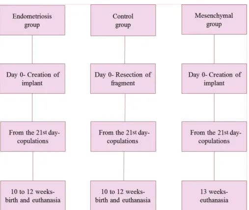
Copulation occurred 4, 6, and 9 weeks after surgery, when the rabbits were fertile. To check fertility, we evaluated the coloring of the vulva daily, and females with red/violet vulva (unlike the normal whitish color)23 were placed in a cage with a male for mating. Abdominopelvic ultrasounds were performed, and the animals were then checked for signs of pregnancy (the presence of a gestational sac or fetus).►Fig. 1 shows aflowchart with a brief description of the chronology of the experiments in the study.
Mesenchymal stem cells were obtained 12 weeks before the application by extracting bone marrow from the long bones of two rabbits. The femur and tibia were removed, and all tissue was scraped away from the bones. The bones were placed in a Petri dish containing phosphate buffered saline (PBS; Gibco Invitrogen, NY, USA). The epiphyses were cut to expose the medullary cavity. The marrow was removed from each bone withflushing medium into a tube (TPP, Trasadin-gen, Switzerland) using a syringe with a needle.
The collected bone marrow was processed using a Ficoll–Hypaque density gradient (d¼1.077 g/mL; Sigma Chemical, St Louis, USA).24The isolated mononuclear cells were plated in 75-cm2 flasks (TPP), and were cultivated using IMDM (Iscove’s Modified Dulbecco’s Media; Gibco Invitrogen) medium containing 15% fetal calf serum (FCS; Gibco Invitrogen) and 1% antibiotics. The cultures were maintained in an incubator, and the culture medium was changed every 72 hours. Upon reaching 80% confluence, the cells were trypsinized using a trypsin–EDTA solution (Gibco Invitrogen), and were subcultured to obtain the required number of cells for potency tests and transplantation.
The MSCs were assessed for potency by inducing their differentiation into adipocytes, osteoblasts, and chondro-cytes (►Fig. 2 A-F). For adipogenic and osteogenic differen-tiation, cells were seeded on glass coverslips (Sarstedt, Newton, USA) in 24-well plates. A commercial differentiation medium (Gibco Invitrogen) was used for 21 days. The MSCs were stained with Oil Red O to analyze adipogenic induction. Large, rounded cells with cytoplasmic lipid-rich vacuoles were observed. Osteogenic differentiation was assessed by visualizing the mineralization of the extracellular matrix with Alizarin Red S staining.
Cells were grown in a micromass culture to promote chondrogenic differentiation.25In the chondrogenic differen-tiation assays, high-density micromass MSC cultures gener-ated cellular nodules, which produced large amounts of cartilage-related extracellular matrix molecules, such as col-lagen. Paraffin sections of the aggregates stained with tolui-dine blue showed condensed structures with cuboidal cells and chondrocyte-like lacunae.
Untreated control cultures, which were grown in regular medium without adipogenic, osteogenic, or chondrogenic
differentiation stimuli, did not exhibit spontaneous adipo-cyte, osteoblast, or chondrocyte formation after 21 days of MSC cultivation.
After 21 days, in the Mesenchymal group, the second surgery for the application of the mesenchymal cells was also performed.
During this reoperation, the size of the lesion formed by the implant was checked. All the implants were considered viable (with an area not exceeding 25 cm2). We used 2
106 cells in each animal (2 ml in a syringe), which were applied on the edge of the implant.
For the histological analysis, endometrial implantsfixed in formalin were prepared in paraffin blocks. These samples were later sectioned at a thickness of 5μm (4 cuts per sample),
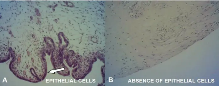
stained with hematoxylin and eosin, and evaluated with an optical microscope. The lesions were classified according to Keenan’s index.26Scores were assigned as follows: score of 3: a well-preserved epithelial layer; score of 2: moderately pre-served epithelium with leukocyte infiltration; score of 1: poorly preserved epithelium (occasional epithelial cells only); and score of 0: no epithelium (►Fig. 3 A-B).
The sample size was based on similarity with other articles; to control Type II Error in the applied tests, test power was used.
Qualitative variables were described as frequencies and percentages, and quantitative variables were described as means, medians, and quartiles. Fisher’s exact test was per-formed to compare dichotomous qualitative variables among the groups. The quantitative variables were compared by the nonparametric Mann-Whitney and Kruskal-Wallis tests. The Mann-Whitney test was used for post-hoc multiple compar-ison with Boniferroni correction. Values of p<0.05 were
considered statistically significant. The data were analyzed with the Statistical Package for the Social Sciences (SPSS; IBM SPSS, Chicago, IL, US) software, version 20.0.
Results
The mesenchymal group had the worst fertility rate (11.1%; p¼0.015; test power: 84.29%); only one animal in this group became pregnant. In contrast, seven rabbits in the control group and seven in the endometriosis group (77.8%) became pregnant. After having executed the surgical procedures and waited 21 days, the time to start fertility was different between the groups. The mesenchymal group had the longest delay when compared with the other groups (p¼0.005; test power: 92.24%) (►Table 1). Considering a significant difference found among the groups, they were compared two by two.
Thepvalues and the test power of these comparisons are shown in the►Table 2.
The endometriosis group had 2 fetal deaths (28.57%) and 5 live births (71.43%). The control group had 7 pregnancies, which resulted in 2 fetal deaths (28.57%), 3 abortions (42.86%), and 2 live births (28.57%).
There was no difference in implant size between the endometriosis (median¼55, 1st quartile¼15, and 3rd quartile¼90) and mesenchymal groups (median¼55, 1st quartile¼25, and 3rd quartile¼80;p¼0.863).
Macroscopically, the formation of a large adhesion in the cavity was noted in the mesenchymal group, even making it difficult to identify the implants (►Fig. 4A). Comparatively, a minor amount of adhesion was noticed in the endome-triosis group (►Fig. 4B). Finality, discrete adhesion forma-tion was observed in the control group. Despite the formation of adhesions, endometriotic lesions were only found in the sites implanted, and no dissemination was observed.
The persistence of epithelial cells in the endometrial implants was assessed semi-quantitatively with Keenan’s index26(►Fig. 3 A-B). The following results were observed among the nine analyzed rabbits in each group. The endo-metriosis group included 3 rabbits with a Keenan index of 0, and 6 with a Keenan index of 3, and the mesenchymal group included 2 rabbits with a Keenan index of 0 and 7 with a Keenan index of 3 (p¼0.730).
Fig. 3 (A) Histological image of the endometrial implant with a Keenan index of 3. (B) Histological picture of the endometrial implant with a Keenan index of 0.
Table 1 Comparison of time until the onset of the fertile period (in days) in the three groups
Groups n Span in days
Endometriosis 9 33 [18–34]
Mesenchymal 9 14 [14–16]
Control 9 40 [33–40]
Note: Nonparametric test of Kruskal-Wallis;p<0.05; median [1st- 3rd
quartiles]; Test power¼92.24%.
Table 2 Test power andpvalues in the inter-group comparison
of time until the onset of the fertile period
Groups p Test power
Control x Endometriosis 0.061 0.3314747
Control x Mesenchymal 0.043 0.7495601
Endometriosis x Mesenchymal <0.001 0.9583571
Discussion
The selection of rabbits as an experimental model was based on their efficacy for developing ectopic endometrial foci in the abdominal wall.8 It is known that the production of various cytokines occurs in endometriosis, which generally stimulates endometrial proliferation and contributes to the production of acute-phase inflammatory proteins, creating a pro-inflammatory environment.22 It is also known that in endometriosis, the endometrial cells exhibit the character-istics of chronicity and expansion, showing capacities for migration, invasion, and apoptosis resistance.9
This scenario has led to new theories exploring the involvement of the immune system, such as immunological mechanisms facilitating the development of endometriosis, or the immune system being activated by it.9The involve-ment of stem cells (particularly of the mesenchymal lineage) in the origin of the disease has also been proposed.11,12,15In contrast with the reports on endometriosis leading to in-fertility,6,27the development of endometriosis in the rabbits did not worsen the fertility rate. However, the rabbits were not a good model for endometriosis-related infertility. The use of the obtained MSCs caused the relative risk of infertility to increase 4-fold in these animals.
One mechanism that may explain the aforementioned result involves the pathophysiology of the disease. The evidence of stem cells in the human endometrium and the suggestion of the presence of stem cells—specifically the mesenchymal lineage8—may be related to the worsening of the fertility rate in the group that received mesenchymal cells, because we used cells that may be involved in the origin and progression of the disease.
Another mechanism that may be involved is the epithe-lial–mesenchymal transition (a process comprising a series of changes in epithelial cell phenotype that causes epithelial cells to assume the characteristics of MSCs, such as having a greater capacity for migration, invasion, and apoptosis resistance).28Because endometriotic cells in the context of
endometriosis also take on this function,8,9,28,29they repre-sent a sum of the effects of both mechanisms.
A question remains regarding whether the mesenchymal group’s delay in entering the fertile period was related to the involvement of MSCs in the origin and progression of endometriosis.
With regard to obstetric outcomes, the endometriosis and control groups showed no statistically significant difference. This result may be because of the small sample size. The mesenchymal group requires further analysis because of its low fertility rate.
The macroscopic evaluation of the pelvic cavity of each animal (during euthanasia) revealed that the mesenchymal group had a greater rate of adhesions than the other groups, in an analogy with the widely used Revised Classification of the American Society for Reproductive Medicine (ASRM).30 Thus, we could consider the mesenchymal group a carrier of stage-IV disease (severe endometriosis with dense andfirm adhesions).
It is known that anatomical distortion and adhesions are some of the mechanisms that generate infertility in the context of endometriosis.4,21,31This mechanism may explain why the group that received MSCs on the endometrial implants may have had a low fertility rate: its higher rate of pelvic adhesions.
Finally, a histological analysis of the implants using Keen-an’s index was performed. This index measures the persis-tence of epithelial cells in endometrial implants.26 A comparison of the two groups revealed no statistically sig-nificant difference in the scores of Keenan’s index.
Declaration of Conflicting Interests
The Authors have no conflicts of interest to declare.
Acknowledgments
We would like to thank the teams from Vivarium, from the Operative Technique Laboratories, and from the Cell Therapy and Pathology Departments of Pontifícia Univer-sidade Católica do Paraná (PUC-PR) for their dedication and support.
References
1 Burney RO, Giudice LC. Pathogenesis and pathophysiology of endometriosis. Fertil Steril 2012;98(03):511–519
2 Nisolle M, Donnez J. Peritoneal endometriosis, ovarian endome-triosis, and adenomyotic nodules of the rectovaginal septum are three different entities. Fertil Steril 1997;68(04):585–596 3 Lafay Pillet MC, Huchon C, Santulli P, Borghese B, Chapron C,
Fauconnier A. A clinical score can predict associated deep infil-trating endometriosis before surgery for an endometrioma. Hum Reprod 2014;29(08):1666–1676
4 Porto BTC, Ribeiro HSAA, Galvão MAL, Sekula VG, Aldrigui JM, Ribeiro PAA. [Histological classification and quality of life in women with endometriosis]. Rev Bras Ginecol Obstet 2015;37(02):87–93 5 Stratton P. The association of clinical symptoms with deep
infiltrating endometriosis: the importance of the preoperative clinical assessment. Hum Reprod 2014;29(08):1627–1628 6 Oliveira R, Musich DS, Ferreira MPSF, Vilarino FL, Barbosa CP. Perfil
epidemiológico das pacientes inférteis com endometrioses. Re-prod Clim 2015;30(01):5–10
7 Eskenazi B, Warner ML. Epidemiology of endometriosis. Obstet Gynecol Clin North Am 1997;24(02):235–258
8 Li T, He H, Liu R, Wang SX, Pu DM. Isolation and identification of epithelial and stromal stem cells from eutopic endometrium of women with endometriosis. Eur J Obstet Gynecol Reprod Biol 2014; 178:89–94
9 Figueira PGM, Abrão MS, Krikun G, Taylor HS. Stem cells in endometrium and their role in the pathogenesis of endometrio-sis. Ann N Y Acad Sci 2011;1221:10–17
10 Jing Z, Qiong Z, Yonggang W, Yanping L. Rat bone marrow mesenchymal stem cells improve regeneration of thin endome-trium in rat. Fertil Steril 2014;101(02):587–594
11 Chan RW, Schwab KE, Gargett CE. Clonogenicity of human endome-trial epithelial and stromal cells. Biol Reprod 2004;70(06):1738–1750 12 Gargett CE. Stem cells in gynaecology. Aust N Z J Obstet Gynaecol
2004;44(05):380–386
13 Deane JA, Gualano RC, Gargett CE. Regenerating endometrium from stem/progenitor cells: is it abnormal in endometriosis, Asherman’s syndrome and infertility? Curr Opin Obstet Gynecol 2013;25(03):193–200
14 Du H, Taylor HS. Stem cells and female reproduction. Reprod Sci 2009;16(02):126–139
15 Vidane AS, Zomer HD, Oliveira BMM, et al. Reproductive stem cell differentiation: extracellular matrix, tissue microenvironment, and growth factors direct the mesenchymal stem cell lineage commitment. Reprod Sci 2013;20(10):1137–1143
16 Dhesi AS, Morelli SS. Endometriosis: a role for stem cells. Womens Health (Lond) 2015;11(01):35–49
17 Mundra V, Gerling IC, Mahato RI. Mesenchymal stem cell-based therapy. Mol Pharm 2013;10(01):77–89
18 Doorn J, Moll G, Le Blanc K, van Blitterswijk C, de Boer J. Therapeutic applications of mesenchymal stromal cells: para-crine effects and potential improvements. Tissue Eng Part B Rev 2012;18(02):101–115
19 do Amaral VF, Dal Lago EA, Kondo W, Souza LC, Francisco JC. Development of an experimental model of endometriosis in rats. Rev Col Bras Cir 2009;36(03):250–255
20 De Miguel MP, Fuentes-Julián S, Blázquez-Martínez A, et al. Immunosuppressive properties of mesenchymal stem cells: advances and applications. Curr Mol Med 2012;12(05): 574–591
21 Howard-Jones N. A CIOMS ethical code for animal experimenta-tion. WHO Chron 1985;39(02):51–56
22 Schor E, Freitas V, Soares Júnior JM, Simões MJ, Baracat EC. Endometriosis: experimental model in rats. Rev Bras Ginecol Obstet 1999;21(05):281–284
23 Risi E. Control of reproduction in ferrets, rabbits and rodents. Reprod Domest Anim 2014;49(Suppl 2):81–86
24 Böyum A. Isolation of mononuclear cells and granulocytes from human blood. Isolation of monuclear cells by one centrifugation, and of granulocytes by combining centrifugation and sedimenta-tion at 1 g. Scand J Clin Lab Invest Suppl 1968;97:77–89 25 Johnstone B, Hering TM, Caplan AI, Goldberg VM, Yoo JU. In vitro
chondrogenesis of bone marrow-derived mesenchymal progeni-tor cells. Exp Cell Res 1998;238(01):265–272
26 Keenan JA, Williams-Boyce PK, Massey PJ, Chen TT, Caudle MR, Bukovsky A. Regression of endometrial explants in a rat model of endometriosis treated with the immune modulators loxoribine and levamisole. Fertil Steril 1999;72(01):135–141
27 Amaral VF, Maestrelli PC, Francisco JC, et al. Bone marrow derived mononuclear stromal cells and experimental model of deep endometriosis. Int J Clin Exp Pathol 2016;9(02):1116–1126 28 Matsuzaki S, Darcha C. Epithelial to mesenchymal transition-like
and mesenchymal to epithelial transition-like processes might be involved in the pathogenesis of pelvic endometriosis. Hum Re-prod 2012;27(03):712–721
29 Du X, Yuan Q, Qu Y, Zhou Y, Bei J. Endometrial mesenchymal stem cells isolated from menstrual blood by adherence. Stem Cells Int 2016;2016:3573846
30 Revised American Society for Reproductive Medicine Classifica-tion of Endometriosis. Fertil Steril 1996;67(05):817–821 31 de Ziegler D, Borghese B, Chapron C. Endometriosis and