Evaluation of contrast media submitted to ionizing
radiation*
Avaliação de meios de contraste submetidos à radiação ionizante
Kátia Elisa Prus Pinho1, Pedro Miguel Gewehr2, Caroline Werner Pereira da Silva3, Andersson Barison4, João Gilberto Tilly Júnior5, Danyel Scheidegger Soboll6
OBJECTIVE: The purpose of the present study was to investigate the influence of ionizing radiation from x-rays and gamma rays on the molecular structure stability of several radiologic contrast media employed in diagnostic imaging by means of 1
H and 13
C nuclear magnetic resonance spectroscopy. MATERIALS AND METHODS: Eight different types of iodinated contrast media (three ionic and five non-ionic) were exposed to x-rays and gamma rays irradiation. Subsequently, the 1
H and 13
C{1
H} nuclear magnetic resonance spectra of these contrast media were collected. RESULTS: The 1H and 13C{1H} nuclear magnetic resonance spectra
of both ionic and non-ionic contrast media irradiated by x-rays or gamma rays demonstrated the absence of any alteration of the contrast media chemical composition. CONCLUSION: There is no problem in keeping contrast media inside examination rooms or close to radiological equipment. It is important to mention that, during the tests, the samples were directly irradiated, while in a radiology examination room, the irradiation is not direct and, therefore, radiation levels in these cases are much lower than those employed in the present study. Keywords: Imaging diagnosis; Contrast media; Ionizing radiation; Nuclear magnetic resonance spectroscopy; X-rays; Gamma rays.
OBJETIVO: O presente estudo consistiu em investigar a influência da radiação ionizante por raios X e raios gama sobre a estabilidade molecular de diversos meios de contraste radiológicos utilizados em exames de diagnóstico por imagem, por meio da espectroscopia de ressonância magnética nuclear de 1
H e 13
C. MATE-RIAIS E MÉTODOS: Oito diferentes meios de contraste iodados (três iônicos e cinco não iônicos) foram expostos à radiação por raios X e raios gama. Em seguida, espectros de ressonância magnética nuclear de 1
H e 13
C{1
H} foram coletados. RESULTADOS: Os espectros de ressonância magnética nuclear de 13C{1H} de ambos os
meios de contraste iônicos e não iônicos irradiados por raios X ou raios gama mostraram que não houve alterações na composição química desses contrastes. CONCLUSÃO: Não há problemas em armazenar as amostras nas salas, ou próximo aos equipamentos em que são realizados os exames. Enfatiza-se que a ra-diação recebida pelas amostras durante os ensaios foi direta, enquanto em uma sala de exames de radiodiag-nóstico a radiação é indireta e, portanto, os níveis de radiações nestes casos são bastante inferiores àqueles empregados neste estudo.
Unitermos: Diagnóstico por imagem; Meios de contraste; Radiação ionizante; Espectroscopia de ressonân-cia magnética nuclear; Raios X; Raios gama.
Abstract
Resumo
* Study developed at Universidade Tecnológica Federal do Paraná (UTFPR), in a partnership with the Laboratory of Nuclear Magnetic Resonance, Department of Chemistry – Universidade Federal do Paraná (UFPR) and Department of Radiotherapy – Hospital Erasto Gaertner, Curitiba, PR, Brazil.
1. M. Sc., Academic Extension Coordinator and Professor, Course of Technology in Radiology, Universidade Tecnológica Federal do Paraná (UTFPR), Curitiba, PR, Brazil.
2. PhD, Professor, Universidade Tecnológica Federal do Paraná (UTFPR), Curitiba, PR, Brazil.
3. Chemist, Fellow Master degree in Chemistry, Universidade Federal do Paraná (UFPR), Curitiba, PR, Brazil.
4. PhD, Professor and Researcher, Department of Chemistry – Universidade Federal do Paraná (UFPR), Curitiba, PR, Brazil.
5. Physicist, Responsible for the Division of General Radiology – Hospital de Clínicas da Universidade Federal do Paraná (UFPR), Curitiba, PR, Brazil.
6. Fellow PhD degree in Biomedical Engineering, Professor, Department of Physics – Universidade Tecnológica Federal do Paraná (UTFPR), Curitiba, PR, Brazil.
Mailing address: Kátia Elisa Prus Pinho. Universidade Tecnológica Federal do Paraná – Diretoria de Extensão. Avenida
prove the quality of radiologic images dates more than half a century(3). Since then,
adverse reactions resulting from the oral or intravenous administration of a foreign substance to the human body have been reported as such compounds are not always harmless, and may alter the blood circula-tion causing unexpected reaccircula-tions(4,5).
Tak-ing this fact into account, several precau-tions must be taken with patients as well as in the preparation and storage of contrast media(6,7).
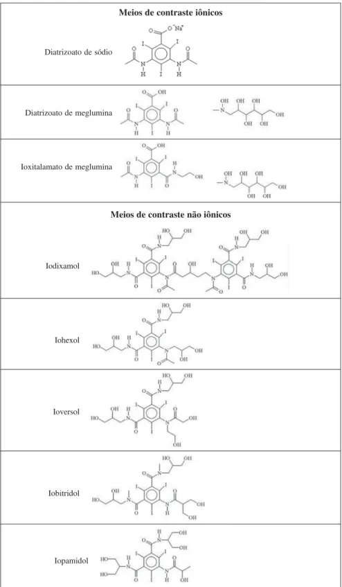
All iodinated contrast media currently in use are derived from 2,4,6-triiodoben-zoic acid (Figure 1). They are classified
ac-Pinho KEP, Gewehr PM, Silva CWP, Barison A, Tilly J, Soboll DS. Evaluation of contrast media submitted to ionizing radia-tion. Radiol Bras. 2009;42(5):309–313.
INTRODUCTION
Radiologic contrast media are com-pounds which are introduced into the or-ganism by different vias, in order to in-crease in the definition of radiographic images due to the contrast improvement caused by such compounds, allowing ac-quisition of high-definition images and as consequence higher accuracy in diagnostic imaging(1,2). The use of compounds to
im-Sete de im-Setembro, 3165, Rebouças. Curitiba, PR, Brazil, 80230-901. E-mail: [email protected]
cording to their physicochemical character-istics, including chemical structure, osmo-larity, viscosity, number of iodine atoms in the structure, biological properties, ioniza-tion capacity in soluioniza-tion, hydrosolubility, lipophilicity and toxicity (6). Ionic contrast
media are those capable of dissociating into cations and anions in aqueous solutions, while the non-ionic ones do not dissociate (Figure 1), but interact with water mol-ecules by means of intermolecular interac-tions(5). Special care in the storage and
asepsis of contrast media is essential, in-cluding storage away from light, as they are photosensitive, and away from the inci-dence of x-rays due to the possibility of ionizing radiations causing molecular deg-radation, thus changing the molecular structure of the contrast media and, as a consequence, its contrast properties on the radiological images. Additionally, it is im-portant to store them under temperatures between 15 and 25°C, as in lower tempera-tures could occurs the crystallization of the contrast media, checking storage time and not using vials after they have been opened for more than 24 hours, due to the risk of contamination by microorganisms(8).
The present study investigates the influ-ence of from x-rays and gamma rays ion-izing radiations on the molecular structure stability of several commercial contrast media widely employed in radiologic im-aging diagnosis by radiography and com-puted tomography by means of 1H and 13C nuclear magnetic resonance (NMR) spec-troscopy. The aim of the work consisted in checking possible changes on the molecu-lar structure of the contrast media due to the incidence of ionizing radiations, once they would alters the physicochemical and bio-logical properties, as well as their toxicity, in order to corroborate with the reported data in the literature(8).
MATERIALS AND METHODS
Samples
Eight different iodinated contrast me-dia, three ionic and five non-ionic (Figure 1) were obtained from four manufacturers available in Brazil. The contrast media, with respective manufacturers were as fol-low: Iopamiron 300 and Pielograf 76% (Bayer Schering Pharma; Berlin, Germany),
Figure 1. Molecular structures of the evaluated iodinated contrast media. Meios de contraste iônicos
Meios de contraste não iônicos Diatrizoato de sódio
Ioxitalamato de meglumina Diatrizoato de meglumina
Iodixamol
Iohexol
Ioversol
Iobitridol
Iopamidol
Ominipaque and Visipaque (Farmasa; São Paulo, Brazil), Henetix and Telebrix (Guer-bet; Paris, France), Optiray 320 and Optiray 350 (Mallinckrodt; Saint Louis, USA). Eight aliquots were taken of each contrast
aliquots of 1.7 mL of each medium were transferred in to microcentrifuge tubes, identified by letters in order to make impos-sible to the instrument operator to known the samples origin. The sample preparation was performed in the Chemistry Labora-tory of the Universidade Tecnológica Fe-deral do Paraná (UTFPR), according to asepsis procedure for each open and aspi-rated vial(9).
X-rays irradiation
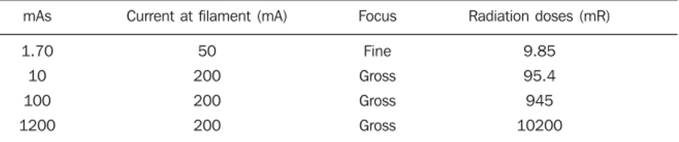
The samples irradiation by x-rays was performed in the Radiotherapy Department of Hospital Erasto Gaertner in the city of Curitiba, PR, Brazil, on a Radcal 9010 ra-diation monitor and a 10X5-6 Radcal ion-ization chamber (Radcal Corp.; Monrovia USA), with a sensitive volume of 6 cm³ calibrated to the radiodiagnosis energy level, thus determining the exact radiation that the samples were being exposed. The source of radiation utilized was an x-ray tube from a RMX 625 R radiotherapy simulator (Raytheon Medical Systems; Melrose Park, USA), with inherent filtra-tion equivalent to 0.5 mm of aluminum. The samples were exposed to different doses of x-rays radiation, ranging from 9.85 to 10200 mR (Table 1), at a distance of 50 cm from the source, and voltage of 90 kV(10).
Gamma rays irradiation
The irradiation of the samples by gamma rays was performed using a mean energy of 1.25 MeV, from a cobalt-60 source installed in a Theratron 780 C tele-therapy unit (MDS Nordion; Ontario, Canada) with a 0.5 cm bolus of gel over the samples. The source-surface distance was 50 cm and the performance at such distance was 5 Gy/min. The samples were exposed to two radiation doses: 0.1 and 10 Gy (10).
In both the cases the samples were irradi-ated in an 8 × 8 cm2 field.
NMR Analysis
The 1H and 13C{1H} NMR spectra were acquired on an Avance 400 (Bruker; Karlsruhe, Germany) NMR spectrometer operating at 9.4 Tesla, installed in the NMR Laboratory of Universidade Federal do Paraná (UFPR), observing the 1H and 13C nuclei at 400.13 and 100.62 MHz,
respec-Table 1 X-rays radiation doses to which the samples were exposed.
mAs
1.70
10
100
1200
Current at filament (mA)
50 200 200 200 Focus Fine Gross Gross Gross
Radiation doses (mR)
9.85
95.4
945 10200
tively, in D2O (deuterated water) at room temperature of approximately 295 K in a 5 mm direct detection multinuclear probe. For this, aliquots of 0.2 ml of each contrast medium were filtered in cotton directly into 5 mm NMR tubes, with the help of Pasteur pipettes, follow by 0.4 ml of D2O contain-ing (3-trimethylsilyl)-2,2’,3,3’-tetradeu-teropropionic acid, sodium salt (TMSP-d4 – internal reference)(11). The 1H NMR spec-tra were acquired with the zg pulse se-quence by accumulating four averages, 64 K points (1 K = 1024) and spectral window of ~13 ppm (11,12). In some samples it was
necessary the pre-saturation of the water signal, using the zgpr pulse sequence. On their turn, the 13C{1H} NMR spectra were acquired with the zgpg30 pulse sequence, accumulating 1024 averages, 32 K points and spectral window of ~255 ppm. Both the 1H and 13C{1H} NMR spectra were processed with aid of TopSpin software (Bruker; Karlsruhe, Germany) by applying exponential multiplications of the free in-duction decay (FID) by factors of 0.3 and 3.0 Hz for the construction of 1H and 13C NMR spectra with 64 K and 32 K points, respectively. All NMR chemical shifts are giving in ppm related to the internal stan-dard signal from TMSP-d4 at 0.0 ppm(11).
RESULTS
In order to evaluating the effected of the application of ionizing radiations on con-trast media utilized in radiology, 13C{1H} and 1H NMR spectra were acquired from the samples after being exposed to ioniz-ing radiations from by x-rays or gamma rays. In a similar way, NMR spectra were obtained from samples of the same contrast media without exposition to ionizing radia-tions, used as references for comparison with those irradiated.
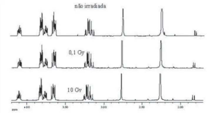
Figure 2 and 3 show the 13C{1H} and 1H NMR spectra comparison, respectively of a ionic contrast medium sample that was exposed to different x-rays radiation doses, while Figures 4 and 5 show the 13C{1H} and 1H NMR spectra comparison, respec-tively of ionic contrast medium sample that was submitted to different gamma ray ra-diation doses.
Figures 6 and 7 present the 13C{1H} and 1H NMR spectra comparison, respectively,
of a non-ionic contrast medium sample submitted to different x-rays radiation doses, while Figure 8 shows the 1H NMR spectra comparison of non-ionic contrast medium sample submitted to different gamma ray radiation doses.
Figure 2. 13C{1H} NMR
Figure 4.13C{1H} NMR spectra comparison (expansion of the region from 20
to 186 ppm) of an ionic contrast medium exposed to different gamma rays radiation doses.
DISCUSSION
The 13C{1H} NMR spectra in both ionic and non-ionic contrast media irradiated by x-rays or gamma rays showed that at the
Figure 7.1H NMR spectra comparison (expansion of the region from 3.2 to
4.2 ppm) of a non-ionic contrast medium exposed to different x-rays radiation doses.
Figure 8.1H NMR spectra comparison (expansion of the region from 1.3 to
4.7 ppm) of a non-ionic contrast medium exposed to different gamma rays radiation doses.
energy levels utilized any changes in the molecular structures of the investigated contrast media occurs, as observed on Fig-ures 2, 4 and 6 for a determined contrast medium. In the same way, the 1H NMR
spectra, which has higher sensitivity than those from 13C{1H}(11), also showed no
changes on the chemical structure of the investigated contrast media, as shown on Figures 3, 5, 7 and 8. In other words, there
Figure 3.1H NMR spectra comparison (expansion of the region from 1.5 to
4.2 ppm) of an ionic contrast medium exposed to different x-rays radiation doses.
Figure 5.1H NMR spectra comparison (expansion of the region from 1.7 to
4.2 ppm) of an ionic contrast medium exposed to different gamma rays radia-tion doses.
Figure 6.13C{1H} NMR spectra comparison (expansion of the region from 20
was no degradation of the analyzed con-trast media, as there were no evidences of new signals in the NMR spectra, which would be an indicative of formation of new compounds, as a consequence of decompo-sition of the contrast media. This becomes evident when the NMR spectra of the con-trol samples which were not submitted to any radiation were compared with those from samples submitted to irradiation, ei-ther by x-rays or gamma rays. In oei-ther words, as all samples presented the same spectral profile at the 1H and 13C{1H} NMR analysis (Figures 2 to 8). Therefore, the molecular structure of the investigated con-trast media is not affected when exposed to irradiation with x-rays or gamma rays, and then there is no problem in storing them in the equipment room or close to the equip-ment in which the examinations are per-formed. Emphasis is given to the fact that the radiation received by the samples dur-ing the trials was direct, while in an actual study the radiation would be indirect and, therefore, the levels of radiation in such cases would be different from those utilized in the present study.
In spite of the fact that some contrast media manufacturers recommend avoiding their storage in the presence of disperse ionizing radiations, the present study did not detect any influence of ionizing radia-tions from x-rays or gamma rays on the
molecular structure of the evaluated iodi-nated contrast media in the described con-ditions. Probably, this observation from manufacturers refers as a general precau-tion in the sense of preserving and manipu-lating only the necessary quantities of con-trast media for the day’s examination needs, while greater quantities might, in some cases, be stored in the examination room being unnecessarily exposed and becoming a constant practice in diagnostic imaging centers.
CONCLUSION
The investigations demonstrated that the ionizing radiation utilized in radiology diagnostic imaging by radiography and computed tomography do not cause any changes in the molecular structures (deg-radation) of the currently utilized contrast media, independently of exposure levels from x-rays or gamma rays radiation in which the contrast media were submitted, and thus allowing the clinical use of such contrast media.
Acknowledgments
The authors are grateful to the Compa-nies Bayer Schering Pharma, Farmasa, Guerbet and Mallinckrodt, that kindly pro-vided contrast media samples as well as to the Chemistry Laboratories of UTFPR and
UFPR and to the Hospital Erasto Gaertner, for supporting the development of the present study with their equipments.
REFERENCES
1. Falgas BJ, Hurlé ADG, Lecumberri VN, et al. Farmacia hospitalaria. Madrid:Sociedad Espa-ñola de Farmacia Hospitalar; 2002.
2. Bontrager LK. Tratado de técnica radiológica e base anatômica. Rio de Janeiro: Guanabara Koogan; 1999.
3. Bettmann MA. Frequently asked questions: iodi-nated contrast agents. Radiographics. 2004;24 Suppl 1:S3–10.
4. Asociación Argentina de Alergia e Imunología Clínica. Reacciones adversas a medios de con-traste radiológicos: criterios y conductas. AAAelC y SAR. 2001;32:101–5.
5. Schild H. Todo sobre medios de contraste: ver o no ver. España: Schering AG; 1995.
6. Pinho KEP. Avaliação de fatores de riscos na uti-lização de contrastes iodados em exames de uro-grafia excretora [dissertação de mestrado]. Curi-tiba: CPGEI/UTFPR; 2006.
7. Colégio Brasileiro de Radiologia. Assistência à vida em radiologia. São Paulo: CBR; 2000.
8. Sugawara AM, Daros KAC. Manual de meios de contraste em raios X. São Paulo: São Camilo; 2004.
9. Nischimura L, Potenza M, Cesaretti I. Enferma-gem nas unidades de diagnóstico por imaEnferma-gem. São Paulo: Atheneu; 1999.
10. Stanton R, Stinson D. Applied physics for radia-tion oncology. Madison: Medical Physics Pub; 1996.
11. Günter H. NMR spectroscopy: basic principles, concepts, and applications in chemistry. Chiches-ter: John Wiley & Sons; 1995.