0103 - 5053 $6.00+0.00
Article
*e-mail: [email protected]
Further Dibromotyrosine-Derived Metabolites from the Marine Sponge
Aplysina caissara
Tatiana O. de Lira,a Roberto G. S. Berlinck,*,a Gislene G. F. Nascimentob and Eduardo Hajduc
a
Instituto de Química de São Carlos, Universidade de São Paulo, CP 780, 13560-970 São Carlos-SP, Brazil
b
Faculdade de Ciências da Saúde, Universidade Metodista de Piracicaba, Rodovia do Açúcar, km 156, 13400-901 Piracicaba-SP, Brazil
c
Museu Nacional, Universidade Federal do Rio de Janeiro, Quinta da Boa Vista, s/n, 20940-040 Rio de Janeiro-RJ, Brazil
A re-investigação química do extrato bruto da esponja Aplysina caissara levou ao isolamento de cinco novos derivados da dibromotirosina, denominados agelocaissarinas A1, A2, B1, B2 e caissarina C, além dos já conhecidos fistularina-3 e 11-hidroxiaerotionina. Os compostos isolados tiveram suas estruturas determinadas pela análise de seus espectros de RMN mono- e bidimensionais, espectro de massas de alta resolução, infravermelho e ultravioleta. A configuração relativa das agelocaissarinas pôde ser estabelecida por análise dos espectros de RMN-1H e modelagem molecular, enquanto que a configuração absoluta do sistema espiroxazolidínico da fistularina-3, da caissarina C e da 11-hidroxiaerotionina pôde ser estabelecida pela análise de seus espectros de dicroísmo circular. A fistularina-3 e a 11-hidroxiaerotionina apresentaram atividade antibiótica moderada contra várias linhagens de bactérias patogênicas.
The re-investigation of the crude extract obtained from the sponge Aplysina caissara led to the isolation of five new dibromotyrosine derivatives, named agelocaissarines A1, A2, B1, B2 and caissarine C, along with the already known fistularin-3 and 11-hydroxyaerothionin. All compounds were identified by analysis of mono- and bidimensional NMR spectra, high resolution mass spectra, infrared and ultraviolet spectra. The relative stereochemistry of agelocaissarines could be established by analysis of 1H NMR spectra and molecular modeling, while the absolute configuration of the spiroxazolidine moieties of fistularin-3, caissarine C and 11-hydroxyaerothionin was established by analysis of 1H NMR and circular dichroism spectra. Fistularin-3 and 11-hydroxyaerothionin displayed moderate antibiotic activity against several pathogenic bacteria.
Keywords: sponge, Aplysina caissara, dibromotyrosine, absolute configuration
Introduction
Marine sponges constitute a remarkable source of novel, potently bioactive secondary metabolites.1,2 In particular,
sponges of the order Verongida have been the source of a variety of biologically active dibromotyrosine-derived modified peptides and alkaloids. Recent examples of compounds belonging to this structural class are the purpurealidins A – D, F – H isolated from the sponge
Psammaplysilla purpurea,3 trisulfide psammalin A, (E,E
)-bromopsammalin A and bispsammalin A from the association of the sponges Jaspis wondoensis and Poecillastra
wondoensis,4 purpuroceratic acids A and B from
Pseudoceratina purpurea,5 several new and known
psammalins active as histone deacetylase and DNA methyltransferase inhibitors, from the sponge Psammaplysilla purpurea,6 the moderately cytotoxic purealidin S and
purpuramine J isolated from the sponge Druinella sp.,7 and
nakirodin A from an unidentified Verongida sponge.8
We have recently reported the isolation of two new members of the dibromotyrosine derivatives, caissarines A (1) and B (2), from the sponge Aplysina caissara. Both
caissarines were identified by analysis of spectroscopic data.9 However, due to the lost of the samples of both 1 and
2, we have been unable to measure their specific rotation
J. Braz. Chem. Soc.
have been interested to re-isolate the compounds in order to complete their characterization and to evaluate their biological activities. Surprisingly, a first recollection of the sponge A. caissara did not provide any dibromotyrosine
derivative.9 Since the recollected sponge was stored in EtOH
at room temperature for several weeks, we considered that the sponge metabolites could suffer degradation under these conditions. It has been indeed reported that dibromotyrosine metabolites of the sponge A. aerophoba can degrade in the
presence of alcohol-H2O mixtures, since the enzymatic activity is not completely suppressed under such conditions.10 Nevertheless, similar experiments carried out
with extracts of the sponges A. insularis and A. archeri did
not presented similar results, since the dibromotyrosine metabolites of these sponges did not suffer degradation when stored in alcohol.11 A second recollection of A. caissara, followed by immediate animal freezing at –20 oC,
rapid transportation to laboratory, freeze-drying and extraction, yielded, after several chromatographic separations, no trace of caissarines A (1) and B (2),9 but
gave fistularin-3 (3),12 11-hydroxyaerothionin (4),13 and five
unprecedented dibromotyrosine derivatives, named agelocaissarines A1 (5), A2 (6), B1 (7), B2 (8) and caissarine
C (9). Herein we report the isolation and structure
determination of the new dibromotyrosine derivatives 5-9
as well as the absolute configuration of the spiroxazolidine moiety of compounds 3, 4 and 9 isolated from A. caissara.
We have also evaluated the antibiotic activity of fistularin-3 (3) and 11-hydroxyaerothionin (4) against several
pathogenic bacteria.
Experimental
General experimental procedures
UV spectra were recorded on a Hitachi U-3210 spectrophotometer. IR spectra (film on Si plate) were recorded on a FT-IR Bomem MB102 infrared spectro-meter. Specific rotations were measured on a Perkin Elmer 241 polarimeter in MeOH. NMR spectra were recorded either on a Bruker ARX 9.4 T instrument, operating at 400 MHz for 1H and 100 MHz for 13C
channels, respectively, or on a Bruker DRX300 7.05 T, operating at 300 MHz for 1H and 75 MHz for 13C,
respectively. All NMR spectra were obtained at 25 oC
Animal collection and identification. The sponge Aplysina caissara (Pinheiro & Hajdu, 2001) was collected during
the summer of 2002 at the São Sebastião channel and immediately frozen at -20 oC. A voucher specimen was
deposited at the Museu Nacional, Universidade Federal do Rio de Janeiro.9
Isolation of compounds 3-9 from Aplysina caissara
The frozen sponge (1278 g) was freeze dried to give 317 g of dry material which was sequentially extracted with MeCN, acetone and MeOH. The MeCN and acetone extracts were separately filtered and evaporated to give brown gums. The MeOH extract was filtered, concentrated to 400 mL of an aqueous suspension, which was partitioned with EtOAc (3 × 400 mL). The organic layer was evaporated, solubilized in MeOH and filtered to eliminate inorganic salts, to yield a dark gum. The MeCN, acetone and EtOAC crude extracts have shown to be virtually identical by TLC analysis (CH2Cl2-MeOH 9:1), and were pooled to a single crude extract (9.25 g). This crude extract was subjected to a series of chromatographic separations by flash chromatography on silica gel (gradients of MeCN in CH2Cl2), by C18 reversed phase column chromatography (gradient of MeOH in H2O), followed by purifications by HPLC with either a C18 reversed phase column (Waters µBondapak 7.8 x 300 mm, 10 m, 100 Å) or a phenyl reversed phase column (Waters µBondapak 7.8 × 300 mm, 10 m, 100 Å) eluting with MeCN-H2O 7:3 or 1:1. The compounds were obtained as amorph solids, caissarine C (9, 23.0 mg), fistularin-3 (3,
40.0 mg), 11-hydroxyaerothionin (4, 127.0 mg),
agelocaissarines A1 and A2 (5 and 6, 4.0 mg) and
agelocaissarines B1 and B2 (7 and 8, 5.0 mg).
11-Hydroxyaerothionin (4).13 Amorphous solid, [α] D28 +
178o (c 0.0014, MeOH). IR (film on Si plate) ν max/cm-1:
3374, 2936, 1659, 1602, 1545, 1436, 1314, 1272, 1108, 992, 918, 775, 664. HRESIMS m/z 854.8400 (Calc. for
C24H2679Br
381BrN4O9Na, 854.8310) corresponding to
[M+Na]+.
Agelocaissarine A1 (5) and agelocaissarine A2 (6).
Amorphous solid. IR (film) νmax/cm-1: 3369, 1701, 1660,
1603, 1544, 1431, 1267, 1108, 1002, 912, 780, 669. HRFABMS m/z 804.81762 (Calc. for C22H2379Br381BrN4O9,
804.81780) corresponding to [M+H]+. 1H and 13C NMR
data: see Tables 1 and 2.
Agelocaissarine B1 (7) and agelocaissarine B2 (8).
Amorphous solid. IR (film) νmax/cm-1: 3348, 1705, 1654,
1602, 1538, 1441, 1268, 1107, 901, 740, 604. HRESIMS
m/z 840.8303 (Calc. for C23H2479Br381BrN4O9Na, 840.8154)
corresponding to [M+Na]+. 1H and 13C NMR data: see
Tables 1 and 2.
Caissarine C (9). Amorphous solid, [α]D28 + 175o (c 0.0021,
MeOH). IR (film) νmax/cm-1: 3351, 2935, 1663, 1596, 1543,
1439, 1309, 1270, 1220, 1106, 988, 911, 766, 739, 606. HRESIMS m/z 868.8544 (Calc. for C25H2879Br381BrN4O9Na,
868.8467) corresponding to [M+Na]+. 1H and 13C NMR
data: see Table 3.
Results and Discussion
Agelocaissarines A1 (5) and A2 (6) were obtained as
an inseparable mixture, which displayed a quasi-molecular cluster [M+H]+ at m/z 803, 805, 807, 809 and 811 by
FABMS, indicating the presence of four bromine atoms in the structures. A HRFABMS measurement at m/z 805
(measured: m/z 804.81762; calculated: 804.81780)
indicated the molecular formula C22H2379Br
381BrN4O9 for
both diastereomers. The 13C NMR spectrum of the
diastereomeric mixture displayed thirteen signals of the major stereoisomer agelocaissarine A1 (5) and eight
signals of the minor stereoisomer agelocaissarine A2 (6)
(Table 1). Five 13C signals were assigned to sp2 carbons:
one α,β-unsaturated ketone carbonyl (C-3 and C-3’) at δ 184.1 and one amide carbonyl (C-9 and C-9’) at δ 160.1 to both 5 and 6, one imine oxide carbon (C-8 and C-8’) at
δ 154.8 to 5 and at δ 155.5 to 6, a vinylic methine (C-5
and C-5’) at δ 149.3 to 5 and at δ 146.2 to 6, as well as a
quaternary bromine-substituted sp2 carbon (4 and
C-4’) at δ 122.7 to 5 (δ 123.0 to 6). The 13C chemical shifts
of the α,β-unsaturated ketone system were clearly reminiscent to those of agelorins A and B isolated from
Agelas oroides,14 of 11,17-dideoxyagelorins A and B
isolated from Suberea aff. praetensa,15 of (1’R, 5’S, 6’S
)- 2-(3’,5’-dibromo-1’,6’-dihydroxy-4’-oxocyclohex-2’-enyl)acetonitrile and its corresponding (1’R, 5’R, 6’S
)-epimer isolated from Aplysinalaevis,16 and of an unnamed
compound isolated from Aplysina archeri.17 Indeed, the
remaining carbon resonances assigned to the 4-(4’,5’-dihydroisoxazolyl)- 2,6-dibromo-5-hydroxycyclohex-2-enone moiety of agelocaissarines A1 and A2 were very similar to the corresponding structural moieties of the preceding mentioned natural products. These included the spiro carbon C-6 at δ 91.5 assigned to 5 (δ 90.5 to 6), the
oximethine carbon C-1 at δ 74.5 for 5 (δ 74.0 for 6) and
the bromine substituted methine C-2 at δ 57.2 for 5 (δ
55.0 for 6). Analysis of the 1H, 13C and HMQC NMR
J. Braz. Chem. Soc.
resonance at δ 4.28 (dd, J 2.2 and 11.4 Hz) of compound
5 to H-1 (at δ 4.39 as a broad singlet in 6), at δ 4.80 (dd,
J 1.3 and 11.5 Hz) of 5 to H-2 (at δ 5.19 also as a broad
singlet in 6), and the 1H resonance at δ 7.51 (s) to H-5 in
compound 5 (δ 7.32, s, in compound 6). The H-7a/H-7b
diastereotopic methylene observed at δ 3.71 (1.3 and 18.0 Hz) and δ 3.15 (dd, J 2.2 and 18.0 Hz) in 5 and at δ 3.76
(dd, 1.3 and 18 Hz) and δ 3.29 (dd, J 2.8 and 18.0 Hz) in
6 completed the 1H assignments of the spiroxazolidine
moiety of both compounds 5 and 6.
Analysis of the 1H-1H COSY spectrum confirmed these
assignments, since H-1 and H-2 were vicinally coupled in
6, while H-1 showed couplings with both H-2 and
H-7a/H-7b (long-range) in compound 5. Additional support for the
spirobicyclic moiety assignments of both 5 and 6 was
obtained by analysis of the HMBC spectra, which showed long range couplings between H-1 and C-2, C-6 and C-7, between H-2 and C-1, C-3 and C-6, between H-5 and C-1, C-3, C-4 and C-7 (weak), as well as between H-7a/H-7b and C-1, C-5, C-6 and C-8 for compound 5. The same
HMBC spectra indicated similar long range couplings between H-5 and C-1, C-3, C-4 and C-7, as well as between H-7a/H-7b and C-1, C-5 and C-6 for compound 6, but no
such couplings were observed for both H-1 and H-2. Considering the molecular formula established by HRFABMS, agelocaissarines A1 (5) and A2 (6) must
present two spirobicyclic moieties joined through a 2-hydroxy-1,4-diaminobutane bridge. The presence of the diamine chain was confirmed by analysis of the NMR data. The methylene group at δ 3.45 (m) and 3.36 (m) in the 1H spectrum (δ
C at 45.9) was assigned to CH2
-10 for both compounds 5 and 6, the oxymethine at δ
3.75 (m) (δC at 68.8) to CHOH-11, the methylene at δ 1.55 (m) and 1.70 (m) (δC at 34.6) to CH2-12, and the methylene at δ 3.25 (m) and 3.31 (m) (δC at 37.0) to CH2-13. The connection of the 2-hydroxy-1,4-diaminobutane chain to the two spiroxazolidine moieties was established through the amide hydrogens, one of them (NH-9a) at δ 7.42 (bt) was vicinally coupled to the methylene CH2-10 (δ 3.45 and 3.36) in the COSY spectrum and showed a long range correlation to the amide carbonyl at δ 160.1. The other amide hydrogen (NH-9a’) at δ 7.26 (bt) showed a vicinal coupling to the methylene CH2-13 at δ 3.25 and 3.31, and also showed a long range coupling to the amide carbonyl at δ 160.1. Since we have not observed a second distinct set of 1H signals of the diamino moiety,
except for the methylene CH2-10, we assumed that the structural difference between 5 and 6 was only the
relative stereochemistry of their respective bicyclic spiroxazolidine moieties.
The relative stereochemistry of the major diastereomer agelocaissarine A1 (5) was established by
analysis of the 1H NMR spectrum and molecular
modeling using both MM2 and MOPAC protocols of the Chem3D software. Considering the presence of three stereogenic centers, it is possible to consider four different relative stereochemistries for the spirobicyclic moieties of 5 and 6. Since the major diastereomer 5
showed H-1 and H-2 vicinally coupled with an 11 Hz coupling constant, the corresponding dihedral angle (φ) H-1/C-1/C-2/H-2 in 5 must be either close to 0o or 180o.
Molecular modeling analysis indicated a dihedral angle φ close to 161o, with a J
calc 10.9 Hz, for the 1(R*), 1’(R*),
2(S*), 2’(S*), 6(S*), 6’(S*) stereoisomer, which indicated
a H-1/H-2 pseudo-trans relationship in compound 5. For
compound 6, both H-1 and H-2 were observed as broad
singlets in the 1H NMR spectrum (see Figure 1),
indicating a dihedral angle close to 90o between these
hydrogens. Molecular modeling analysis indicated a dihedral angle close to 80o, with a J
calc 1.0 Hz,
corresponding to the 1(R*), 1’(R*), 2(R*), 2’(R*), 6(S*),
6’(S*) stereoisomer, in which H-1 and H-2 in 6 must
present a pseudo-axial/pseudo-equatorial relationship.
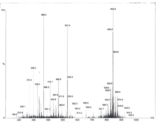
Agelocaissarines B1 (7) and B2 (8) were also isolated as
an unseparable mixture which displayed a quasi molecular ion cluster at m/z 838, 840, 842, 844 and 846. A HRESIMS
measurement at m/z 840.8303 (calc. 840.8154) indicated the
formula C23H2479Br
381BrN4O9Na of the corresponding sodium
adduct. Therefore, compounds 7 and 8 were assigned as the
higher homologues of compounds 5 and 6 at the 2-hydroxy
diamino chain. This hypothesis was confirmed by analysis of NMR data of 7 and 8 (Tables 1 and 2), indicating the
CH2-14 at δ 3.35 (m) and 3.24 (m) (δC 37.0) in the COSY spectrum. The remaining NMR data of compounds 7 and 8
was essentially identical to the corresponding assignments of 5 and 6, including the relative stereochemistries of the
bicyclic spiroxazolidine systems.
The relative stereochemistry of agelocaissarines A1 (5) and A2 (6) as well as of agelocaissarines B1 (7) and
B2 (8) are in agreement to the relative stereochemistry
established for agelorines A and B,14
11,17-dideoxy-agelorins A and B,15 and the monocyclic nitriles isolated
from A. laevis.16 These compounds have a similar bicyclic
spiroxazolidine system, or a monocyclic substituted ring, in which the CH2-7 methylene and the C-1 hydroxyl group have a cis relationship. The hypothesis that the
two bicyclic spiroxazolidine moieties within 5 and 6 or
in 7 and 8 may not have an identical relative
stereo-chemistry was considered, but rulled out since the 1H
NMR signals of each single diastereomer had consistent
intensities (measured by the integration of 1H signals).
In the spectrum of agelocaissarines B1 (7) and B2 (8),
the minor diastereomer 8 corresponds to ca. 26% the
amount of 7. In the case of agelocaissarines A1 (5) and
A2 (6), the amount of the minor diastereomer 6
corresponds to ca. 50% of compound 5. Since in the 1H
NMR spectra of compounds 5 and 6 and of compounds 7 and 8 the 1H signals of the bicyclo spiroxazolidine
moiety with the 1(R*), 1’(R*), 2(R*), 2’(R*), 6(S*),
6’(S*) stereochemistry are consistently of lower intensity,
it is clear that each compound of the pairs 5 and 6 as
well as 7 and 8 did not present two bicyclic
spiroxazolidine systems with different relative stereo-chemistry each. This fact is relevant, since it would be possible to consider these compounds as artifacts of isolation generated by acid-catalyzed hydrolysis of the methoxyl group. However, such a hypothesis is questionable due to the following reasons. Firstly, a related metabolite with a 11-keto functionality was isolated from the sponge Aplysina archeri as a single
diastereomer.17 Secondly, if the bicyclo spiroxazolidine
system present in agelocaissarines A1, A2, B1 and B2 and related metabolites isolated from other sponges14-17
would be chemically generated in vitro, we would expect
to isolate compounds with each of the two bicyclo spiroxazolidine moieties presenting distinct relative stereochemistries. We have been unable to detect such compounds, which are likely to present a very close retention time to the agelocaissarines in the HPLC analysis. The mixture of 5 and 6 as well as the mixture
of 7 and 8 have proven to be inseparable under several
different HPLC separation conditions using reversed phase with C18 or phenyl bonded columns, or using normal phase separation conditions using silica gel or
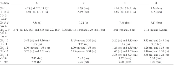
Table 2. 1H NMR data (300 MHz, MeCN-d
3) obtained for agelocaissarines A1 (5), A2 (6), B1 (7) and B2 (8)
Position 5 6 7 8
CH-1, 1’ 4.28 (dd, 2.2, 11.4)* 4.39 (brs) 4.14 (dd, 3.0, 11.6) 4.24 (brs) CH-2, 2’ 4.80 (dd, 1.3, 11.5) 5.19 (brs) 4.65 (dd, 1.0, 11.6) 5.05 (brs) C-3, 3’
C-4,4’
CH-5, 5’ 7.51 (s) 7.32 (s) 7.36 (brs) 7.17 (brs)
C-6, 6’
CH2-7, 7’ 3.71 (dd, 1.3, 18.0) and 3.15 (dd, 2.2, 18.0) 3.76 (dd, 1.3, 18.0) and 3.29 (2.8, 18.0) 3.01 (m) and 3.5 (m) 3.72 (m) and 3.28 (m) C-8, 8’
C-9, 9’
CH2-10 3.45 (m) and 3.36 (m) 3.45 (m) and 3.36 (m) 3.20 (m) and 3.13 (m) 3.33 (m) and 3.49 (m)
CH-11 3.75 (m) 3.75 (m) 3.15 (m) 3.15 (m)
CH2-12 1.70 (m) and 1.55 ( m) 1.70 (m) and 1.55 (m) 1.26 (m) and 1.35 (m) 1.26 (m) and 1.35 (m) CH2-13 3.25 (m) and 3.31 (m) 3.25 (m) and 3.31 (m) 1.46 (m) and 1.55 (m) 1.46 (m) and 1.55 (m)
CH2-14 3.35 (m) and 3.24 (m) 3.35 (m) and 3.24 (m)
NH-9a 7.42 (brt) 7.42 (brt) 7.37 (brm) 7.37 (brm)
NH-9a’ 7.26 (brt) 7.26 (brt) 7.20 (brm) 7.20 (brm)
*Multiplicity and coupling constant (Hz) in parentheses. Table 1. 13C NMR data (75 MHz, MeCN-d
3) obtained for agelocaissarines A1 (5), A2 (6), B1 (7) and B2 (8)
Position 5 6 7 8
C-1, 1’ 74.5 74.0 74.5 68.5
C-2, 2’ 57.2 55.0 57.2 54.6
C-3, 3’ 184.1 184.1 184.2 183.0 C-4,4’ 122.7 123.0 122.7 123.2 C-5, 5’ 149.3 146.2 149.4 146.3
C-6, 6’ 91.5 90.5 91.4 90.5
C-7, 7’ 38.2 41.4 38.2 41.4
C-8, 8’ 154.8 155.5 154.8 155.5 C-9, 9’ 160.1 160.1 160.1 159.7
C-10 45.9 45.9 45.9 45.9
C-11 68.8 68.8 70.3 70.3
C-12 34.6 34.6 32.4 32.4
C-13 37.0 37.0 26.0 26.0
J. Braz. Chem. Soc.
phenyl bonded HPLC columns. Therefore, it seems that these compounds are true secondary metabolites, although it has been recently mentioned that prolonged standing of 11-epi-fistularin-3 at -20 oC led to the
formation of both agelorins A and B.18
A biogenetic pathway to the formation of the bicyclo spiroxazolidine system present in agelocaissarines A1, A2, B1 and B2, as well as in the related metabolites mentioned previously,14-17 is proposed in Scheme 1. A possible
enzyme-catalyzed methyl extrusion, followed by proton capture concomitantly to the enol ketolyzation gives the bicyclic spiroxazolidine system unique to this class of secondary metabolites, of which only seven related compounds are known up to the present.14-17 The enzymatic
formation of two different stereoisomers in different proportions suggest that this mechanism has a low stereospecificity, indicating that proton capture may not be enzymatically controlled.
The isolation of 11-hydroxyaerothionin (4) and
caissarine C (9) from A. caissara during the present
investigation is a support that 4 and 9 are the biogenetic
precursors of each of the diastereomeric pairs, 5 and 6 and 7 and 8, respectively. Caissarine C (9) was obtained as an
amorphous solid which gave a quasi-molecular sodium adduct [M+Na]+ ion cluster at m/z 866/868/870/872/874.
A HRESIMS measurement at m/z 868.8544 (calculated:
868.8467) indicated the formula C25H2879Br
381BrN4O9Na,
corresponding to the 11-hydroxyaerothionin higher homologue (or to the caissarine B lower homologue). This hypothesis was fully supported by analysis of the 1H and 13C NMR (Table 3), 1H-1H COSY and HMQC spectra, as
well as by comparison with data reported for 11-hydroxyaerothionin13 and for caissarine B.9
11-Hydroxyaerothionin (4) also was identified by
analysis of spectroscopic data, including HRESIMS, 1H, 13C, 1H-1H COSY, HMQC and HMBC NMR spectra, as
well as by comparison with literature data.19 The absolute
stereochemistry of 4 isolated from A. caissara was
established by analysis of 1H NMR and circular dichroism
spectra. The 1H spectrum of 4 displayed the signals of
H-1 (H-H-1’) and H-6 (H-6’) as broad singlets in MeCN-d3.
Molecular modeling indicated that a 1R*, 6R*
confi-Scheme 1. Postulated biogenetic pathway of the agelocaissarines.
Table 3. 1H (400 MHz) and 13C (100 MHz) NMR data (DMSO-d 6) ob-tained for caissarine C (9)
Position δ13C δ1H (multiplicity, J in Hz) C-1, 1’ 72.4
C-2, 2’ 119.6 C-3, 3’ 145.9 C-4,4’ 111.9
C-5, 5’ 130.1 6.58 (s) C-6, 6’ 89.0 (88.9)a
C-7, 7’ 38.5 3.20 (d, 18.2) and 3.62 (d, 18.2) C-8, 8’ 153.3 (153.4)b
C-9, 9’ 157.4
C-10 44.2 3.30 (m) and 3.00 (m) C-11 67.2 (65.7)c 3.54
C-12 30.6 1.40 (m) and 1.24 (m) C-13 24.0 1.60 (m) and 1.45 (m) C-14 35.0 (32.5)c 3.15 (m)
OCH3 58.4 3.64 (s)
NH-9 8.51 (t, 5.5) [8.46 (t, 5.8)]c NH-9a 8.27 (t, 5.5) [8.32 (5, 5.8)]c
guration would present a W long range coupling between H-1 (H-1’) and H-6 (H-6’), which was not observed in both 1H and COSY spectra. Therefore, the relative
configuration should be 1R*, 6S*, which was confirmed
by analysis of the CD spectrum of 4 {[θ]284max + 68,000,
[θ]248max + 78,000}, virtually identical to those of
11-epi-fistularin-3 and aerothionin, both of which have the 1R,
1’R, 6S, 6’S absolute configuration.14,19 Therefore, the
absolute configuration of both spiroxazolidine bicyclic moieties of 4 was assigned as 1R, 1’R, 6S, 6’S. Since the
specific rotation of 11-hydroxyaerothionin (4) previously
isolated from the sponge Pseudoceratina durissima was
[α]D +189o (c = 0.15, solvent not specified)13 and the
specific rotation of 4 isolated from A. caissara was [α]D +178o (c = 0.0014, MeOH), we can suggest the same
absolute configuration for the spiroxazolidine bicyclic moieties of both compounds.
The same 1(R), 1’(R), 6(S), 6’(S) absolute
stereo-chemistry of the spiroxazolidine moieties was assigned for fistularin-3 (3) and caissarine C (9) isolated from A. caissara, based on the analysis of the 1H NMR and CD
spectra of 3 {[θ]290max + 67,500, [θ]
248max + 76,000} and
of 9 {[θ]290max + 90,000, [θ]
248max + 87,000}. It is worth of
mention that the absolute stereochemistry at C-1, C-1’, C-6 and C-6’ of fistularin-3 (3) isolated from A. caissara
is identical to the same compound recently reported from
A. cauliformis.18 Therefore, if we consider
11-hydroxy-aerothionin (4) as the direct biogenetic precursor of
agelocaissarines A1 (5) and A2 (6), and caissarine C (9)
the precursor of agelocaissarines B1 (7) and B2 (8), the
relative stereochemistry assigned for each component of the diastereomeric pairs may be considered as their respective absolute configurations. No attempts have been made to establish the absolute stereochemistry at C-11 of compounds 4-9.
The dibromotyrosine derivatives fistularin-3 (3) and
11-hydroxyaerothionin (4) displayed moderate
antibiotic activity against Escherichia coli ATCC 25922
(MIC at 300 µg mL-1 for 3, while 4 was inactive),
Pseudomonas aeruginosa ATCC 27853 (MIC at 300
µg mL-1 for 4, while 3 was inactive), oxacillin resistant Staphylococcus aureus strain 8 (MIC at 600 µg mL-1
for both 3 and 4), oxacillin resistant S. aureus strain
108 (MIC at 50 µg mL-1 and 80 µg mL-1 for 3 and 4,
respectively), and no activity at all when tested against
Pseudomonas aeruginosa and Candida albicans.
Compounds 5, 6, 7 and 8 were not tested since they
were isolated as mixtures.
In conclusion, we described five new dibromo-tyrosine derivatives from the sponge A. caissara,
agelocaissarines A1 (5), A2 (6), B1 (7) and B2 (8),
isolated as pairs of diastereomers, and caissarine C (9).
The relative stereochemistry of the bicyclic spiroxazo-lidine moiety of compounds 5-8 could be assigned by
1H NMR and molecular modeling analysis. The
concurrent isolation of fistularin-3 (3) and
11-hydroxyaerothionin (4) enabled us to establish the
absolute configuration of the spiroxazolidine moiety in 4 and 9 by 1H NMR and circular dichroism analysis.
Compounds 3 and 4 displayed moderate antibiotic
activity against different pathogenic bacteria.
Acknowledgments
The authors thank to Prof. José Carlos de Freitas and the technical staff of CEBIMar-USP for the many facilities during the sponge collection. The authors are also gratefully indebted to Dr. Norrie Pearce and Prof. Brent R. Copp (University of Auckland, Auckland, New Zealand) and to Dr. David E. Williams and Dr. Prof. Raymond J. Andersen (University of British Columbia, Vancouver, Canada) for their assistance in the MS analyses, as well as to Prof. Gil V. J. Silva and Virginia H. B. Glass (Departamento de Química, Faculdade de Filosofia, Ciências e Letras de Ribeirão Preto, Universidade de São Paulo) in obtaining the NMR spectra. Financial support to RGSB was funded by FAPESP grant 01/03095-5. EH received grants and/or fellowships from CNPq and FAPERJ. T.O.L. thanks CAPES for a MSc scholarship.
Supplementary Information
Supplementary Information is avaliable free of charge at http://jbcs.sbq.org.br, as PDF file.
References
1. Faulkner, D. J.; Nat. Prod. Rep. 2002, 19, 1.
2. Blunt, J. W.; Copp, B. R.; Munro, M. H. G.; Northcote, P. T.; Princep, M. R.; Nat. Prod. Rep. 2006, 23, 26.
3. Tilvi, S.; Rodrigues, C.; Naik, C. G.; Parameswaran, P. S.; Wahidhulla, S.; Tetrahedron 2004, 60, 10207.
4. Park, Y.; Liu, Y.; Hong, J.; Lee, C. O.; Cho, H.; Kim, D. K.; Im, K. S.; Jung, J. H.; J. Nat. Prod. 2003, 66, 1495.
5. Kijjoa, A.; Bessa, J.; Wattanadilok, R.; Sawangwong, P.; Nascimento, M. S. J.; Pedro, M.; Silva, A. M. S.; Eaton, G.; Van Soest, R.; Herz, W.; Z. Naturforsch., B: Chem. Sci. 2005, 60, 904.
6. Pina, I. C.; Gautschi, J. T.; Wang, G. Y. S.; Sanders, M. L.; Schmitz, F. J.; France, D.; Cornell-Kennon, S.; Sambucetti, L. C.; Remiszewski, S. W.; Perez, L. B.; Bair, K. W.; Crews, P.; J.
Nat. Prod. 2004, 68, 3866.
J. Braz. Chem. Soc.
8. Tsuda, M.; Endo, T.; Watanabe, K.; Fromont, J.; Kobayashi,
J.; J. Nat. Prod. 2002, 65, 1670.
9. Saeki, B. M.; Granato, A. C.; Berlinck, R. G. S.; Magalhaes, A.; Schefer, A. B.; Ferreira, A. G.; Pinheiro, U. S.; Hajdu, E.; J. Nat. Prod. 2002, 65, 796; Saeki, B. M.; Granato, A. C.; Berlinck, R.
G. S.; Magalhaes, A.; Schefer, A. B.; Ferreira, A. G.; Pinheiro, U. S.; Hajdu, E.; J. Nat. Prod. 2003, 66, 1039.
10. Ebel, R.; Brenzinger, M.; Kunze, A.; Gross, H. J.; Proksch, P.;
J. Chem. Ecol. 1997, 23, 1451.
11. Puyana, M.; Fenical, W.; Pawlik, J. R.; Mar. Ecol. Prog. Ser.
2003, 246, 127.
12. Gopichand, Y.; Schmitz, F. J.; Tetrahedron Lett. 1979, 3921.
13. Kernan, M. R.; Cambie, R. C.; Bergquist, P. R.; J. Nat. Prod.
1990, 53, 615.
14. König, G. M.; Wright, A. D.; Heterocycles 1993, 36, 1351.
15. Kijjoa, A., Watanadilok, R., Sonchaeng, P., Silva, A. M. S., Eaton, G., Herz, W.; Z. Naturforsch., C: J. Biosci. 2001, 56, 1116.
16. Capon, R. J.; MacLeod, J. K.; Aust. J. Chem. 1987, 40, 341.
17. Ciminiello, P.; Dell’Aversano, C.; Fattorusso, E.; Magno, S.; Carrano, L.; Pansini, M.; Tetrahedron 1996, 52, 9863.
18. Rogers, E. W., Oliveira, M. F., Berlinck, R. G. S., König, G. M., Molinski T. F.; J. Nat. Prod. 2005, 68, 891; Oliveira, M. F.,
Oliveira, J. H. H. L., Galetti, F. C. S., Souza, A. O., Silva, C. L., Hajdu, E., Peixinho, S., Berlinck, R. G. S.; Planta Med. 2006, 72, 437, in which the absolute configuration of
(+)-fistularin-3 and 11-deoxyfistularin-(+)-fistularin-3 was correctly drawn but inversely indicated as 1(S), 1’(S), 6(R), 6’(R).
19. McMillan, J. A.; Paul, I. C.; Goo, Y. M.; Rinehart, Jr., K. L.; Krueger, W. C.; Pschigoda, L. M.; Tetrahedron Lett. 1981, 22, 39.
Received: February 28, 2006 Published on the web: August 11, 2006
0103 - 5053 $6.00+0.00
Supplementary Information
*e-mail: [email protected]
Further Dibromotyrosine-Derived Metabolites from the Marine Sponge
Aplysina caissara
Tatiana O. de Lira,a Roberto G. S. Berlinck,*,a Gislene G. F. Nascimentob and Eduardo Hajduc
a
Instituto de Química de São Carlos, Universidade de São Paulo, CP 780, 13560-970 São Carlos-SP, Brazil
b
Faculdade de Ciências da Saúde, Universidade Metodista de Piracicaba, Rodovia do Açúcar, km 156, 13400-901 Piracicaba-SP, Brazil
c
Museu Nacional, Universidade Federal do Rio de Janeiro, Quinta da Boa Vista, s/n, 20940-040 Rio de Janeiro-RJ, Brazil
Figure S1. 1H NMR spectrum (300 MHz, MeCN-d
J. Braz. Chem. Soc.
Figure S2. 13C NMR spectrum (75 MHz, MeCN-d
3) of agelocaissarines A1 (5) and A2 (6).
Figure S4. 1H NMR spectrum (MeCN-d
3, 400 MHz) of agelocaissarines B1 (7) and B2 (8).
Figure S5. 13C NMR spectrum (75 MHz, MeCN-d
J. Braz. Chem. Soc.
Figure S6. ESI+ mass spectrum of agelocaissarines B1 (7) and B2 (8).
Figure S7. 1H NMR spectrum (DMSO-d
Figure S8. 13C NMR spectrum (100 MHz, DMSO-d
6) of caissarine C (9).