*e-mail: [email protected]
Hydrothermal Method for Preparing Calcium Phosphate Monoliths
Raúl García Carrodeguasa*, Loreley Morejón Alonsoa, José Angel Delgado García-Menocala, Lizette Morejón Alonsoa, María Pau Ginebra Molinsb, Salvador Martínez Manentc,
Javier Gil Murb, Jorge Toledo Péreza, José Antonio Planell Estanyb
aCentro de Biomateriales, Universidad de La Habana,
A.P. 6130, 10600 C. Habana, Cuba
bCentre de Recerca en Enginyeria Biomèdica,
Diagonal 647, pabelló E. 08028 Barcelona, Spain
cDepartament de Cristallografia, Mineralogia i Dipòsits Minerals, Facultat de Geologia,
Universitat de Barcelona, Martí i Franquès s/n, 08028 Barcelona, Spain
Received: August 30, 2002; Revised: April 5, 2003
A new hydrothermal route for preparing biphasic calcium phosphate monoliths is proposed. Firstly, a slurry of β-tricalcium phosphate/ortho-phosphoric acid (β-TCP/H3PO4) is cast into the desired final shape and size to obtain a block composed of dicalcium phosphate dihydrate (DCPD) and β-TCP. This block is then treated in 1.0 M Na2HPO4 at 60 °C in order to hydrolyze the DCPD into Ca10-x(HPO4)x(PO4)6-x(OH)2-x (CDHA) and Ca8H2(PO4)6.5H2O (OCP). The result is a mono-lithic piece which preserves the initial shape and size, but which is composed instead of CDHA, OCP, and β-TCP. During the initial stage, when the pH is slightly alkaline, the product of DCPD hydrolysis is CDHA. However, when a neutral or slightly acidic pH is reached OCP is formed. Test samples processed by this method showed complete conversion of DCPD into CDHA and OCP after 112 h of hydrolysis, and with a compressive strength of 16.2 MPa, similar to cancellous bone.
Keywords:dicalcium phosphate dihydrate, calcium deficient hydroxyapatite, octacalcium phosphate, β-tricalcium phosphate, calcium phosphate cement, calcium phosphate
large cranial, maxillary or facial bone defects.
On the other hand, slurries or fluid pastes of calcium phosphate cements can be easily formed into practically any desired shape by cast moulding before setting, without sig-nificant shrinkage or expansion. One of the more simple and inexpensive calcium phosphate cements is that based on β-TCP/H3PO4, formerly developed and studied by Bohner et al.13 The setting occurring in this kind of cement results from the dissolution of β-TCP and the precipitation of an entanglement of dicalcium phosphate dihydrate (DCPD) crystals, which bridge the remaining particles of
β-TCP, as represented in Eq. 113-15.
β-Ca3(PO4)2(s) + H3PO4(aq) + 6H2O → 3CaHPO4.2H2O(s) (1)
The main limitations of β-TCP/DCPD cement for clini-cal applications are its excessively high in vivo resorption rate, and the low initial pH and strength16.
1. Introduction
Nowadays, calcium phosphate monoliths of varying chemical and phase compositions and different porosities are widely used in dental, maxilla-facial, and orthopaedic surgeries for bone repairs and remodelling. The standard procedure for their preparation is to mould a powder of cal-cium phosphate, with the desired composition, into the re-quired shape and size, followed by sintering at tempera-tures over 950 °C1. Pressing (uniaxial or isostatic)2-5, slip-6-10
or gel-casting11,12 are the moulding methods commonly used.
Another way of converting powders and coatings of DCPD into apatite is by hydrolysis in aqueous CaCO317,
NH4OH 17,18, KOH 18, NaOH 18, ammonium and alkaline
fluorides19, Na
2HPO420, and in modified and un-modified
Hank balanced solutions21,22. An example of this is the work
of Fulmer and Brown where finely divided DCPD was con-verted into calcium deficient hydroxyapatite (CDHA) by hydrolysis over 16 h in 1 M Na2HPO4 at 60 °C, as showed by Eq. 220.
(10-x)CaHPO4.2H2O(s) + (4-x)Na2HPO4(aq)→
Ca10-x(HPO4)x(PO4)6-x(OH)2-x(s) + (8-2x)NaH2PO4(aq) + (18-x)H2O (2) The hydrolysis of DCPD, if conducted in NaCOOCH3 solution or in solutions containing low levels of Ca2+ ions,
at pH 7 or 7.5, and at 25, 37, or 60 °C, may also produce octacalcium phosphate (OCP), probably in accordance with Eq. 323.
8CaHPO4.2H2O(s) + 2Na2HPO4(aq)→
Ca8H2(PO4)6.5H2O(s) + 4NaH2PO4(aq) + 11H2O (3)
OCP is a well-recognized precursor of apatite, which gradually transforms into apatite under the conditions of pH, ionic concentration, and temperature existing in physi-ological fluids. Both, CDHA and OCP are more insoluble and have a lower resorption rate in vivo than DCPD. Be-sides, they produce an almost neutral pH in an aqueous en-vironment, differently than DCPD which can induce some necrosis because of its acidic character23.
Taking into account the above points, it should be pos-sible to employ β-TCP/H3PO4 cement as a starting material for the preparation of calcium phosphate monoliths of DCPD/β-TCP by mould casting and setting, and then to transform the DCPD constituent of the cement into a more biostable and biocompatible form of calcium phosphate, such as CDHA or OCP, by hydrolysis in Na2HPO4 aqueous solution. In this way it would be possible to manufacture a wide range of monolithic calcium phosphate bone implants with complex shapes and large sizes. The preliminary de-velopment and results of such a procedure are presented in this work.
2. Experimental
2.1 Materials
β-TCP was synthesized by titration at room tempera-ture of 0.62 mol CaO (P.A., Reachim), which had been pre-viously heated at 800 °C for 2 h, in 1 l of commercial su-crose (20 wt.%/vol.), with 1 l of 0.40 M H3PO4 (P.A., Merck, 0.50 l/h). The precipitate was aged in the mother liquor for 24 h, filtered, washed with water, oven dried, and heated at 1000 °C for 8 h. The product was divided into two portions. One portion (coarse) was sieved using 160 µm mesh. The
other portion (fine) was ball-milled in ethanol to a mean particle size of 2.5 µm.
The liquid (L) employed for the preparation of the ce-ments was a solution of H3PO4 2.0 M and Na3C6H5O7.2H2O (P.A., Riedel-de Häen) 0.08 M14,15.
Na2HPO4 (P.A., Merck) 0.5 M and 1.0 M were employed as hydrolysing solutions20.
2.2 Preparation of cement samples
The liquid-to-powder ratios (L/P) employed were 0.8 and 1.0 ml/g. The β-TCP powder was kneaded with L for 30 s, and the slurry was cast into discs (of height 6 mm and diameter 12 mm) or cylinders (of height 12 mm and diam-eter 6 mm). The samples were left to set and age at 100% relative humidity and room temperature for 24 h, and then dried at room temperature in air.
2.3 Hydrolysis experiments
Discs or cylinders of dried cement were placed into glass flasks and 0.5 M or 1.0 M Na2HPO4 solution was added in a ratio of 5 ml/g of cement. The flasks were immediately tightly sealed and placed in a thermostatic bath at 60 ± 1 °C, over different periods of time. After the required period of time expired, the samples were removed from the flasks, immersed in fresh acetone for 30 min to stop the hydroly-sis, and then dried in air at room temperature. In some ex-periments the pH of the hydrolysing solution was continu-ously monitored.
2.4 Methods of characterisation
The mineralogical composition of starting powder, set cement, and hydrolysis products was studied by means of a Siemens D-500 X-ray Diffractometer, using CuKα, Ni-fil-tered radiation. An angular sweeping rate of 0.05 °(2θ)/s was employed in the case of the patterns shown in Figs. 1 and 2, and step size of 0.02 °(2θ) and counting time of 10 s for patterns in Fig. 3.
Compressive strength was measured using a servo-hy-draulic universal testing machine MTS Bionix 858 with a crosshead speed of 1 mm/min. At least six cylinders were tested for each specimen.
The microstructure of fracture surface of set and hydro-lysed cements was observed with a Jeol JS 6300 scanning electron microscope. Prior to analysis the samples were coated with a gold layer.
3. Results and Discussion
The XRD pattern of the starting powder (see Fig. 1) corresponded to well-crystallized β-TCP (JCPDF 9-169) this being the main crystalline phase present. A minor amount of Ca2P2O7 (JCPDF 9-346) was also found.
irrespectively of the P/L employed, set into solid bodies in approximately 3-4 min. This time permitted the mixing and casting of cements into moulds. In this work 0.08 M so-dium citrate was used as retardant additive, in accordance with previous reports by Bohner et al., who exhaustively studied the effect of citrate ion and other setting retardants on this cement14,15. The use of a higher concentration of
citrate ion in L would increase the setting time even more. However, in this work this concentration was kept to a mini-mum in order to reduce the inhibitory effect of citrate ion on the crystallization of apatite23.
The X-ray diffraction pattern of cement prepared from
β-TCP (fine) with L/P 1.0 ml/g is presented in Fig. 2. Be-side the characteristic peaks of β-TCP and Ca2P2O7, already identified in the starting powder, new peaks of DCPD (JCPDF 9-77) appeared as result of the reaction represented in Eq. 1. Cements prepared with other particle sizes and L/P used in this work presented very similar X-ray diffrac-tion patterns.
The hydrolysis of DCPD present in a block of cement is a heterogeneous process; therefore its rate should depend on the area of the liquid/solid interface and on the diffusion rate of fresh hydrolysing solution through pores and chan-nels into the interior of the block. Thus, high cement poros-ity should favour the rate of hydrolysis. The powder parti-cle size and L/P ratio are factors affecting the cement po-rosity24. The concentration of the hydrolysing species, i.e.
Na2HPO4, may also affect the reaction rate.
To establish the effect on the conversion of DCPD to CDHA or/and OCP by the particle size of the starting β-TCP powder, hydrolysis experiments were carried out on discs of cements prepared from coarse and fine β-TCP. The L/P used was 1.0 ml/g and the concentration of the Na2HPO4 hydrolysing solution was 1.0 M. The hydrolysis was stopped after 16 h, and the outer layer (≈1.5 mm) of the discs was sliced off. The X-ray diffraction patterns of the powdered central portion of the discs were recorded, and the integrated intensities of peaks at 20.9° (2θ) for DCPD (d = 4.24 Å, I100, 0 2 1), and 31.0° (2θ) for β-TCP (d = 2.88 Å, I100, 0 2 10) were measured. The intensity ratios I100DCPD/I100 β-TCP were calculated for both of the hydrolysed cements, as shown in Fig. 3a. As the intensity of β-TCP may be consid-ered to be invariable during the hydrolysis in Na2HPO4, the ratio I100DCPD/I100 β-TCP can be used as an index of the amount of DCPD present in the sample25.
Similar experiments were carried out in which the ef-fect of L/P and the concentration of Na2HPO4 on the trans-formation of DCPD was studied. The results are shown in Figs. 3b and 3c.
It can be seen in Fig. 3 that the most rapid depletion of DCPD as result of hydrolysis through reactions of Eqs. 2 and/or 3 occurs in the case of those cements made of fine
β-TCP with L/P of 1.0 ml/g and hydrolysed in 1.0 M
Figure 1. X-ray powder diffraction pattern of the starting β-TCP pow-der (fine).z: β-TCP (JCPDF 9-169); : Ca2P2O7 (JCPDF 9-346).
Figure 3. Intensity ratios of the I100β-TCP and I100 DCPD X-ray diffraction peaks for cements hydrolysed at 60 °C for 16 h. (a) pre-pared with coarse and fine β-TCP, L/P = 1.0 ml/g, and hydrolysed in 1.0 M Na2HPO4; (b) prepared with fine β-TCP, L/P of 0.8 and 1.0 ml/g, and hydrolysed in 1.0 M Na2HPO4; (c) prepared with fine β-TCP, L/P = 1.0 ml/g, and hydrolysed in 0.5 and 1.0 M Na2HPO4.
Figure 2. X-ray powder diffraction pattern of the resulting cement
Na2HPO4. These results can be explained in terms of the higher surface area of the DCPD crystals when a smaller particle size is employed, the increasing of porosity with L/P ratio, and the diminution of ∆G for the global hydroly-sis reaction with increase of concentration of the hydrolys-ing species. These optimised conditions were employed in all subsequent experiments.
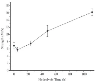
Cement samples were hydrolysed for periods of 5, 24, 48, 112 and 192 h in order to study the kinetics of the con-version of DCPD. After each period, hydrolysis was stopped and the compressive strength of the dried hydrolysed pieces was determined. The results of the strength determinations are shown in Fig. 4.
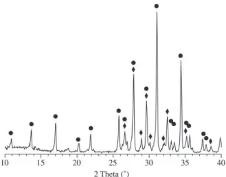
The material remaining from the strength tests was tritu-rated (without removing the outer layer) and the resulting powder was examined by X-ray diffraction. The correspond-ing X-ray diffractograms are shown in Fig. 5.
Compressive strength fell slightly from 6.9 to 5.8 MPa after 5 h of hydrolysis, and increased again on extending the hydrolysis time, reaching 16.2 MPa at a reaction time of 112 h. The compressive strength of the hydrolysed materials is similar to that of cancellous bone (2-12 MPa) and coralline macroporous hydroxyapatite (2.5-5.4 MPa)1. The initial drop
of strength with hydrolysis could be attributed to partial dis-solution of the large DCPD crystals originally present in the cement before hydrolysis, as shown in Fig. 6a.
As the hydrolysis progressed, the intensities of the X-ray diffraction peaks of DCPD decreased while new peaks corresponding to apatite, presumably CDHA according to Fulmer and Brown20, and OCP appeared (Fig. 5). After 5 h
(Fig. 5a), only CDHA and the phases already identified in
the starting cement (Fig. 2) were present. After 24 h peaks of OCP appeared and the intensities of the apatite peaks increased, while the DCPD signals weakened. After 112 h, DCPD had been completely transformed into CDHA or OCP, and no additional formation of CDHA or OCP was observed at 192 h. The NaH2PO4 formed by reactions of Eqs. 2 and 3 could not be completely removed by washing with acetone and was detected in all hydrolysed samples as NaH2PO4.H2O.
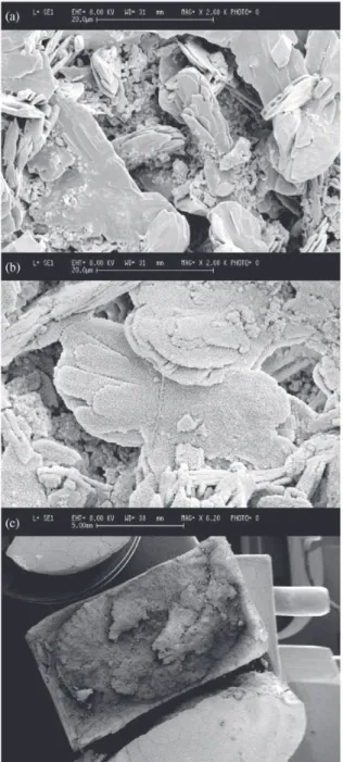
The micrograph of the fracture surface after 5 h of hy-drolysis is shown in Fig. 6b. This is similar to the micro-graph of the fracture surface before hydrolysis (Fig. 6a), with large plate-like crystals of DCPD embedding the re-maining granules of β-TCP. However, in Fig. 6b it can be seen that nanometric crystals of CDHA have completely
Figure 5. X-Ray diffraction patterns of cement (fine β-TCP; L/P = 1.0 ml/g; 1.0 M Na2HPO4) hydrolysed for different periods of time: (a) 5 h; (b) 24 h; (c) 48 h; (d) 112 h; (e) 192 h.
z: β-TCP (JCPDF 9-169), : Ca2P2O7 (JCPDF 9-346),
: CaHPO4.2H2O (JCPDF 9-77), UUUUU: Ca10(PO4)6(OH)2 (JCPDF 9-432), V
V V V
V: Ca4H(PO4)3.2.5H2O (JCPDF 44-778), ZZZZZ: NaH2PO4.H2O (JCPDF 11-651).
Figure 4. Variation of compressive strength with hydrolysis time.
Figure 6. SEM micrographs of the fracture surface of cement:
(a) before hydrolysis; (b) after 5 h of hydrolysis; (c) b at low mag-nification. (Cement: fine β-TCP; L/P = 1.0 ml/g; 1.0 M Na2HPO4).
coated the partially dissolved DCPD crystals. At this stage, the transformation of DCPD into CDHA has only reached the outer layer of the sample, as shown in the low magnifi-cation micrograph of Fig. 6c. This confirms the results of X-ray diffraction where CDHA was the only new phase formed after 5 h of reaction (Fig. 5a).
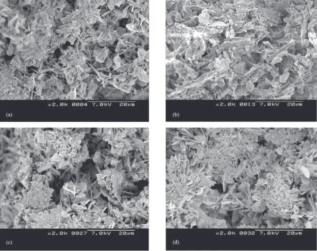
At 48 h the microstructure of the fracture surface of hydrolysed cement is quite different from that observed at 5 h, as can be observed in Figs. 7a and 7b. Aggregates of short plates, very similar to those described for OCP by LeGeros23, are found near to the surface of the sample after
hydrolysis for 48 h (Fig. 7a). However, at the centre of the sample (Fig. 7b) the same microstructure as described for the non-hydrolysed cement in Fig. 7a, is observed. This find-ing suggests that hydrolysis proceeds from the outer to the inner portion of the piece, and that the rate depends on rate of diffusion of the hydrolysing solution into the solid mass. The formation of OCP instead of CDHA can be explained by the variation in pH of the hydrolysis media shown in Fig. 8. In the first stage of hydrolysis, pH is clearly alkaline or neutral; hence hydrolysis via the reaction of Eq. 2 is fa-voured. However, pH drops below 7 after 24 h of hydroly-sis, and formation of OCP through the reaction of Eq. 3 becomes predominant under such conditions of slightly acidic pH and low concentration of Ca2+ ions23.
After 112 h of hydrolysis the fracture surfaces near to the outer layer (Fig. 7c) and in the inner core of the sample (Fig. 7d) have the same appearance and both consist of the aggregates of OCP short-plate crystals. This indicates that the hydrolysis front reached the centre of the sample.
An attempt to relate microstructure to strength changes during hydrolysis suggests that entanglement of crystals is the main responsible for strength. In this way, the large plate-like DCPD crystals embedding β-TCP granules that exist in the set cement are very entangled and provide the mate-rial with higher strength than the smaller CDHA crystals grown onto partially dissolved DCPD with a low entangle-ment degree, formed in the first stage of hydrolysis. The strengthening observed with hydrolysis time is a result of the more clogged microstructure provided by the aggregates of plates of OCP effectively entangled, and developed in the medium and final stages of hydrolysis.
According to Bohner et al., a completion of 92.6% of the reaction of Eq. 1 is obtained during the setting of a ce-ment of β-TCP/H3PO4 containing 2 M H3PO4 and 0.08 M citric acid in the liquid15. Assuming the same conversion
for the cement studied in this work, and considering that half of the DCPD hydrolyses into CDHA, and the remain-ing into OCP, the approximate composition of the mono-liths so obtained is 30 wt.% β-TCP, 49 wt.% OCP, and 21 wt.% CDHA.
Figure 8. Variation of pH with hydrolysis time. (Cement: fine
β-TCP; L/P = 1.0 ml/g; 1.0 M Na2HPO4).
Figure 7. SEM micrographs of the fracture surface of cement after 48 and 112 h of hydrolysis in different parts of the sample: (a) 48 h, near to the outer surface; (b) 48 h, at the centre; (c) 112 h, near to the outer surface; (d) 112 h, at the centre. (Cement: fine β-TCP; L/P = 1.0 ml/g; 1.0 M Na2HPO4).
4. Conclusions
By following the simple and low cost methods described above it is possible to obtain monoliths of biocompatible calcium phosphates with practically any desired shape and size. Implants prepared in this way possess composition and strength which make them suitable for bone remodelling or replacement in low bearing applications.
Acknowledgements
References
1. Hench, L.L.; Wilson, J. (Eds.), An Introduction to Bioceramics, World Scientific, Singapore, Malaysia, 1993.
2. Raynaud, S.; Champion, E.; Lafon, J.P.; Bernache-Assollant, D. Biomaterials, v. 23, n. 4, p. 1081-1089, 2002.
3. Slózarczyk, A.; Paskiewicz, Z. Euro Ceramics VII, PT 1-3, v. 206-2, p. 1621-1624, 2002.
4. Rodriguez-Lorenzo, L.M.; Vallet-Regí, M.; Ferreira, J.M.F. Biomaterials, v. 22, n. 6, p. 583-588, 2001. 5. Zyman, Z.Z.; Ivanov, I.G.; Glushko, V.I.; Kijko, S.M.;
Surov, Y.N.; Chmutov, V.M. J. Biomed. Mater. Res., v. 46, n. 2, p. 135-140, 1999.
6. Rao, R.R.; Kannan, T.S. J. Am. Ceram. Soc., v. 84, n. 8, p. 1710-1716, 2001.
7. Rodriguez-Lorenzo, L.M.; Vallet-Regí, M.; Ferreira, J.M.F. Biomaterials, v. 22, n. 13, p. 1847-1852, 2001. 8. Carotenuto, G.; Spagnuolo, G.; Ambrosio, L.; Nicolais,
L. J. Mater. Sci. Mater. Med., v. 10, n. 10-11, p. 671-676, 1999.
9. Vaz, L.; Lopes, A.B.; Almeida, M. J. Mater. Sci. Mat. Med., v. 10, n. 4, p. 239-242, 1999.
10. Liu, D.M., Ceram. Int., v. 24, n. 6, p. 441-446, 1998. 11. de Campos, M.; Bressiani, A.H.; Bioceramics14, Key
Eng. Mat., v. 218-2, p. 171-174, 2002.
12. Sepulveda, P.; Binner, J.G.P.; Rogero, S.O.; Higa, O.Z.; Bressiani, J.C. J. Biomed. Mater. Res., v. 50, n. 1, p. 27-34, 2000.
13. Bohner, M.; Lemaître, J.; Ring, T.A. in: Proceedings of the Third Euro-Ceramics Conference, Madrid, Septem-ber 1993, Durán, P.; Fernández, J.F. (Eds.), Faenza
Editrice Ibérica, Arganda del Rey, Spain, p. 95, 1993. 14. Bohner, M.; Lemaître, J.; Ring, T.A. J. Am. Ceram. Soc.,
v. 79, n. 6, p. 1427-1434, 1996.
15. Bohner, M.; Merkle, H.P.; Van Landuyt, P.; Trophardy, G.; Lemaître, J. J. Mater. Sci. Mater. Med., v. 11, p. 111-116, 2000.
16. Driessens, F.C.M.; Planell, J.A.; Boltong, M.G.; Khairum, I.; Ginebra, M.P. Proc. Inst. Mech. Engrs., v. 212(Part H), p. 427-435, 1998.
17. Kim, S.R.; Park, S.J. in: Ceramic Transactions Vol.22: Ceramic Powder Science III, Messing, G.L.; Hirano, S.; Hausner, H. (Eds.), American Ceramic Society, Westerville, OH, p. 201, 1991.
18. Prado da Silva, M.H.; Lima, J.H.C.; Soares, G.A.; Elias, C.N.; de Andrade, M.C.; Best, S.M.; Gibson, I.R. Surf. Coat. Technol., v. 137, p. 270-276, 2001.
19. Martin, R.I.; Brown, P.W. J. Crystal Growth, v. 183, p. 417-426, 1998.
20. Fulmer, M.T.; Brown, P.W. J. Mater. Sci. Mater. Med., v. 9, p. 1997-2002, 1998.
21. Kumar, M.; Xie, J.; Chittur, K.; Riley, C. Biomaterials, v. 20, p. 1389-1399, 1999.
22. Kumar, M.; Dasarathy, H.; Riley, C. J. Biomed. Mater. Res., v. 45, p. 302-310, 1999.
23. LeGeros, R.Z. Calcium Phosphates in Oral Biology and Medicine, Karger, Basel, Switzerland, 1991.
24. Ishikawa, K.; Asaoka, K. J. Biomed. Mater. Res., v. 29, p. 1537-1543, 1995.