Abstract
Background: The fractures of the comminuted type have a prevalence of 30 to 50% when related to the ones affecting the mandibular bone. They are characterized by the presence of multiple bone fragments in-volving several lines of fracture, resulting in small fragments within the same area. Usually resulting from high-energy trauma, they cause large displacements, tooth loss, as well as associated lesions in soft tissues.
Cases Reports: This article aimed to report two cases addressing the simplification method in the reduction of comminuted fractures treated by the method of open reduction and functionally stable fixa-tion, which emphasized the importance of establishing a sequence of reduction maneuvers and application of osteosynthesis for a functio-nal, occlusal and aesthetic result of the lower facial third.
Conclusion: The simplification of mandibular comminuted fractures proves to be an excellent ally when one intends to reduce difficulties of reduction of the fracture and the dental occlusion on the transoperati-ve, as well as to facilitate the application of 2.4 reconstruction plates.
Simplification on the Reduction of Comminuted
Mandibular Fractures for Stable Internal Fixation
ORIGINAL
Ivo Cavalcante Pita-Neto1, Jeférson Martins Pereira Lucena Franco2, Milana Drumont RamosSantana1, Hermes Melo Teixera Batista1, Eduardo Costa Studart Soares3, David Gomes de Alencar Gondim4, Francisco Aurélio Lucchesi Sandrini4, Luiz Carlos De Abreu1, Luciano Miller Reis Rodrigues1
1 Laboratory of Study Design and Scientific Writing, ABC Faculty of Medicine.
2 Academic of dentistry course, Leão Sampaio Faculty.
3 Professor of Maxillofacial Surgery, Ceará Federal University.
4 U Oral and Maxillofacial Surgery Departament of Cariri Regional Hospital.
Contact information:
Ivo Cavalcante Pita Neto.
[email protected]Keywords
Fracture fixation; mandible; facial injuries.
Background
bone that requires a relatively strong trauma to have it fractured and may also be a result of practicing sports, injury caused by firearm or a weapon, phy-sical assault, tooth extraction, workplace accidents, metabolic diseases and tumors [4, 5, 6, 7, 8].
Regarding the gender, young men aged between 21 to 30 years old are mostly involved, since this group participates in the most dangerous exercises and sports, drives less carefully and is more pro-ne to engage in episodes of interpersonal violence. However, there is a worldwide trend to the increase of incidence in women, who are increasingly more exposed to the risk factors of this type of trauma [9, 10, 11, 12].
The location and the pattern of mandibular fractures are determined by the mechanism of the injury and the direction of the force vector. In addition, other factors such as age of the patient, the presence or absence of teeth, the presence of pathologies and the physical properties of the etiologic agent also have a direct effect on the characteristics and results of the fracture [11]. Ac-cording to the anatomical region, the distribution of the main fractured sites have been reported as 16% to 33% involving the body; 23.1% to 27.3%, the angle; 19.5 to 29.3%, the condyle; 8.4% to 22%, the symphysis; 16% to 33% parasymphysis; 1.7% to 2.4%, the ramus; 0.2% to 4.8%, the co-ronoid process and 1.4% to 3.1% in the alveolar process [11, 12, 13].
The comminuted fractures are characterized by the presence of multiple bone fragments involving several lines of fracture, resulting in small bone
One of the major goals of its treatment is focu-sed on the union of fractured segments restoring the pre-injury strength and function. This requires a proper anatomical reduction and immobilization that will consolidate the fractured segments [16, 17].
The functionally stable internal fixation is the treatment of choice for the vast majority of mandi-bular fractures by the rapid return of the functions of the masticatory system [8, 16]
This study aims to address the simplification method in the reduction of the comminuted frac-tures for a stable internal fixation with two reported cases of comminuted mandible fractures, treated by the method of open reduction and rigid internal fixation, emphasizing the importance of a correct diagnosis and treatment.
Case Reports
Case Report 1
comminu-llary fixation screws (IMF®) and steel wire n° 1 (Aci-flex®), guided by dental occlusion. For fixation of the mandible, the simplification of the fracture was done joining the smaller fragments with 2.0 mm titanium
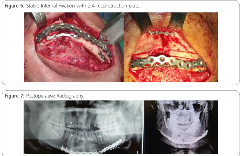
plates system, facilitating the definitive fixation with a “load bearing” reconstruction plate of the 2.4 sys-tem. The mandibular contour was reconstructed and occlusion was restored (Figure 4, 5 and 6).
Figure 2: Surgical access with display of fractured stumps.
Figure 4: Alignment of the fracture and fixation following the oblique jaw line with a 2.0 mm titanium plate system.
Figure 3: Surgical access with visualization of intrao-ral and contintrao-ralateintrao-ral fractured stumps .
Figure 5: Simplification of fractured stumps with mini-plates.
The stability of fixed fragments and the obtained occlusion were tested with a clinically satisfactory result and the essig-type splint was removed, and the intermaxillary fixation was released with main-tenance of the IMF screws.
Functional approach was adopted for the undis-placed subcondylar fractures with the use of Class II elastic bands 7 weeks in the IMF screws, , and the patient was instructed to perform physiotherapy exercises 4 times a day.
The alignment of the repositioned fragments
found with facial trauma with partially obstruc-ted airways, conscious, orienobstruc-ted and pallor. The-re was evidence of open fractuThe-re of the anterior mandible region with facial swelling and loss of chin prominence. Examination revealed an open fracture of the mandible, sublingual hematoma and lingual ptosis. Computed tomography (CT), revealed a highly comminuted fracture of anterior mandibular region with multiple bone fragments
(Figure 8, 9).
An immediate intervention for reduction and
Figure 6: Stable Internal Fixation with 2.4 reconstruction plate.
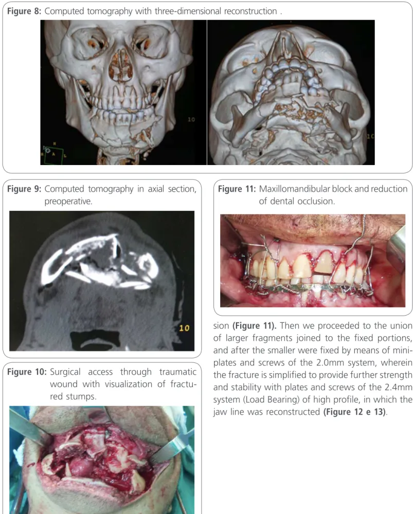
sion (Figure 11). Then we proceeded to the union of larger fragments joined to the fixed portions, and after the smaller were fixed by means of mini-plates and screws of the 2.0mm system, wherein the fracture is simplified to provide further strength and stability with plates and screws of the 2.4mm system (Load Bearing) of high profile, in which the jaw line was reconstructed (Figure 12 e 13).
Figure 8: Computed tomography with three-dimensional reconstruction .
Figure 9: Computed tomography in axial section, preoperative.
Figure 11: Maxillomandibular block and reduction of dental occlusion.
Figure 12: Simplification of the reduction with union of the smaller fragments.
Figure 13: Stable internal fixation with mandi-bular reconstruction with 2.4 plates.
The clinical and radiographic analysis (CT) in the postoperative, found stable occlusion, satisfactory mouth opening and the alignment of properly re-positioned fragments with well adapted plates (Fi-gure 14 and 15)
Discussion
Cranio-maxillofacial trauma can be considered one of the most devastating aggressions found in trau-ma centers because of its emotional consequences, the possibility of deformity and also the economic impact caused in the public and private health sys-tem [18]. Treatment is multidisciplinary , involving mainly the specialties of traumatology, speech the-rapy, ophthalmology, plastic surgery, neurosurgery and physiotherapy [12, 18].
guidance, function is restored; other times, the surgeon performs an intermaxillary fixation [20, 21].
In general, the authors recommend that the treatment of mandibular fractures without dis-placement or with minimum disdis-placement should be closed or conservative treatment, obeying the principles of immobilization through the fixation of Erich bars, by essig-type splint and intermaxillary fixation with elastic bands for 45 to 90 days [10, 11, 16, 19, 21, 22].
In the case number 1 reported here, we opted for a closed treatment with elastic bands, for the consolidation of subcondylar fractures, given the presence of minimum displacement of the fractu-red segments.
On the other hand, in the presence of displace-ment of the bone fragdisplace-ments, the treatdisplace-ment must be surgical or open, , with stable rigid internal fixation in order to get better results [10, 22, 23].
The simplification of the comminuted fragments with mini-plates and screws are used as an aid in
Figure 14: Postoperative computed tomography..
reducing fractures, for subsequent fixation with re-construction plates [19].
In the reported cases, because they are commi-nuted fractures of the mandible, the fractures when simplified, provide strength and stability to the use of reconstruction plates. The main advantages of this technique are: to provide reductions with grea-ter precision and stability, to eliminate the need for post operative prolonged intermaxillary fixation , ra-pidly rehabilitate and restore the function in the im-mediate postoperative period, contributing greatly to the overall health status of the patient [12, 24, 25].
The use of simplification and stable internal fixa-tion in the reducfixa-tion of comminuted fractures of the mandible is an effective treatment, provided that the oral and maxillofacial surgeons have knowledge of mandibular anatomy and physiology as well as knowledge of the fixation principles and surgical te-chniques of applying the system of osteosynthesis. The most important fact is that they should possess "common sense" when they are using this system [12, 21].
Conclusion
The simplification of mandibular comminuted frac-tures proves to be an excellent ally when one intends to reduce difficulties of reduction of the fracture and the dental occlusion on the transoperative, as well as to facilitate the application of 2.4 recons-truction plates.
Ethics questions
The ethical committee of the Regional Hospital of Cariri, for case presentation, approved the current study.
Competing interests
The authors declare no conflicts of interest. All re-search was conducted with their own resources.
Financial resources
The authors state that they used their own resour-ces and did not receive financial aid from any insti-tution. The authors state that they did not receive financial aid from any institution (political, personal, religious, ideological, academic, intellectual, com-mercial, or any other).
References
1. Patrocínio, GP. et al . Mandibular fracture: analysis of 293 patients treated in the Hospital of Clinics, Federal University of Uberlândia. Rev Bras Otorrinolaringol 2005, V.71, n.5, 560-65, sep./oct.
2. Wulkan, M.; Parreira, JR.; Botter, DA. Epidemiologia do trauma facial. Rev. Assoc. Med. Bras 2005 v. 51, n. 5, p. 290-5.
3. Sassi, LM .; Dissenha., JL . et al. Fratura da mandibular: revisão de 82 casos. Rev. Bras. Cir. Cabeça Pescoço 2010, v.39, no 3, p. 190-192.
4. Busuito, MJ.; Smith, JRDJ.; Robson, MC. Mandibulary fractures in na urban trauma center. J Trauma 1986, v. 26(9), p. 826-0.
5. Gassner, R.; Tuli, Tarkan.; Hachl, O.; Rudisch, A.; Ulmer, H. Cranio-maxillofacial trauma: a 10 year review of 9543 cases with 21067 injuries. Jounal of Cranio-Maxillofacial Surgery 2003, v. 31, p. 51-61.
10. Fonseca, RJ. Oral and Maxillofacial Trauma, 3 ed. Philadelphia: W.B. Saunders Company, v. 3, 500p, 2000.
11. Miloro, M. Peterson’s Principals of Oral and Maxillofacial Surgery. 2 ed. Philadelphia: BC Decker. 2004.
12. Camargo, IB.; Oliveira, DM.; Fernandes, AV.; farias, EM. Fratura parassinfisária em Mulher Vítima de Violência Doméstica: Relato de Caso. Rev. Cir. Traumatol. Buco-Maxilo-Fac 2012, Camaragibe v.12, n.1, p. 9-16, jan./março.
13.Hupp, JR.; III, EE.; tucker, MR. Cirurgia oral e maxilofacialcontemporânea. São Paulo, 5ed., Elservier, 2009. 704p.
14. Finn, RA. Treatment of comminuted mandibular fractures by closed reduction. J Oral Maxillofac Surg 1996, v54, p. 320-327.
15. Dingman, RO.; natvig, P. Cirurgia das fraturas faciais. São Paulo:
Ed. Santos, 3a Reimpressão, 2004.
16. Futran ND. Management of comminuted mandible fractures.
Operative Techniques in Otolaryngology 2008, Vol 19, No 2.
17. Vasconcellos, RJH.; Oliveira, DM.; Santos KPC.; Calado, MV. Métados de tratamento as fraturas mandibulares. Rev. Cir. Traumat. Buco-Maxilo-Facial 2001, v.1, n2, p.21-27.
18. Wulkan, M.; Parreira Jr, J.G.; Botter, D.A. Epidemiologia do Trauma Facial. Rev Assoc Med Bras 2005 v.51, n.5, p.290-5.
1 9. Bagheri, SC.; Bell, RB.; Khan, HA.; Terapias atuais em Cirurgia Bucomaxilofacial. 1. ed., Rio de Janeiro: Elsevier. 2013.
20. Toledo Filho, JL. et al. Utilização De Miniplacas No Tratamento De Fraturas Da Mandíbula. Revista da APCD 1998, v. 52, n. 1.
21. Caubi, AF. et al. Fratúra de mandíbula em pacientes geriátricos: Relato de caso clínico. Revista de Cirurgia e Traumatologia Buco-Maxilo-Facial 2004, v.4, n.2, p. 115-120.
22. Gomes, ACA. et al. Tratamento Das Fraturas Mandibulares: Relato de Caso Clínico. Rev. Cir. Traum. Buco-Maxilo-Facial
2001, v. 1, n. 2, p. 31-38.
23. Souza, LCM.; Lucca, MES. Fratura de mandíbula: análise de 282 pacientes. Revista Paulista de Odontologia 1992, n.1, p. 2-4.
24. Schmidt, BL.; Kearns, G.; Gordon, N.; Kaban, LB. A financial analysis of maxillomandibular fixation versus rigid internal fixation for treatment of mandibular fractures. J Oral Maxillofac Surg 2000, v. 58, n. 1206-10.
25. Zronba, H.; Lutz, JC.; Zinhk, S.; Wilk, A. Epidemiology and treatment outcome of surgically treated mandibular condyle fractures. A five years retrospective study. Jornal of Cranio-Maxillo-Facial Surgery 2014, v. 42, p. 879-884.
Where Doctors exchange clinical experiences, review their cases and share clinical knowledge. You can also access lots of medical publications for free. Join Now!
http://medicalia.org/
Comment on this article:
International Archives of Medicine is an open access journal publishing articles encompassing all aspects of medical scien-ce and clinical practiscien-ce. IAM is considered a megajournal with independent sections on all areas of medicine. IAM is a really international journal with authors and board members from all around the world. The journal is widely indexed and classified Q1 in category Medicine.
Publish with iMedPub