*Correspondência:
Medical University of Graz Department of Internal Medicine
Division of Endocrinology and Nuclear Medicine
Auenbruggerplatz 15, A-8036 Graz, Austria
Summary
Obesity has become a very frequent condition with important consequences for the health of affected individuals. Current evidence shows that the excess of adipose tissue as observed in obesity is responsible for secreting inlammatory mediators in a deregulated manner, thus inducing a chronic state of systemic low-grade inlammation that underlies the metabolic and cardiovascular outcomes in these populations. This article reviews the state of the art regarding mediators produced in the adipose tissue, their roles in the pathophysiology of obesity-associated insulin resistance and diabetes, and inally, tries to build a bridge between these mechanistically oriented insights and clinical practice.
Keywords: Inlammation.Obesity.Insulin resistance.Metabolic Syndrome X.Cardiovascular Diseases. Diabetes Mellitus. Type 2.
adipose
tissue
,
inflammation
and
cardiovascular
disease
dimas ikeoka1*, Julia k. mader2, thomas r. pieber3
Trabalho realizado na Division of Endocrinology and Nuclear Medicine, Medical University of Graz
1. Ph.D. - Fellow Scientist of the Division of Endocrinology and Nuclear Medicine, Medical University of Graz 2. MD. - Fellow Scientist of the Division of Endocrinology and Nuclear Medicine, Medical University of Graz
3. Prof. MD. - Head of the Division of Endocrinology and Nuclear Medicine, Medical University of Graz
i
ntroductionRecent epidemiological studies report an alarming increase in the prevalence of obesity in modern societies (1-6). Visceral and subcutaneous adipose tissue accumulation, mainly as a result of poor nutritional habits and largely associated with lack of physical activity, is clearly responsible for increased body weight, which in turn underlies an elevated risk of diabetes and cardiovascular complications (7-12). Although it seems that individuals with higher body mass index (BMI) are prone to cardiovascular events, reasons for this connection are not entirely clear. Over the last two decades some mecha-nisms of disease, especially in association with recently described functions of the adipose tissue were discovered, shedding light on the pathophysiology of obesity-associated cardiovascular diseases.
Initially, important observations demonstrated that fat metabolism is importantly modified by excess body weight. Non-esterified fatty acids (NEFA) delivered from adipose tissue by enzymatic cleavage of triglycerides are found at increased concentrations in blood of obese individuals and associated with a higher risk of developing type-2-diabetes (T2DM) (13, 14). In addition, experimental elevations of NEFA have been shown to induce insulin resistance in animal models and humans (15). More recently, a very active secre-tory function of adipose tissue was disclosed. Adipocytes, infiltrating macrophages and mesenchimal cells produce a number of cytokines, hormones and other substances with distinct effects on the control of glucose tolerance and vascular function. These substances secreted in the adipose
tissue were collectively denominated adipokines(7, 9, 16-19). Deregulated production of adipokines in adipose tissue seems to determine a state of low-grade chronic inflammation that plays a role in the generation of insulin resistance and vascular complications of obesity. It has been consistently observed that inflammatory pathways activated in type-2-diabetic and obese subjects appear in close association with a cluster of distinct clinical manifestations, including high blood pressure, hypertriglyceridemia, low HDL-cholesterol, and endothelial dysfunction, which were grouped under the common denomination of metabolic syndrome (MS) (20-23). In addition to the cardiovascular consequences of obesity, the chronic state of low-grade inflammation was also linked to a disturbed function of hepatocytes, which resulted in the recently described forms of hepatic disease related to obesity, namely the nonalcoholic steatohepatitis (NASH) and the nonalcoholic fatty liver disease (NAFLD) (24).
correlate these pathophysiological insights with the clinical consequences to metabolism and to the cardiovascular system. It is not intended to exhaustively review all mediators in this specific regard, therefore the reader is referred to other more detailed information on the specific topics.
adipose tissue as an endocrine organ
At the microscopic level, two types of adipocytes can be distinguished, namely the brown adipocytes that have a high intra-cellular content of large mitochondria, and the white adipocytes, with far less mitochondrial content. Similarly, two different types of adipose tissue can be identiied in mammals according to their macroscopic aspect: brown adipose tissue (BAT), which contains predominantly brown adipocytes and a pattern of a largely distributed capillary network and white adipose tissue (WAT) that contains mostly white adipocytes and is poorly vascularized. The main function described for BAT is to maintain body temperature, especially in childhood, although some publications have raised the possibility of an active partici-pation in the pathophysiology of the metabolic complications of obesity (25, 26). WAT is the largest fraction of adipose tissue and is responsible for triglyceride accumulation and adipokine secretion. WAT can be further divided, according to the body distribution, into visceral adipose tissue (VAT) and subcutaneous adipose tissue (SAT) (27). The relative importance of each sub-type
of WAT in producing low-grade chronic inlammation and insulin resistance is under dispute in current literature (8, 28, 29), although
strong clinical evidence indicates that central (android) obesity, a condition where adipose tissue preferentially accumulates in the peri-visceral region, is in general more related to higher cardiovascular risk and the features of MS than the peripheral (gynecoid) type (11).
The irst and best described physiological function of adipose tissue was as an energy depot. As mentioned before, WAT stores triglycerides within the large intra-cellular vesicles of adipocytes during the post-prandial phase of digestion and releases them after enzymatic cleavage by lipoprotein lipase (LPL), in the form of fatty acids and glycerol during periods of starvation. Initial reports of the secretory and endocrine functions of adipose tissue come from estrogen conversion, a very important step to produce the activated sexual hormone (30, 31). Later on, tumor necrosis factor-alpha
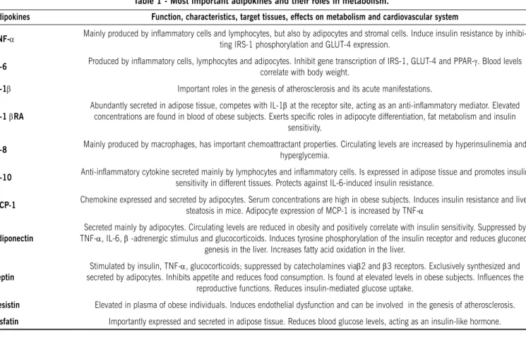
(TNF-α), a cytokine with very important functions in the stimulation of inlammatory cells and in the immune system, was described as a product of adipose tissue secretion, playing a possible role in the development of insulin resistance in humans (17). During subsequent years, following this primary description of a cytokine produced in adipose tissue, a number of other molecules were found and studied, most of them involved in the regulation of energy metabolism and control of appetite. Cytokines, small proteins with previously well deined functions in the regulation of the immune system, consti -tute the largest group of biologically active substances produced in adipose tissue. Other adipokines belong to different molecular fami-lies, such as chemokines and growth factors. A list of currently recognized adipokines is provided in table 1, together with their
Table 1 - most important adipokines and their roles in metabolism.
adipokines Function, characteristics, target tissues, effects on metabolism and cardiovascular system
TNF-α Mainly produced by inlammatory cells and lymphocytes, but also by adipocytes and stromal cells. Induce insulin resistance by inhibi
-ting IRS-1 phosphorylation and GLUT-4 expression.
IL-6 Produced by inlammatory cells, lymphocytes and adipocytes. Inhibit gene transcription of IRS-1, GLUT-4 and PPAR-γ. Blood levels
correlate with body weight.
IL-1β Important roles in the genesis of atherosclerosis and its acute manifestations.
IL-1βra
Abundantly secreted in adipose tissue, competes with IL-1β at the receptor site, acting as an anti-inlammatory mediator. Elevated concentrations are found in blood of obese subjects. Exerts speciic roles in adipocyte differentiation, fat metabolism and insulin
sensitivity.
IL-8 Mainly produced by macrophages, has important chemoattractant properties. Circulating levels are increased by hyperinsulinemia and
hyperglycemia.
IL-10 Anti-inlammatory cytokine secreted mainly by lymphocytes and inlammatory cells. Is expressed in adipose tissue and promotes insulin
sensitivity in different tissues. Protects against IL-6-induced insulin resistance.
mCP-1 Chemokine expressed and secreted by adipocytes. Serum concentrations are high in obese subjects. Induces insulin resistance and liver
steatosis in mice. Adipocyte expression of MCP-1 is increased by TNF-α
adiponectin
Secreted mainly by adipocytes. Circulating levels are reduced in obesity and positively correlate with insulin sensitivity. Suppressed by TNF-α, IL-6, β -adrenergic stimulus and glucocorticoids. Induces tyrosine phosphorylation of the insulin receptor and reduces
gluconeo-genesis in the liver. Increases fatty acid oxidation in the liver.
Leptin
Stimulated by insulin, TNF-α, glucocorticoids; suppressed by catecholamines viaβ2 and β3 receptors. Exclusively synthesized and secreted by adipocytes. Inhibits appetite and reduces food consumption. Is found at elevated levels in obese subjects. Inluences the
reproductive functions. Reduces insulin-mediated glucose uptake.
resistin Elevated in plasma of obese individuals. Induces endothelial dysfunction and can be involved in the genesis of atherosclerosis.
most important features and regulatory pathways.
In a quantitative comparison, adiponectin, plasminogen acti-vator inhibitor-1 (PAI-1) and heparin binding endothelial growth factor-like growth factor are the most abundantly expressed genes in adipose tissue. Plasma concentrations of PAI-1 seem to strictly correlate with the adipose tissue expression, suggesting adipose tissue as the primary source of this speciic molecule
(32). A variety of genes encoding for proteins with important
func-tions in the control of the immune system, growth factors and other molecules are also expressed differently in adipose tissue of obese and non-obese individuals, and most of them are still to be investigated in detail (28, 33).
adipokines, insulin resistance and type 2 diabetes
Interleukin-6 (IL-6) and TNF-α are among the best known and well investigated cytokines produced in adipose tissue. The expres-sion of TNF-α in adipose tissue from both humans and rodents is increased in conditions of obesity and closely correlates with hyperinsulinemia (9, 34). Moreover, weight loss promotes a signiicant decrease in the TNF-α expression in adipose tissue (34). It could also be demonstrated that TNF-α inhibits insulin-dependent glucose uptake in human cultured adipocytes, which correlates with a signiicant reduction of the density of insulin-regulated glucose transporter-4 (GLUT-4) protein at the cellular membrane (35, 36).
In addition, insulin receptor substrate-1 (IRS-1) phosphorylation via c-jun N-terminal kinase (JNK) is reduced by TNF-α, leading to suppression of insulin signaling downstream (17). Levels of IL-6 in blood correlate positively with overweight (37) and negatively with insulin sensitivity (38). Weight loss induced by very low-caloric diet reduces IL-6 levels and improves insulin sensitivity in obese subjects (39). IL-6 concentrations in subcutaneous adipose tissue are increased by local insulin infusion (40). At cellular level, IL-6 has demonstrated that it exerts inhibitory effects on gene transcription of IRS-1, GLUT-4, and peroxisome proliferator-activated receptor gamma (PPAR-gamma). These mechanisms are the most likely explanation for the glucose intolerance induction effect (41, 42). Both TNF-α and IL-6 have demonstrated lipolytic properties and promote lipid release from adipocytes (36, 43). TNF-α and IL-6 are both secreted by adipocytes, although only IL-6 seems to be released from adipose tissue to the systemic circulation (44). The concentrations of TNF-α, IL-6, IL-1beta and IL-8 in adipose tissue seem to be unrelated to their systemic concentrations, acting more likely as paracrine mediators (40, 44).
Adiponectin is a protein secreted by adipocytes which retains signiicant homology with collagen X and VII, as well as with the complement factor C1q (45). It is found at low concentra-tions in plasma of obese subjects, negatively correlating with body mass index and with central (visceral) adiposity (46, 47). Moreover, plasma levels of adiponectin strongly correlate with insulin sensitivity and an enhanced insulin receptor tyrosine phosphorylation, a key pathway in the insulin-induced glucose uptake (48). In addition, adiponectin exerts a suppressive effect on gluconeogenesis (49) and increases fatty acid oxidation, reducing triglyceride accumulation in the liver, which also contributes to improve glucose tolerance (50). Individuals at lower serum levels of adiponectin are subject to endothelial dysfunction, the primary pathophysiological manifestation of atherosclerosis (51). More-over, they are exposed to a higher risk of diabetes and its vascular
complications (52, 53), present an increased severity of coronary
artery disease and also higher degrees of plaque calciication (54).
Several other cytokines, such as IL-1beta, IL-1beta receptor antagonist (IL-1betaRA), IL-8, IL-10, and MCP-1, to cite only the most important ones, have shown to be synthesized in adipose tissue (7, 29, 33, 55, 56). IL-1beta is a cytokine produced mainly by
inlammatory (monocytes, macrophages and foam cells) and endothelial cells, playing a very important role in the patho-physiology of atherosclerosis (57, 58) . It was further demonstrated that IL-1beta induces cytotoxic effects in pancreatic beta-cells, being possibly involved in the pancreatic beta-cell insuficiency as seen mainly in type-1 but also in the late phases of type-2-diabetes (59). The expression of IL-1beta in adipose tissue is of minor importance in quantitative terms, except under conditions of acute inlammation (40). On the other hand, adipose tissue is a major source of IL-1betaRA, a cytokine that functions as a competitive inhibitor of IL-1beta at the receptor site. This cyto-kine belongs to the IL-1 family and acts as a counter-regulatory mediator of inlammation in different diseases, such as sepsis and autoimmune disorders (60, 61). In the obese it is found at
elevated concentrations in blood and has demonstrated speciic roles in the control of adipocyte differentiation, fat metabolism and insulin sensitivity (62-64).
Interleukin-8 can be better classiied as a chemokine (IL-8 or CXCL-8), given its predominant chemoattractant properties. It is produced mainly by macrophages and can be used as a clinical predictor of coronary artery disease (65). Moreover, the IL-8 levels in blood are increased by both hyperinsulinemia and hyperglycemia, indicating a possible participation of IL-8 in carbohydrate metabolism (66).
Monocyte chemoattractant protein-1 (MCP-1) is another chemokine that has shown important metabolic effects. It is expressed and secreted by adipose tissue of obese individuals and lean controls at similar levels, but serum concentrations are increased with obesity (67). Different studies could demonstrate
that MCP-1 contributes to increase the iniltration of inlam -matory cells, induces insulin resistance and liver steatosis in animal models (68-70). The expression of MCP-1 by adipocytes is
signiicantly increased by the presence of TNF-α in cell cultures (69). Despite these evidences, the role of MCP-1 in the
pathophysi-ology of human type-2-diabetes is not completely determined. Interleukin-10 is another cytokine synthesized by immune and inlammatory cells, and also by adipocytes (71). The expres-sion of IL-10 in the adipose tissue is increased in obese humans and rodents (71, 72). Since this is a cytokine with anti-inlammatory activity, its over-expression in adipose tissue may represent a counter-regulatory mechanism. Indeed, IL-10 is positively associ-ated with insulin sensitivity and has a protective effect in skeletal muscle against IL-6-induced insulin resistance (73). Additionally, in adipocyte cell cultures, IL-10 expression is down-regulated by palmitic acid, an 18-carbon non-esteriied fatty acid that is found at elevated concentrations in plasma of obese individuals (74).
also secreted by adipose tissue, mainly visceral fat, and presents insulin-like effects, reducing blood glucose concentrations (76).
In summary, it can be postulated that over-secretion of inlam -matory cytokines both systemically and in the adipose tissue contributes to generate insulin resistance, a key factor in the pathophysiology of T2DM. The contribution of anti-inlammatory cytokines offering protective effects against insulin resistance and diabetes is dificult to summarize, given that from one side adipo -nectin, the major anti-inlammatory and anti-diabetic cytokine is up-regulated and, on the other, IL-10 seems to be depressed in conditions of obesity. Therefore, obesity can well be characterized as a condition where adipose tissue production of cytokines is deregulated, since a marked excess of pro-inlammatory cyto -kines is one of its most remarkable features. Pro-inlammatory effects prevail against the counter-regulatory mediators, leading obese individuals to severe metabolic and vascular complica-tions. Adipokines can be measured in blood and might indicate increased risk of diabetes and cardiovascular complications when found at elevated concentrations. However, a few cytokines exert their effects at a very short distance in the adipose tissue, in a paracrine or autocrine manner and therefore are not useful as outcome predictors for evaluation in clinical practice.
The control of appetite and body weight by adipokines Some agents produced by adipose tissue are not primarily involved in the genesis of vascular damage, but instead, are responsible for weight excess by controlling food ingestion. Leptin is a 16kDa glycoprotein expressed and secreted almost exclusively in adipose tissue (77). Leptin is well described both in rodents and humans and produces an important action at hypo-thalamic level, promoting satiety and inhibiting ingestion of food (77-79). The phenotype of congenitally leptin deicient individuals is characterized by normal body weight at birth, but hyperphagia and food-seeking behavior is promptly manifest, promoting a rapid fat accumulation and increase of body weight already within the irst weeks or months of life (78). Leptin is therefore essential to the control of satiety at the level of the central nervous system, contributing to regulation of body weight in humans. Obese and overweight individuals present leptin resistance which is char-acterized by elevated levels of circulating leptin and signiicant weight loss when supplemental recombinant leptin is provided (37, 80-82). In addition, leptin has demonstrated inhibitory effects
on insulin-mediated glucose uptake in muscle and adipose cells, which seems to be determined by reduced insulin signaling at the level of the insulin receptor substrate-1 phosphorylation, thus contributing to regulate energy metabolism in a paracrine and autocrine manner. Such effects have raised the hypothesis that elevated levels of leptin in circulation of obese individuals could at least partially explain the mechanisms of insulin resistance.
Non-esteriied fatty acids, glucose metabolism and inlammation Non-esteriied fatty acids (NEFA) have the property to induce insulin resistance which was observed both in humans and animal models, as cited above. Indeed, recent evidence has demonstrated that NEFA activate important intracellular signaling molecules, such as c-jun N-terminal kinase and nuclear factor kappa-B in mice cultured adipocytes which are both mechanistically implicated in inlammation and also in the reduction of insulin-stimulated
glucose uptake (17, 83). More recently, NEFA have also demonstrated
to produce pro-inlammatory actions, contributing to increase secretion of inlammatory cytokines and reduce anti-inlammatory cytokines in cultured adipocytes (74). However, contribution of NEFA for in the induction of cytokines in vivo and the relevance of such mechanism in humans remains to be conirmed.
The role of adipokines in the development of cardiovascular diseases
Different inlammatory mediators are implicated in the induction of endothelial dysfunction, plaque formation and plaque instability which constitute the main mechanisms of vascular damage in atherosclerotic disease. The individual roles of different cytokines in atherosclerosis are extensively described by previous publications and will not be considered in detail in the present review (84, 85). Of note, the endothelial layer of arteries may display a pathological behavior when stimulated by speciic cytokines as well as produce inlammatory mediators, such as IL-1beta, IL-6, IL-8, TNF-α and MCP-1, that have important actions on the initiation and ampliica -tion of the inlammatory process within the atherosclerotic plaque
(85-88). The combined effect of the interaction between these
cyto-kines and the vascular wall can be summarized as an increased leukocyte and monocyte recruitment and activation in the vessel wall, disturbance of the nitric-oxide mediated mechanisms for local regulation of blood low, promotion of smooth muscle cell migra -tion and differentia-tion, and induc-tion of a prothrombotic state (85).
These pathophysiological features are important in the irst phases of plaque formation and also in the induction of plaque rupture and thrombosis during the acute vascular events.
Myocardial dysfunction and heart failure are other important clinical features of obesity. Cytokines, as well as other inflam-matory mediators (soluble IL-6-receptor, IL-6 and C-reactive protein [CRP]) might also be involved in the generation of systolic and diastolic ventricular dysfunction, independent of coronary obstruction, as indicated by echocardiography in normotensive obese individuals (22).
Now, while waiting for the development of these new agents, clinicians must be encouraged to stimulate the practice of exer-cise and indicate speciic and target-oriented strategies for weight reduction to patients with high BMI, and especially in those individuals with a detected, augmented cardiovascular risk or, evidence of insulin resistance. Bariatric surgery should be care-fully considered for all patients at elevated cardiovascular risk. In addition, to the routine parameters to be measured, and in order to better estimate the actual risk of these patients, measure-ment of markers such as high sensitive C-reactive protein as an indicator of low-grade chronic inlammation, and perhaps in the future other speciic cytokines may be helpful in clinical practice.
r
esumotecidoadiposo, inflamaçãoedoençacardiovascular
Obesidade é uma condição frequente com importantes consequencias para a saúde dos indivíduos acometidos. Evidência atual tem demonstrado que o excesso de tecido adiposo, tal qual observado na obesidade, é responsável pela secreção de mediadores inflamatorio de forma descontrolada, levando assim a um estado crônico de inflamação sistêmica de baixa intensidade que está por trás das consequências metabólicas e cardiovasculares em tais populações. Este artigo revisa o estado da arte referente aos mediadores produzidos no tecido adiposo, seus papeis na fisiopatologia da resistência insulínica relacionada à obesidade e ao diabetes, e por fim, tenta estabelecer uma ponte de ligação entre estes conceitos mecanisticamente orientados e a prática clínica. [Rev Assoc Med Bras 2010; 56(1): 116-21]
Unitermos: inlamação. Obesidade. Resistência à Insulina.Sín -drome X Metabólica.Doenças cardiovasculares.Diabetes Mellitus
Tipo 2.
r
eferences1. Branca F, Nikogosian H, Lobstein T. The challenge of obesity in the WHO Euro-pean Region and the strategies for response. In: Branca F, Haik Nikogosian
H, Lobstein T, editors. Data WLCiP. Copenhagen: WHO Regional Ofice for
Europe; 2007.
2. Elizabeth DW, Baur LA. Adolescent obesity: making a difference to the epidemic. Int J Adolesc Med Health. 2007;19(3):235-43.
3. Flegal KM, Carroll MD, Kuczmarski RJ, Johnson CL. Overweight and obesity
in the United States: prevalence and trends, 1960-1994. Int J Obes Relat
Metab Disord. 1998;22(1):39-47.
4. Jackson-Leach R, Lobstein T. Estimated burden of paediatric obesity and co-morbidities in Europe. Part 1. The increase in the prevalence of child obesity in Europe is itself increasing. Int J Pediatr Obes. 2006;1(1):26-32.
5. Knai C, Suhrcke M, Lobstein T. Obesity in eastern Europe: an overview of its
health and economic implications. Econ Hum Biol. 2007;5(3):392-408.
6. Swinburn BA. The obesity epidemic in Australia: can public health interventions work? Asia Pac J Clin Nutr. 2003;12(Suppl):S7.
7. Bastard JP, Maachi M, Lagathu C, Kim MJ, Caron M, Vidal H, et al. Recent
advances in the relationship between obesity, inlammation, and insulin
resistance. Eur Cytokine Netw. 2006;17(1):4-12.
8. Dolinkova M, Dostalova I, Lacinova Z, Michalsky D, Haluzikova D, Mraz M,
et al. The endocrine proile of subcutaneous and visceral adipose tissue of
obese patients. Mol Cell Endocrinol. 2008;291(1-2):63-70.
9. Hotamisligil GS, Arner P, Caro JF, Atkinson RL, Spiegelman BM. Increased
adipose tissue expression of tumor necrosis factor-alpha in human obesity and insulin resistance. J Clin Invest. 1995;95(5):2409-15.
10. Mathieu P, Pibarot P, Larose E, Poirier P, Marette A, Despres JP. Visceral obesity and the heart. Int J Biochem Cell Biol. 2008;40(5):821-36.
11. Wajchenberg BL. Subcutaneous and visceral adipose tissue: their relation to
the metabolic syndrome. Endocr Rev. 2000;21(6):697-738.
12. Wellen KE, Hotamisligil GS. Obesity-induced inlammatory changes in adipose
tissue. J Clin Invest. 2003;112(12):1785-8.
13. Charles MA, Eschwege E, Thibult N, Claude JR, Warnet JM, Rosselin GE, et al.
The role of non-esteriied fatty acids in the deterioration of glucose tolerance in Caucasian subjects: results of the Paris Prospective Study. Diabetologia.
1997;40(9):1101-6.
14. Gordon ES. Non-esteriied fatty acids in the blood of obese and lean subjects.
Am J Clin Nutr. 1960;8(7)740-7.
15. Roden M, Price TB, Perseghin G, Petersen KF, Rothman DL, Cline GW, et al. Mechanism of free fatty acid-induced insulin resistance in humans. J Clin Invest. 1996;97(12):2859-65.
16. Bergman RN, Van Citters GW, Mittelman SD, Dea MK, Hamilton-Wessler M, Kim SP, et al. Central role of the adipocyte in the metabolic syndrome. J Invest
Med. 2001;49(1):119-26.
17. Hotamisligil GS, Peraldi P, Budavari A, Ellis R, White MF, Spiegelman BM.
IRS-1-mediated inhibition of insulin receptor tyrosine kinase activity in TNF-alpha-
and obesity-induced insulin resistance. Science. 1996;271(5249):665-8. 18. Mohamed-Ali V, Pinkney JH, Coppack SW. Adipose tissue as an endocrine and
paracrine organ. Int J Obes Relat Metab Disord. 1998;22(12):1145-58.
19. Senn JJ, Klover PJ, Nowak IA, Mooney RA. Interleukin-6 induces cellular
insulin resistance in hepatocytes. Diabetes. 2002;51(12):3391-9. 20. Reaven GM. Banting lecture 1988. Role of insulin resistance in human disease.
Diabetes. 1988;37(12):1595-607.
21. Day C. Metabolic syndrome, or what you will: deinitions and epidemiology.
Diabetes Vasc Dis Res. 2007;4(1):32-8.
22. Malavazos AE, Corsi MM, Ermetici F, Coman C, Sardanelli F, Rossi A, et al. Proinlammatory cytokines and cardiac abnormalities in uncomplicated
obesity: relationship with abdominal fat deposition. Nutr Metab Cardiovasc Dis. 2007;17(4):294-302.
23. Silveira IL, Maranhao TM, Azevedo GD. Metabolic syndrome in postmenopausal
women: higher prevalence in the Northeastern Region of Brazil than in other
Latin American countries and the inluence of obesity and socioeconomic
factors. Climacteric. 2007;10(5):438-9; author reply 40.
24. Clark JM. The epidemiology of nonalcoholic fatty liver disease in adults. J Clin
Gastroenterol. 2006;40(Suppl 1):S5-10.
25. Cannon B, Nedergaard J. Brown adipose tissue: function and physiological
signiicance. Physiol Rev. 2004;84(1):277-359.
26. Himms-Hagen J. Does brown adipose tissue (BAT) have a role in the physiology or treatment of human obesity? Rev Endocr Metab Disord. 2001;2(4):395-401.
27. Avram AS, Avram MM, James WD. Subcutaneous fat in normal and diseased
states: 2. Anatomy and physiology of white and brown adipose tissue. J Am Acad Dermatol. 2005;53(4):671-83.
28. Gomez-Ambrosi J, Catalan V, Diez-Caballero A, Martinez-Cruz LA, Gil MJ,
Garcia-Foncillas J, et al. Gene expression proile of omental adipose tissue in human obesity. FASEB J. 2004;18(1):215-7.
29. Murdolo G, Herder C, Wang Z, Rose B, Schmelz M, Jansson PA. In situ proiling of adipokines in subcutaneous microdialysates from lean and obese
individuals. Am J Physiol Endocrinol Metab. 2008;295(5):E1095-105.
30. Grodin JM, Siiteri PK, MacDonald PC. Source of estrogen production in post -menopausal women. J Clin Endocrinol Metab. 1973;36(2):207-14.
31. MacDonald PC, Rombaut RP, Siiteri PK. Plasma precursors of estrogen. I.
Extent of conversion of plasma delta-4-androstenedione to estrone in normal males and nonpregnant normal, castrate and adrenalectomized females. J Clin Endocrinol Metab. 1967;27(8):1103-11.
32. Alessi MC, Juhan-Vague I. PAI-1 and the metabolic syndrome: links, causes, and consequences. Arterioscler Thromb Vasc Biol. 2006;26(10):2200-7. 33. You T, Yang R, Lyles MF, Gong D, Nicklas BJ. Abdominal adipose tissue
cytokine gene expression: relationship to obesity and metabolic risk factors. Am J Physiol Endocrinol Metab. 2005;288(4):E741-7.
34. Kern PA, Saghizadeh M, Ong JM, Bosch RJ, Deem R, Simsolo RB. The
expression of tumor necrosis factor in human adipose tissue. Regulation by obesity, weight loss, and relationship to lipoprotein lipase. J Clin Invest. 1995;95(5):2111-9.
35. Stephens JM, Lee J, Pilch PF. Tumor necrosis factor-alpha-induced insulin
resistance in 3T3-L1 adipocytes is accompanied by a loss of insulin receptor substrate-1 and GLUT4 expression without a loss of insulin receptor-mediated signal transduction. J Biol Chem. 1997;272(2):971-6.
36. Hauner H, Petruschke T, Russ M, Rohrig K, Eckel J. Effects of tumour necrosis factor alpha (TNF alpha) on glucose transport and lipid metabo-lism of newly-differentiated human fat cells in cell culture. Diabetologia. 1995;38(7):764-71.
37. Maachi M, Pieroni L, Bruckert E, Jardel C, Fellahi S, Hainque B, et al. Systemic low-grade inlammation is related to both circulating and adipose tissue
TNFalpha, leptin and IL-6 levels in obese women. Int J Obes Relat Metab Disord. 2004;28(8):993-7.
38. Heliovaara MK, Teppo AM, Karonen SL, Tuominen JA, Ebeling P. Plasma
IL-6 concentration is inversely related to insulin sensitivity, and acute-phase proteins associate with glucose and lipid metabolism in healthy subjects. Diabetes Obes Metab. 2005;7(6):729-36.
adipose tissue of obese women after weight loss. J Clin Endocrinol Metab. 2000;85(9):3338-42.
40. Pachler C, Ikeoka D, Plank J, Weinhandl H, Suppan M, Mader JK, et al. Subcutaneous adipose tissue exerts proinlammatory cytokines after minimal
trauma in humans. Am J Physiol Endocrinol Metab. 2007;293(3):E690-6.
41. Rotter V, Nagaev I, Smith U. Interleukin-6 (IL-6) induces insulin resistance
in 3T3-L1 adipocytes and is, like IL-8 and tumor necrosis factor-alpha, overexpressed in human fat cells from insulin-resistant subjects. J Biol Chem. 2003;278(46):4577-84.
42. Stith RD, Luo J. Endocrine and carbohydrate responses to interleukin-6 in vivo. Circ Shock. 1994;44(4):210-5.
43. Greenberg AS, Nordan RP, McIntosh J, Calvo JC, Scow RO, Jablons D. Inter -leukin 6 reduces lipoprotein lipase activity in adipose tissue of mice in vivo and in 3T3-L1 adipocytes: a possible role for interleukin 6 in cancer cachexia. Cancer Res. 1992;52(15):4113-6.
44. Mohamed-Ali V, Goodrick S, Rawesh A, Katz DR, Miles JM, Yudkin JS, et al. Subcutaneous adipose tissue releases interleukin-6, but not tumor necrosis
factor-alpha, in vivo. J Clin Endocrinol Metab. 1997;82(12):4196-200.
45. Maeda K, Okubo K, Shimomura I, Funahashi T, Matsuzawa Y, Matsubara K. cDNA cloning and expression of a novel adipose speciic collagen-like factor,
apM1 (AdiPose Most abundant Gene transcript 1). Biochem Biophys Res Commun. 1996;221(2):286-9.
46. Arita Y, Kihara S, Ouchi N, Takahashi M, Maeda K, Miyagawa J, et al. Parado
-xical decrease of an adipose-speciic protein, adiponectin, in obesity. Biochem
Biophys Res Commun. 1999;257(1):79-83.
47. Cote M, Mauriege P, Bergeron J, Almeras N, Tremblay A, Lemieux I, et al. Adipo-nectinemia in visceral obesity: impact on glucose tolerance and plasma lipo-protein and lipid levels in men. J Clin Endocrinol Metab. 2005;90(3):1434-9.
48. Stefan N, Vozarova B, Funahashi T, Matsuzawa Y, Weyer C, Lindsay RS, et
al. Plasma adiponectin concentration is associated with skeletal muscle insulin receptor tyrosine phosphorylation, and low plasma concentration precedes a decrease in whole-body insulin sensitivity in humans. Diabetes. 2002;51(6):1884-8.
49. Berg AH, Combs TP, Du X, Brownlee M, Scherer PE. The adipocyte-secreted
protein Acrp30 enhances hepatic insulin action. Nat Med. 2001;7(8):947-53.
50. Yamauchi T, Kamon J, Minokoshi Y, Ito Y, Waki H, Uchida S, et al. Adiponectin
stimulates glucose utilization and fatty-acid oxidation by activating AMP-activated protein kinase. Nat Med. 2002;8(11):1288-95.
51. Ouchi N, Ohishi M, Kihara S, Funahashi T, Nakamura T, Nagaretani H, et al.
Association of hypoadiponectinemia with impaired vasoreactivity. Hyperten-sion. 2003;42(3):231-4.
52. Kumada M, Kihara S, Sumitsuji S, Kawamoto T, Matsumoto S, Ouchi N, et
al. Association of hypoadiponectinemia with coronary artery disease in men. Arterioscler Thromb Vasc Biol. 2003;23(1):85-9.
53. Zoccali C, Mallamaci F, Tripepi G, Benedetto FA, Cutrupi S, Parlongo S, et al.
Adiponectin, metabolic risk factors, and cardiovascular events among patients
with end-stage renal disease. J Am Soc Nephrol. 2002;13(1):134-41. 54. Maahs DM, Ogden LG, Kinney GL, Wadwa P, Snell-Bergeon JK, Dabelea D,
et al. Low plasma adiponectin levels predict progression of coronary artery
calciication. Circulation. 2005;111(6):747-53.
55. Mohamed-Ali V, Goodrick S, Bulmer K, Holly JM, Yudkin JS, Coppack SW.
Production of soluble tumor necrosis factor receptors by human subcutaneous adipose tissue in vivo. Am J Physiol. 1999;277(6 Pt 1):E971-5.
56. Wellen KE, Hotamisligil GS. Inlammation, stress, and diabetes. J Clin Invest.
2005;115(5):1111-9.
57. Dinarello CA, Wolff SM. The role of interleukin-1 in disease. N Engl J Med.
1993;328(2):106-13.
58. Tipping PG, Lowe MG, Holdsworth SR. Glomerular interleukin 1 production is dependent on macrophage iniltration in anti-GBM glomerulonephritis. Kidney
Int. 1991;39(1):103-10.
59. Major CD, Wolf BA. Interleukin-1beta stimulation of c-Jun NH(2)-terminal kinase activity in insulin-secreting cells: evidence for cytoplasmic restriction. Diabetes. 2001;50(12):2721-8.
60. De Benedetti F, Pignatti P, Massa M, Sartirana P, Ravelli A, Martini A. Circulating
levels of interleukin 1 beta and of interleukin 1 receptor antagonist in systemic juvenile chronic arthritis. Clin Exp Rheumatol. 1995;13(6):779-84. 61. Van Deuren M, Van der Ven-Jongekrijg J, Demacker PN, Bartelink AK, Van
Dalen R, Sauerwein RW, et al. Differential expression of proinlammatory
cytokines and their inhibitors during the course of meningococcal infections. J Infect Dis. 1994;169(1):157-61.
62. Beutler BA, Cerami A. Recombinant interleukin 1 suppresses lipoprotein lipase activity in 3T3-L1 cells. J Immunol. 1985;135(6):3969-71.
63. Juge-Aubry CE, Somm E, Chicheportiche R, Burger D, Pernin A, Cuenod-Pittet
B, et al. Regulatory effects of interleukin (IL)-1, interferon-beta, and IL-4 on the production of IL-1 receptor antagonist by human adipose tissue. J Clin Endocrinol Metab. 2004;89(6):2652-8.
64. Matsuki T, Horai R, Sudo K, Iwakura Y. IL-1 plays an important role in lipid
metabolism by regulating insulin levels under physiological conditions. J Exp Med. 2003;198(6):877-88.
65. Gerszten RE, Garcia-Zepeda EA, Lim YC, Yoshida M, Ding HA, Gimbrone MA,
Jr., et al A. MCP-1 and IL-8 trigger irm adhesion of monocytes to vascular endothelium under low conditions. Nature. 19992;398(6729):718-23. 66. Straczkowski M, Kowalska I, Nikolajuk A, Dzienis-Straczkowska S, Szelacho
-wska M, Kinalska I. Plasma interleukin 8 concentrations in obese subjects with impaired glucose tolerance. Cardiovasc Diabetol. 2003;2(1):5.
67. Murdolo G, Hammarstedt A, Sandqvist M, Schmelz M, Herder C, Smith U, et
al. Monocyte chemoattractant protein-1 in subcutaneous abdominal adipose tissue: characterization of interstitial concentration and regulation of gene expression by insulin. J Clin Endocrinol Metab. 2007;92(7):2688-95.
68. Kamei N, Tobe K, Suzuki R, Ohsugi M, Watanabe T, Kubota N, et al.
Overexpression of monocyte chemoattractant protein-1 in adipose tissues causes macrophage recruitment and insulin resistance. J Biol Chem. 2006;281(36):26602-14.
69. Sartipy P, Loskutoff DJ. Monocyte chemoattractant protein 1 in obesity and insulin resistance. Proc Natl Acad Sci USA. 2003;100(12):7265-70. 70. Kanda H, Tateya S, Tamori Y, Kotani K, Hiasa K, Kitazawa R, et al. MCP-1
contributes to macrophage iniltration into adipose tissue, insulin resistance,
and hepatic steatosis in obesity. J Clin Invest. 2006;116(6):1494-505.
71. Juge-Aubry CE, Somm E, Pernin A, Alizadeh N, Giusti V, Dayer JM, et al. Adipose
tissue is a regulated source of interleukin-10. Cytokine. 2002;29(6):270-4. 72. Esposito K, Pontillo A, Giugliano F, Giugliano G, Marfella R, Nicoletti G, et al.
Association of low interleukin-10 levels with the metabolic syndrome in obese women. J Clin Endocrinol Metab. 2003;88(3):1055-8.
73. Straczkowski M, Kowalska I, Nikolajuk A, Krukowska A, Gorska M. Plasma
interleukin-10 concentration is positively related to insulin sensitivity in young healthy individuals. Diabetes Care. 2005;28(8):2036-7.
74. Bradley RL, Fisher FF, Maratos-Flier E. Dietary fatty acids differentially regulate production of TNF-alpha and IL-10 by murine 3T3-L1 adipocytes. Obesity. 2008;16(5):938-44.
75. Gomez-Ambrosi J, Fruhbeck G. Evidence for the involvement of resistin in inlamma -tion and cardiovascular disease. Curr Diabetes Rev. 2005;1(3):227-34.
76. Sethi JK, Vidal-Puig A. Visfatin: the missing link between intra-abdominal obesity
and diabetes? Trends Mol Med. 2005;11(8):344-7.
77. Zhang Y, Proenca R, Maffei M, Barone M, Leopold L, Friedman JM. Posi-tional cloning of the mouse obese gene and its human homologue. Nature. 1994;372(6505):425-32.
78. Farooqi IS, Matarese G, Lord GM, Keogh JM, Lawrence E, Agwu C, et al. Bene
-icial effects of leptin on obesity, T cell hyporesponsiveness, and neuroendocrine/ metabolic dysfunction of human congenital leptin deiciency. J Clin Invest.
2002;110(8):1093-103.
79. Farooqi IS, ORahilly S. Leptin: a pivotal regulator of human energy homeostasis. Am J Clin Nutr. 2009;89(3):980S-4S.
80. Heymsield SB, Greenberg AS, Fujioka K, Dixon RM, Kushner R, Hunt T, et al.
Recombinant leptin for weight loss in obese and lean adults: a randomized, controlled, dose-escalation trial. JAMA. 1999;282(16):1568-75.
81. Schwartz MW, Prigeon RL, Kahn SE, Nicolson M, Moore J, Morawiecki A, et al.
Evidence that plasma leptin and insulin levels are associated with body adiposity via different mechanisms. Diabetes Care. 1997;20(9):1476-81.
82. Takahashi M, Funahashi T, Shimomura I, Miyaoka K, Matsuzawa Y. Plasma leptin
levels and body fat distribution. Horm Metab Res. 1996;28(12):751-2.
83. Gao Z, Zhang X, Zuberi A, Hwang D, Quon MJ, Lefevre M, et al. Inhibition of insulin
sensitivity by free fatty acids requires activation of multiple serine kinases in 3T3-L1 adipocytes. Mol Endocrinol. 2004;18(8):2024-34.
84. Gustafson B, Hammarstedt A, Andersson CX, Smith U. Inlamed adipose tissue: a
culprit underlying the metabolic syndrome and atherosclerosis. Arterioscler Thromb Vasc Biol. 2007;27(11):2276-83.
85. Koler S, Nickel T, Weis M. Role of cytokines in cardiovascular diseases: a focus on endothelial responses to inlammation. Clin Sci (Lond). 2005;108(3):205-13. 86. Bhagat K, Vallance P. Inlammatory cytokines impair endothelium-dependent
dilatation in human veins in vivo. Circulation. 1997;96(9):3042-7.
87. Ross R. Atherosclerosis--an inflammatory disease. N Engl J Med. 1999;340(2):115-26.
88. Mantovani A, Garlanda C, Introna M, Vecchi A. Regulation of endothelial cell function
by pro- and anti-inlammatory cytokines. Transplant Proc. 1998;30(8):4239-43. 89. Foster-Schubert KE, Cummings DE. Emerging therapeutic strategies for obesity.
Endocr Rev. 2006;27(7):779-93.
90. Hansen EN, Torquati A, Abumrad NN. Results of bariatric surgery. Annu Rev Nutr. 2006;26:481-511.
91. Okuno A, Tamemoto H, Tobe K, Ueki K, Mori Y, Iwamoto K, et al. Troglitazone increases the number of small adipocytes without the change of white adipose tissue mass in obese Zucker rats. J Clin Invest. 1998;101(6):1354-61.