AtS
23,
n
A Guide
for
the Identification of the
Snail Intermediate Hosts of
in
the Americas
PAN AMERICAN HEALTH ORGANIZATION
A
Guide
for the Identification of the
Snail Intermediate Hosts of
Schistosomiasis
in
the Americas
CONTENTS
Page
FoREwoRD v
Life Cycle of Schistosoma mansoni vi
INTRODUCTION vii
1. Notes on the Ecology of the Planorbid Intermediate Hosts 1
2. Basic Malacological Concepts i 4
3. Groups of Freshwater Snails of the Neotropics 17
4. Key to Neotropical Biomphalaria Species 30
5. Synopsis of the Biomphalaria Species 33
APPENDIX I. Collection, Preservation, and Storage of Snails 89
APPENDIX II. Laboratory Rearing of Snails 96
APPENDIX III. Techniques for Studying Morphology 98 APPENDIX IV. Infection of Snails in the Laboratory 103
APPENDIX V. Shipment of Snails 105
GLOSSARY 107
ABBREVIATIONS USED IN THE FIGURES AND KEY 112
FOREWORD
This Guide was prepared by the Pan American Health Organization/ World Health Organization Working Group for the Development of Guidance for Identification of American Planorbidae, which is com-posed of the following members:
Dr. Frederico S. Barbosa, Instituto de Higiene, Universidade Federal de Pernambuco, Caixa Postal 1626, Recife, Pernambuco, Brazil Dr. Elmer G. Berry, Museum of Zoology, University of Michigan,
Ann Arbor, Michigan
Dr. Harold W. Harry, Research Associate, Rice University, Houston, Texas, U.S.A.
Dr. Bengt Hubendick, Director, Naturhistoriska Museet, G5oteborg, Sweden
Dr. Emile A. Malek, Associate Professor of Parasitology, Department of Tropical Medicine and Public Health, Tulane Medical School, New Orleans, Louisiana, U.S.A.
Dr. Wladimir Lobato Paraense, Centro Nacional de Pesquisas Mala-cológicas, Instituto Central de Biologia, Universidade de Brasilia, Brasilia, Brazil
Secretariat:
Dr. Earl C. Chamberlayne, Communicable Diseases Branch, Pan American Sanitary Bureau, Regional Office of the World Health Organization, Washington, D.C., U.S.A. (until April 1964) Dr. Louis J. Olivier, Communicable Diseases Branch, Pan American
Sanitary Bureau, Regional Office of the World Health Organiza-tion, Washington, D.C., U.S.A. (from April 1966)
Life Cycle of
Schistosoma mansoni
The eggs of Schistosoma mansoni leave the human host with the feces.
If the feces (1) are deposited or washed into fresh water, the eggs (2) hatch and release a tiny, ciliated, free-swimming larva, known as miracidium (3). To survive, the latter must enter a snail within a few hours. If it encounters a suitable snail (4) it will penetrate the surface and enter the tissue, where it quickly loses its ciliated plates and becomes a large elongated sac, called a primary sporocyst. This sporocyst forms within it numerous, small, secondary sporocysts that eventually leave the primary sporocyst and move to the digestive gland of the snail. There they become very long, thin-walled sacs threaded into the snail's tissues. Each secondary sporocyst forms within its cavity numerous fork-tailed larvae, called cercariae (5). Some of these cercariae leave the sporocyst each day through a pore, migrate to the free surface of the snail, and escape into the water, usually around midday. Cercariae can be produced continuously by the sporocysts for weeks or even months. They swim actively, and if they come in con-tact with the human skin they can penetrate it with the aid of secre-tions from gland cells producing lytic enzymes (6). The worms enter the blood stream, travel to the heart, are pumped to the lungs, and then migrate to the branches of the hepatic portal vein in the liver. After the worms become adults they pair (7) and migrate to the mesenteric venules (8) of the portal system, where the females deposit large quantities of eggs. Some of the eggs pass through the wall of the intestine and are carried out of the body in the feces. Thus the life cycle
of the worm is completed.
Fig. 1
INTRODUCTION
Medical malacology is the study of snails and other molluscs that
serve as intermediate hosts of parasitic diseases of man and animals.
According to present knowledge, almost all diseases known to be
trans-mitted by snails are caused by parasitic worms of the class Trematoda,
or flukes. Nearly all snails that harbor diseases of medical importance
live in fresh water. In the Neotropics of the Western Hemisphere, the
major disease of man caused by a trematode is Manson's schistosomiasis,
or bilharziasis. This disease is not only gradually extending over a
wider geographic area; it is also infecting a progressively larger number
of people each year. Facioliasis, another trematode disease of the
Neotropics, is gaining importance among cattle and other ruminants,
and it is also of some importance in man. Paragonimiasis is still
an-other snail-borne disease of the Neotropics that affects both animals
and humans. Probably the most reasonable methods of control, or
preferably prevention and eradication, of these diseases will be
cen-tered on control of the snails that are essential for the completion of
the life cycles of the trematodes that cause them.
At present, control of the snail vectors of schistosomiasis is being
accomplished by the use of molluscicides (chemicals that kill the
snails) and to a lesser extent by introducing predators or competitors
of the snails (biological control), or by weeding or draining the bodies
of water containing the snail and other ecological or environmental
approaches. Other efforts include fencing or posting of contaminated
waters so that human access to them is limited and educating the public
to avoid polluting the water or becoming infected with the disease.
The details of these procedures have been extensively discussed in
various publications of the World Health Organization. Suffice it to
say that as of now they all have limitations, either because they are
only temporarily effective or relatively expensive, or both.
for reference purposes. The incompleteness of existing knowledge must be recognized. Workers in the areas in which schistosomiasis is pres-ent can do much to improve currpres-ent knowledge. Some of the more technical literature is listed in the bibliography for those who wish to pursue the subject in greater detail.
In most Neotropical areas the snails that are vectors of schistosomiasis are not present in every kind of freshwater environment. Accurate knowledge of the distribution and habitat selection of the snails is useful in control programs, for bodies of water without vector snails would, naturally, not require attention. There are usually several species of planorbid snails (the group that contains the intermediate hosts of Schistosoma mansoni) in any given area, though most of them
can not serve as vectors of the fluke. It is essential to the conduct of efficient programs to know which snails can and which can not transmit the disease, particularly if they live in different kinds of freshwater habitats, as is frequently the case.
Unfortunately, owing to lack of sufficient external characteristics, it is often very difficult to distinguish closely related species of planorbid snails. Even the specialists have not reached universal agreement on differentiation of some of the planorbid species, but it is hoped that the confusion can be resolved by accumulating more data through the aid of many workers, whom this Guide may encourage. Problems in taxonomy are plainly stated as such in this manual, so that even begin-ning workers in medical malacology can recognize them and help to seek their solution. If they do not have the opportunity to pursue the problems intensively, at least they can watch for material to send to more experienced investigators who are able to undertake such work. Many new and unexpected developments are likely to occur in the Neotropics that may be of interest to the medical malacologist. For in-stance, Rey (1959) claimed that a species of the freshwater amnicolid genus Oncomelania occurs in Mato Grosso, Brazil, in an area inhabited
by immigrants from Japan and Okinawa. He stressed the possibility that another blood fluke, S. japonicum, might become established in the
Americas. It has subsequently been observed that this snail has a wide distribution.
Another problem that might well be worthy of greater attention is cercarial dermatitis, or "swimmer's itch"-a disease of the skin caused by the cercariae of blood flukes that normally live in water fowl and certain mammals. These penetrate the skin of humans and cause an irritating rash, but the flukes do not reach maturity in man. This malady may be more extensive in the Neotropics than realized.
estab-lished in Belo Horizonte, Brazil, in 1963. The Center has the following
specific functions:
* To receive and collect preserved and live material of snails of
known or potential medical importance from interested workers
and institutions.
* To store this material in such a way that it can be used for research
at the Center or by other institutions or workers.
* To distribute information at reasonable intervals, and according
to a gradually developed practical mailing list, on material
avail-able at the Center for use by interested workers and institutions.
* To distribute available snail and shell material on request from
institutions and workers.
* To carry out identification of snails that are medically important,
or suspected to be so, received by the Center from institutions,
agencies, or workers.
· To carry out research on taxonomy, variability, ecology, population
dynamics, and susceptibility of the snails concerned.
· To receive trainees and visiting research workers in such numbers
as the facilities will permit.
Services of the Center can be obtained by writing to:
Schistosomiasis Snail Identification Center
for the Americas
Instituto Central de Biologia
Universidade de Brasilia
Brasília, Brazil
1. NOTES ON THE ECOLOGY OF THE
PLANORBID INTERMEDIATE HOSTS
Snail Ecology
The planorbid intermediate hosts have become adapted to a wide range of environmental conditions. They are found in freshwater bodies-large and small, flowing and standing; in waters with pH vary-ing between 5.8 and 9.0; in tropical forest regions and in arid situations; at low or at high altitudes; and at water temperatures from 20°to 30°C. Usually they inhabit the shallow waters-either still or only slightly flowing-of streams with moderate organic content, moderate light penetration, little turbidity, a muddy substratum rich in organic mat-ter, submergent or emergent aquatic vegetation, and abundant micro-flora. They have been collected from rivers, lakes, marginal pools along streams, borrow-pits, marshes, flooded areas, irrigation canals, aque-ducts, and water cress fields. They are not found in environments with high tidal fluctuations or in reaches of streams having a fall steeper than 20 meters per 1,000 meters of length, though protected pools or swampy areas alongside these steep streams often harbor them. They are generally not found in situations close to the sea, but in some areas fair-sized colonies have been discovered only about 200 meters up-stream.
Often the snails are found in isolated freshwater environments quite independent of major drainage systems. This would indicate that the snails or their eggs are sometimes carried to these habitats passively by man and other animals.
Sometimes it is difficult to explain the absence of snails from habitats that are seemingly favorable. The combined effects of several factors rather than an extreme of any one factor may account for this phe-nomenon. It has become evident that aside from the known physical deterrents, such as stream gradient, the water quality in certain habitats may be largely responsible for their spotty distribution in otherwise suitable habitats. Studies on the copper content of natural waters in-dicate that this metal, and perhaps others, might be limiting agents in waters low in total dissolved solids. High concentrations of bicarbo-nate, carbobicarbo-nate, sulphate, chloride, magnesium, and calcium ions in natural fresh waters do not exclude these snail hosts. Lethal
trations of these ions as determined by laboratory studies are not
usually encountered in nature.
One of the notable characteristics of the planorbid intermediate
hosts is their ability to withstand drought for long periods of time.
It has been found that if desiccation takes place gradually in a habitat
where the humidity in the immediate vicinity (microhabitat) of the
snails is high, the level of subsoil water is high, and an adequate growth
of weeds and accumulation of debris is present, a good proportion of
them survive. Snails exhibit species and strain differences in their
ability to survive during dry periods, and these are of significance from
the standpoint of epidemiology and transmission of the disease. In
certain areas, snail habitats are without water throughout the annual
dry season, which may last from five to seven months. Some snails are
able to survive the dry season by estivating in sheltered spots, under
vegetation on mud, or in mud crevices. When the water returns, the
survivors are able to repopulate the area within a short period, usually
one to two months.
In certain permanent habitats where the water level fluctuates, snails
may be stranded on the banks. Such snails can survive as long as they
are on the wet mud and among vegetation. If the water recedes
grad-ually, some of them are able to move down with the water. Rains also
wash them into the water.
In many endemic areas of the Americas that have permanent
habi-tats, the prevailing temperatures favor continuous reproduction of the
snails throughout the year and thus the maintenance of large colonies.
Ecology Related to Transmission
The presence of a large colony of snails does not necessarily mean
that transmission of the disease is taking place. On the other hand, a
few infected snails may be capable of maintaining a high endemicity
among humans. Snails exhibit species and strain variations in their
susceptibility to infection with the schistosome. For example,
Biom-phalaria straminea (=B. centimetralis)
in Brazil shows a much lower
susceptibility to the infection than
B. glabrata,
yet it is responsible for
high infection prevalence among the human population.
normally resumed in the wet season about two months after the water
returns to the snail habitats. Under permanent water conditions,
trans-mission usually continues throughout the year.
2. BASIC MALACOLOGICAL CONCEPTS:
MORPHOLOGY OF FRESHWATER SNAILS
The Shell
All freshwater snails secrete an external shell, which is composed of calcium carbonate. On its external surface is a thin cuticle of organic material. The natural color of the shell usually comes from the color of the cuticle and may vary from light tan to dark brown or even black. In some families-for example, Pilidae, Chilinidae, Neritidae, some of the Amnicolidae, and others-pigments are deposited in the calcareous shell. Often the shells are covered with extraneous deposits of diatoms, marl, or iron, and these must be removed to observe the natural color and texture of the shell. The texture of the shell may be smooth, pol-ished, malleolate, beaded, or with spiral grooves or pits. Larger textural features-for example, grooves and spines-are called sculpture. The sculpture of the apex is often important for species determination, but it may be obliterated through erosion of the spire. Young shells are therefore more reliable for determining the apical sculpture.
The animal of freshwater snails is attached to the shell in one or only a few (Ancylidae, Neritidae) places by means of a strong columellar retractor muscle. The muscle runs from a layer of epithelial cells adja-cent to the shell into the foot and head of the animal, where it branches out into many endings. By contracting this muscle, the snail can draw the head and foot completely inside the shell, thus escaping adversities in its environment. Some snails (the prosobranchs) have an operculum ("trap door") attached to the upper part of the foot. When they con-tract, the operculum just fits into the opening of the shell, thus provid-ing even greater protection for these snails.
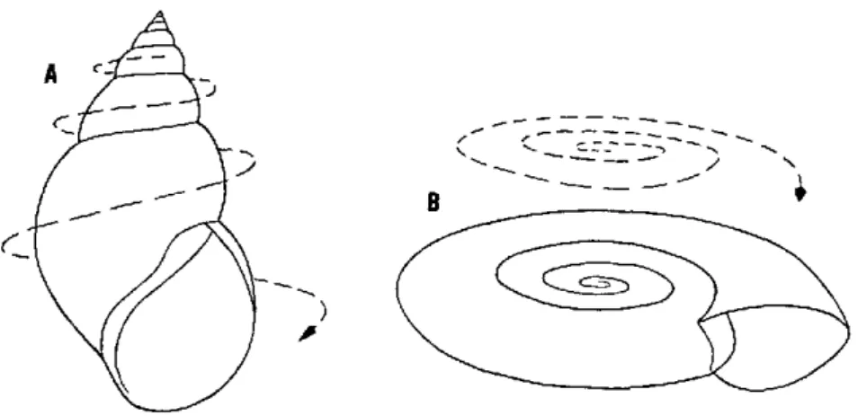
refer-will be flat-or discoidal or planispiral, as in Biomphalaria. If the tube
increases rapidly in diameter and the coils are located outside and below each other, a shell with an appearance somewhere between these two extremes will result (Fig. 2A). If the tube is coiled very little and increases rapidly in diameter, the shell can be pyramidal or patel-loid, as in the Ancylidae (Fig. 20).
The turning of the coil may be clockwise (dextral), in which case the shell aperture is directed to the right when it is facing the observer and the apex is held upwards (Fig. 2A). If it is counterclockwise (sin-istral), the aperture is directed to the left when it is facing the observer and the apex is held upwards (Fig. 18). In discoidal shells the apical side may be hard to recognize. Planorbid shells are in fact sinistral. Some species have the spire secondarily pushed down through the peripheral whorls towards the originally umbilical side and are called pseudodextral ( Fig. 3).
When a snail hatches from an egg mass, in the case of oviparous snails (Fig. 4A), or is born, in the case of ovoviviparous snails (Fig. 4B), it already has an apical whorl, and on rare occasions (some of the Thiaridae) even several. All subsequent growth of the shell is by addi-tion of material at the edge of the aperture. Many species have each subsequent whorl spaced progressively farther from the axis, thus leav-ing a hole in the base, which is called the umbilicus (Fig. 5). The umbilicus varies in size in different species, according to how far from the axis each whorl is formed. If there is no opening in the base of the shell, the shell is called imperforate.
Freshwater snails increase the size of their shells throughout life, and it is often difficult to tell when examining a specimen of a species new to the observer whether it is of maximum size, since the shell main-tains a relatively constant general shape regardless of the stage of growth. Such shells may be said to have indeterminate growth. A few freshwater snails of the Neotropics, among them certain populations of some species of the planorbids, have determinate growth-that is to say, they deflect the aperture when they have reached maximum size (Fig. 55).
-- - Nz
- C : -B
Fig. 2. Direction of growth in a gastropod shell (A, shell with a well-developed spire; B, discoidal shell)
A
B
E
F
B
Imm
!mm ' O.5mm
-Fig. 4. Egg mass (A) and embryonic shells (B) of Plesiophysa from Brazil
---
apex
spire
---suture
body
whorl
inner lip
columell -.
-1
NSA
/
l~-peristome
umbilicu .- |/..
-/---aperture
u--- basal
lip
LEFT SIDE
(in shell books often called under side]
left lip-... .. .
aperture
-right
RIGHT SIDE
(in shell books often called upper side)
apical
.region
body whorl or
...
last
whorl
inner
whorls
...
penultimate
whorl
...---
suture
VENTRAL VIEW
Fig. 7. Counting of whorls on a planorbid shell (left or right side is chosen according to convenience)
The Animal
It is possible to recognize several major parts of the snail besides the shell (Fig. 8). The part that fills the shell when the animal is extended and crawling is the visceral mass, and the part that extends beyond the aperture is the head and foot, or cephalopedal mass. There is no sharp boundary between them. The visceral mass fills all the whorls of the shell and normally conforms to the coiling and shape of the whorls. The cephalopedal mass is joined to the visceral mass by a constricted neck or body stalk.
The cephalopedal mass has a foot with a flat sole and a mouth in front, both of which are applied to the substrate when the snail crawls. On each side of the mouth is a labial palp, usually quadrate in form. In the Pilidae each is drawn out into a thin filament resembling a tentacle. Labial palps are absent in the Thiaridae. In many proso-branchs the front end of the head is distinctly separated from the foot and forms a proboscis.
On the dorsal (top) part of the head is a pair of appendages known as tentacles. There is also a pair of eyes, which may be in the head proper near the tentacles (Pulmonata), on the tentacles near their bases (some Prosobranchia), or on short stalks beside the tentacles
(Pilidae).
The part of the body wall covering the visceral mass is called the mantle. The cavity inside the body is the hemocoel, and it is filled with body fluid, or blood. In most freshwater snails of the Neotropics the blood is colorless, though in some genera of the Planorbidae (but not
Drepanotrema) it is red, owing to the presence of hemoglobin.
.1-.
E- a
CD CD <2 4u> Co 4-a 4' 'a a
m
to =u a> .o4 co Y> . b-oD -_. CD a 4--E -e eo c o E uo I co o o .E co =
respiratory system is constituted in part by the body wall, which is
exposed to the external environment, and by special and varied
modi-fications of the wall in different families.
The external structures of the cephalopedal mass and the pulmonary
cavity of freshwater snails are not entirely symmetrical in their location.
Thus, the reproductive opening or openings, the anus, and the
pneu-mostome open only on one side of the midline and have no counterpart
on the other. If these structures are on the right side of the
cephalo-pedal mass, the animal is said to have dextral anatomy; if they are on
the left, it has sinistral anatomy. Ordinarily, dextral shells contain
dextral animals, and sinistral shells contain sinistral animals, but there
are exceptions. Thus,
Acrorbis
and some varieties of
Biomphalaria
andecola
(both Planorbidae) have sinistral animals in dextral shells
(Fig. 3). Snails of the Planorbidae with disc-shaped shells have
sin-istral animals, but snails of the Pilidae and Amnicolidae, which also
have disc-shaped shells
(Marisa,
in Fig. 12, and
Cochliopa,
respec-tively), have dextral animals.
When reference is made in the following descriptions to left- or
right-hand position in the morphology of the snails, the positions are
always in accordance with the left and right sides, as indicated in
Figure 6.
.The Organ Systems
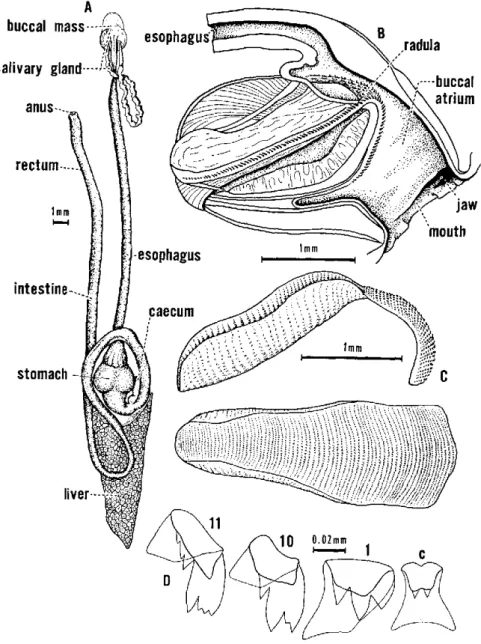
Digestive System
The digestive system (Fig. 9) consists of a single tube that expands
in various areas to form special organs. Several large glands (the
salivary gland and the liver lobes) are attached to the tube.
Imme-diately behind the mouth the digestive tube expands into a large sac,
known as the buccal mass, the outer side of which is mostly covered
with muscles.
The anterior end of the buccal mass (buccal atrium) is lined with a
thin, acellular cuticle, and its anterior margin is thickened to form a
jaw. The jaw may consist of many small platelets, or these platelets may
be fused into one or more major pieces, which in turn have distinctive
location, shape, and sculpture, depending on the family or genus.
A
buccal
mass--salivary
gland---anus--...,
rmm
intestine-..
stomach
jaw
C
D
Fig. 9. Internal structures of Biomphalaria
The esophagus emerges from the top of the buccal mass, and the
duct of a salivary gland is attached to each side of its origin. The
salivary glands may be branched to varying degrees, and they are often
attached to each other and to the esophagus at their posterior end,
which scarcely extends to the upper end of the cephalopedal hemocoel.
The esophagus passes through this hemocoel and then expands in the
apertural end of the upper visceral complex into a stomach.
In the freshwater pulmonates, the stomach is a spherical expansion
of the digestive tract. Its anterior and posterior ends (the cardiac and
pyloric parts, respectively) are membranous and separated by a
mus-cular region between. The esophagus enters the cardiac end, and the
intestine leaves the pyloric end. At the junetion of the pyloric end and
the intestine, the upper lobe of the liver enters the digestive tube
proper, and opposite it is a small gastric caecum of unknown function.
Whether it has a crystalline style is debatable. The intestine curves
around the lower (apertural) end of the stomach, forming a lower loop,
then swings apicad above the stomach, forming a much larger upper
loop. It then passes to the roof of the mantle cavity and traverses that
as the rectum. The anus opens near the pneumostome.
The stomach of most freshwater pulmonates, unlike prosobranchs,
always contains sand grains.
Reproductive System
The reproductive system (Fig. 10) is one of the most important
indicators for classifying freshwater snails, possibly because it is one
of the most complex, stable, and diversified of all the organ systems.
Even so, the funetion of many of the reproductive structures is still
unknown.
The gonad is associated with the upper lobe of the liver and consists
of many follicles, which drain into a sacculate gonadal atrium. In most
prosobranchs, the sexes are separate. The gonad of the male is called
the testis, that of the female the ovary. The pulmonates are
hermaph-roditic, and the gonad is called the ovotestis.
In the males of most prosobranchs (but not Thiaridae), the
gono-duct, or vas deferens, leads to an external appendage called the penis,
or verge. This structure is variously located in different families. Some
female prosobranchs of the Thiaridae and Amnicolidae are
ovovivi-parous-that is, they retain the fertilized eggs inside the body until they
hatch. Most prosobranchs, however, and all the freshwater pulmonates
lay eggs and are therefore oviparous.
fe-u>
cri <a
c v
._,C)
¢
Q -~ m
':-.cu
Q
o o,
- . -, C
C]
_
o~"-Pti
aC°
EH
UDv,
i;,
a>e cu
a> ca
ol
u
E
male genital pore. A spermathecal sac opens into the vagina generally through a duct. The simple or complex prostate gland opens into the vas deferens either directly or through a separate prostate duct. The upper portion of the male duct is called the sperm duct. The part below the prostate leading to the male copulatory organ is the vas deferens. The male copulatory organ is differentiated into a penis sac containing the penis, or verge, and the preputium, which opens at the male genital pore. The great diversity in the size, shape, and structure of the male organ is of systematic importance. In certain genera accessory struc-tures are associated with the male copulatory organ-for example, flagella (Fig. 26) and preputial organs.
Nervous System
In freshwater snails the central nervous system is composed of sev-eral ganglia, or swollen masses of nerve cells, connected to each other by strands of nerve tissue called commissures and connectives. Nerves lead to various parts of the body from the ganglia. The ganglia and their joining strands are arranged to form two rings, the most impor-tant one encircling the frontal portion of the esophagus.
A small, spherical statocyst rests on each pedal ganglion. This con-tains particles that are displaced as the snail moves, giving it a sense of balance.
Excretory System
In its simplest form, the kidney of freshwater snails consists of a large, flattened sac, the wall of which is formed of a single layer of epithelial cells. The sac is situated in the roof of the mantle cavity, at its upper end, and it touches the pericardium.
In the pulmonates, the kidney (Figs. 8 and 38) is restricted to the roof of the pulmonary cavity and does not touch the hemocoel of the upper visceral complex. In larger species the epithelium of the wall is pushed inward to form lamellae, or kidney folds, which are variously formed and situated in different genera. Except in Chilina, there is a
true ureter at the apertural end of the kidney. This is a narrow tube, usually without folds, that conducts the excretory products to a pore near the pneumostome. The shapes of the kidney and the ureter vary in different families or genera.
Circulatory System
The anterior portion, the auricle, pumps the blood into the posterior
portion, the ventricle. Muscle fibers running obliquely across each
chamber cause the heart chambers to contract alternately. A valve
be-tween the chambers and another at the entry into the auricle prevent
the backward flow of blood.
At the point where the pericardium and kidney touch there is a small
opening, the renopericardial passage, which connects the pericardial
cavity with the cavity of the kidney.
Blood leaves the cephalopedal, isthmian, and upper visceral parts of
the hemocoel by passing into the roof of the pulmonary cavity. There
it seeps through the loosely packed connective tissue that fills all the
space not occupied by the pericardium, kidney, and rectum. A few
tubular channels, mere recessions in the connective tissue, carry most
of the blood to the kidney, where it flows across the outer surface
through another smaller set of channels. It then collects in a tubular
sinus, the pulmonary vein, along the side of the kidney that is toward
the heart. This prominent vessel carries the blood directly to the
auricle.
The vessels of the pulmonary roof are veins, since they conduct the
blood toward the heart. None is lined with an epithelium. As the blood
passes through the roof of the mantle cavity it also exchanges carbon
dioxide for oxygen, and thus the roof serves a respiratory function.
Respiration is assisted by a gill in the prosobranchs, and by a
pseudo-branch in most of the Planorbidae, Ancylidae, and Chilinidae.
3. KEY TO THE GROUPS OF FRESHWATER SNAILS
OF THE NEOTROPICS
In applying the following key to find the genus or family of a species,
the investigator should always have a series of specimens. This will
help assure that adult shells are being used. Immature shells are easily
misidentified. It is best to work with living material, or at least with
shells containing the animal preserved in alcohol, rather than with
empty shells.
Determination Key to Families of Neotropical
Freshwater Snails
Main Groups
With operculum; mantle opening di-rected more or less forward (opercu-late snails) . - ---Without operculum; mantle opening directed more or less left or right side of the body (lung snails) ...
Families of Prosobranchia
Spire reduced to a minute structure on the side of the body whorl, making the whole shell more or less subspherical (20 mm or less) ... Shell large (20 mm or more) and more or less globose or thickly discoidal; spire not more than one third of shell height; labial palps drawn out into threads
---Shell medium-sized and globose, sim-ilar to last family; no thread-like labial palps (known only in Cuba and North America)
Shell small (less than 10 mm high), generally with well-developed spire .-..
Shell medium-sized (more than 10 mm high), with well-developed or,
Prosobranchia
Pulmonata
Neritidae (Fig. 11)
Pilidae
(Fig. 12)
(syn. Ampullariidae)
(Fig. 13) Viviparidae
(Fig. 14) Amnicolidae
tOmm
Fig. 11. Shell and operculum
of Neritina punctulata
La-marck from a Puerto Rican rivér
B
Fig. 12. A: Shell and operculum of Ampullaria
B: Shell ofMarisa cornuarietis (L) from Trinidad, ventral view
C: Right side view of the same species D: Left side view of the same species
Fig. 13. Shell of Lioplax pilsbryi Walker from
Florida-a representative of the Viviparidae
B· :`
::i>? :
·- ·
Fig. 14. A: Shell of Littoridina andecola (Orbigny) from Lake Titicaca, Peru
B: Shell, with operculum, of Littoridina languiensis Haas from Lake
Langui, Peru
A B
A
10mm
Fig. 15. A: Shell and operculum of Tarebia granifera (Lamarck), a melaniid
snail introduced into Puerto Rico-a potential intermediate host of
Paragonimus westermani
B: Shell of Pachychilus laevissimus Sowerby, a melaniid snail from
generally, high spire ...-. . (sy
Families of Pulmonata
Shell dextral with distinct spire; columella without any sort of callosity; shell uniformly colored ...
Shell dextral with distinct spire; columella with callus and with one or two folds (only in southern South America); shell often with brownish
stripes or bands ... Shell sinistral with distinct spire; shell usually glossy; juvenile shell not hir-sute; animal without pseudobranch; mantle border digitate or lobed ... Shell sinistral with distinct spire, tex-tured, with raised, beaded spiral lines; juvenile shell hirsute; animal with pseudobranch; mantle border even ....
(subfamily Shell discoidal (rarely spired and pseudodextral), e.g. Drepanotrema
(Acrorbis) petricola and certain forms
of B. andecola . ... (exce
Shell cap-shaped; spire reduced ...
Thiaridae (Fig. 15) yn. Melaniidae, in part) *
(Fig. 16) Lymnaeidae
Chilinidae (Fig. 17)
Physidae (Fig. 18)
(Fig. 19) Plesiophysinae of Planorbidae)
Planorbidae ept Plesiophysinae)
Ancylidae (Fig. 20)
Diagnostic Characteristics of the Neotropical Genera of Planorbidae
Biomphalaria
Among the Planorbidae, there is only one genus, Biomphalaria,**
that is important at present to the problem of schistosomiasis in the
Neo-' A few so-called pleurocerids (Goniobasis, Pleurocera) occur in the southern
lmm
Immn
Fig. 16. A: Shell of Lymnaea columella Say from
Puerto Rico
B: Shell of Lymnaea cubensis Pfeiffer
from Puerto Rico
Fig. 17. Shell of Chilina major Sowerby
from Chile
lOmm
q--
----b oE
o,
-I
.p
tropics. A key and a detailed synopsis are therefore provided in later sections of this Guide.
The embryonic and the first few postembryonic whorls of the shell never have a sharply angled shoulder. The adult shell is nearly always discoidal, though some variants of B. andecola are pseudodextral with
the spire slightly elevated. In a few species-for example, B. tenagophila
-the shoulder is anglied in the intermediate whorls and occasionally in the final whorl of larger specimens. In other species the shoulder is evenly rounded at all stages of growth. Some species never grow larger than 10 mm in diameter, with 41/2 whorls; others reach a maximum diameter of more than 30 mm and have about 6 whorls.
There is neither a preputial organ in the preputium nor a set of flagellar glands attached externally at the upper tip of the penis sac. The opening of the penis is terminal and the tip of the penis has no stylet.
The jaw is T-shaped, with the dorsal part composed of a single crescent-shaped piece. The central tooth of the radula has only two cusps, with no small accessory denticles on their shoulders.
The blood is red in all living specimens that have been observed. The Biomphalaria genus ranges from the southern part of North
America, through Middle America, to the southern part of South America.
Drepanotrema (Figs. 21-26)
Several species in this genus occur throughout the area in which
Biomphalaria is found. They differ from Biomphalaria in their small
shell size, which is generally less than 5 mm in diameter at 4 whorls (though a few species may grow about twice that size, with, of course, more whorls).
The jaw is composed of many minute platelets in both the lateral and dorsal parts. There are always glandular flagella at the tip of the penis sac. The penis has a terminal opening and no stylet on its tip. There is no penial gland in the preputium.
The blood is colorless in the species that have been studied.
The generic names Drepanotrema, Acrorbis, Antillorbis, and Fos-sulorbis have been used in the literature for species of this group.
Helisoma (Fig. 27)
Throughout North America, Middle America, and South America (from Venezuela to Southwestern Peru) snails are found that closely resemble some species of Bionmphalaria but in reality belong to the
genus Helisoma. Their embryonic and early postembryonic whorls
EE
-1
E o
2=
ri S .
IZÓ
cu·
o-,
CnE
o> - 4z
s
S >
o S,
U
bD $'
Lroi t
-~ -l
'E-:
cf'i
¢
o-h
-Qa>
Q; o
"oc'
.- o
Q
' ^4
j
CN;
.
eA, _ eq Yib,
>C
Im m
Fig. 25. Shell of Drepanotrema (Antillorbis) aeruginosum (Morelet) from
fl
lmm
Fig. 26. Reproductive system of Drepanotrema anatinum (Orbigny) from Brazil.
lOmm
Fig. 27. Shell of Helisoma foveale (Menke) from an irrigation tank in southern
throughout the next whorls. The shells are usually discoidal and may grow to 25 mm in diameter with about 5 whorls. There are some rare cases of populations in which the spire is elevated but sinistral or pseudodextral.
The jaw and central teeth of the radula are like those of Biompha-laria. There are no flagellar glands at the tip of the penis sac, but a
large preputial organ is present with an external duct that connects with the penis sac. The tip of the penis has a stylet, and the seminal opening is lateral.
Plesiophysa (Fig. 19)
Some rare snails known from limited localities in Brazil and from a few locations in the Antilles are related to the planorbid genera cited above, though superficially they resemble species of the Physidae. The shell is sinistral with an elevated spire. It differs from Physa in that it
has spiral lines of small beads or nodules and the shell of younger specimens is hirsute.
Species of Undetermined Genera
4. KEY TO NEOTROPICAL BIOMPHALARIA SPECIES*
1. Ventral surface of the renal tube with a pigmented longitudinal ridge (Fig. 38, rer), or a piginented line in
young specimens ... glabrata
Ventral surface of the renal tube smooth and barely pigm ented (Fig. 62, ret) ... ... 2 2. Vaginal wall with a pouch or swelling to the right of
the spermathecal duct (Fig. 63, vp) ... 3 Vaginal wall without a pouch or swelling to the right of the spermathecal duct (Fig. 58, va) ... 13
3. Ovotestis diverticula over 200 (Fig. 63, ot) ... tenagophila
Ovotestis diverticula under 150 (Fig. 50, ot) ... 4 4. Vaginal pouch scarcely extending beyond the level of
the insertion of the spermathecal duct, the latter
empty-ing outside and caudad to the pouch (Fig. 65, vp) ... trigyra
Vaginal pouch normally extending well beyond the level of the insertion of the spermathecal duct, the lat-ter emptying inside the pouch (Fig. 50, vp) ... 5 5. Maximum width of the distal half of the vas deferens
about the same as that of the middle part of the penis sac (Fig. 29, vd, ps) ... 6
Maximum width of the distal half of the vas deferens distinctly narrower than the middle part of the penis sac (Fig. 54, vd, ps) ... .... 7
6. Clearly distinct and bulgy vaginal pouch (Fig. 29, vp) amazonica
Poorly developed vaginal pouch, lacking in some speci-mens or coexisting with a little-developed corrugation
to the left of the spermathecal duct (Fig. 46 A) [See 14] intermedia
8. Ovotestis diverticula under 30 (Fig. 31, ot) ... andecola
Ovotestis diverticula over 30 (Fig. 54, ot) ... peregrina
9. Vaginal pouch conspicuous (Fig. 54, vp); prostatic di-verticula arborescent, with terminal branches bent to the right and left sides and clustering together into a
compact cauliflower-like surface (Fig. 54, pr) ... prona
Vaginal pouch little to moderately developed (Fig. 41, vp); prostatic diverticula subdivided into branches with tips arranged more or less into a row (Fig. 41, pr) 10
10. Vaginal pouch slightly projecting but distinct (Fig.
41, vp) ... havanensis
Vaginal pouch extremely low, sometimes nearly indis-tinct (Fig. 48, vp) ... 11
11. Prostatic diverticula over 13 (Fig. 48, pr) ... obstructa
Prostatic diverticula under 13 (Fig. 35, pr) ... 12
12. Seminal vesicle with conspicuous diverticula (Fig. 35, sv); vagina with a median constriction (Fig. 35, va); vaginal pouch only vaguely indicated as a flattened elevation hardly delimited from the surrounding
sur-face (Fig. 35, vp) ... ... ... ....cousini
Seminal vesicle with poorly developed diverticula (Fig. 52, sv); vagina cylindric (Fig. 52, va); vaginal pouch
apparently lacking in some specimens [see 18] ... philippiana
13. Vaginal wall with corrugations to the left of the sperm athecal duct (Fig. 60, ve) ... . 14 Vaginal wall without corrugations to the left of the spermathecal duct (Fig. 58, va) ... 15
14. Vaginal wall strongly corrugated to the left of the
spermathecal duct (Fig. 60, vce) ... ... straminea
Vaginal wall weakly corrugated (sometimes merely swollen) to the left of the spermathecal duct (Fig. 45, ve; Fig. 46 A); little to moderately developed vaginal pouch to the right coexisting with the corrugation in
some specimens (Fig. 46 A) [see 6] ... ... intermedia
15. Vaginal wall smooth (Fig. 58, va); ovotestis diverticula
over 200 (Fig. 58, ot) ... ... sericea Vaginal wall smooth (Fig. 43, va); ovotestis diverticula
16. Very long penial complex and spermatheca (Fig. 56, ps-pp, sp); penis sac more than 4 times as long as the preputium (Fig. 56, ps, pp); spermatheca more than twice as long as the preputium (Fig. 56, sp, pp) ... Comparatively short penial complex and spermatheca
(Fig. 43, ps-pp, sp); penis sac clearly less than 4 times as long as the preputium (Fig. 43, ps, pp); spermatheca less than twice as long as the preputium (Fig. 43, sp, pp) ... 17. Seminal vesicle with conspicuous diverticula (Fig.
33, sv) ... ... Seminal vesicle with poorly developed diverticula (Fig. 43, sv) ... 18. Prostatic diverticula usually 0-4, occasionally 5-6 (Fig.
52, pr); little-developed vaginal pouch in some speci-m ens [see 12] ... ...
Prostatic diverticula usually over 6, exceptionally 4-5 (Fig. 43, pr) ...
schrammi
17
chilensis
18
philippiana
5. SYNOPSIS OF THE BIOMPHALARIA SPECIES
Animal species are not static entities; they are subject to genetic changes. The accumulation of these changes over time brings about animal evolution. If populations of one species in a certain geographic area are by some means isolated from the populations of the same species from another area, independent evolution in the two areas may result in a splitting of the species into two geographical races. Further independent evolution of the two races may transform the races into distinct species. Though as a rule true distinct species do not inter-breed spontaneously in nature, specimens of different races of the same species do, and they produce fertile progeny. This difference between races of a single species and entirely distinct species is of course not always clear-cut; it develops gradually over the course of evolution. It is therefore difficult and sometimes even impossible to distinguish as races or distinct species forms that have recently evolved from a common ancestor. Even if they should rightly be regarded as species, hybridization might occasionally occur.
Forms that have obscure taxonomic status often occupy neighboring geographical areas and sometimes create hybrid populations where the areas meet. They may or may not be morphologically distinct. However, as a principle, the concept of species is biologically based. At present the investigators are not in position to judge whether some of the taxonomic units distinguished here correspond to true biological species. For example, the forms dealt with under the names B. pere-grina, B. chilensis, B. stramainea, B. intermedia, B. obstructa, and B. havanensis might well have a recent common origin.
Biomphalaria amazonica
Paraense, 1966
The Shell (Fig. 28)
The shell reaches a maximum of about 8 mm in diameter and 2.5 mm in width, with about 5 whorls increasing moderately in diameter.
The whorls are rounded on both sides, except the body whorl, which is somewhat flattened in full-grown shells. The periphery is rounded. The aperture is egg-shaped, sometimes rounded, a little oblique, and
more or less bent to the left in most full-grown specimens.
whorl is deeply sunken. The left side is broadly and shallowly concave
and shows the surface of the whorls more plainly than the right side.
The Soft Parts (Fig. 29)
The pigmentation of the normally exposed parts is grayish brown,
with the mantle pigment distributed in dark patches, tending to
coalesce on the roof and right side of the pulmonary cavity.
There are about 50 ovotestis diverticula, which are pear- or
club-shaped and predominantly unbranched though sometimes bifurcate
and exceptionally trifurcate. The seminal vesicle has conspicuous
di-verticula. The vagina is cylindric, with a bulging pouch to the right
of the spermathecal duct. The spermatheca is usually club-shaped,
with a duct less than half as long as the spermathecal body and not
well delimited from the latter. The prostate has 8 to 12 diverticula,
most of which are divided into two or three short branches, which may
have small ovoid or elongate bud-like subdivisions. The distal half of
the vas deferens is nearly as wide as the penis sheath, and the latter is
about half as long as the preputium, which at the caudal end is only
a little wider than the penis sheath and gradually expands to become
about twice as wide at the cephalic end.
There is no renal ridge.
The lateral radular teeth have dagger-like or triangular cusps.
Geographical Distribution
The species is known only from Manaus (Amazonas) Brazil.
Ecological Features
It is found in slowly running waters that have abundant
Eichornia
and other aquatic and semiaquatic plants and pH 6.0 to 6.3.
Genetics
No data have been recorded.
Epidemiological Importance
¡?/~i~?~!,~i~i~~I ~
3mm
i~-- I
Fig. 28. Shell of Biomphalaria amazonica Paraense from Manaus, Brazil
Fig. 29. Reproductive system of Biomphalaria amazonica Paraense from Manaus,
Biomphalaria andecola
(Orbigny, 1935)
The Shell (Fig. 30)
The shell rarely exceeds a diameter of 21.9 or a width of 9.8 mm.
Specimens 16 mm in diameter and 6 mm in height are regarded as large
shells. Normal dimensions are about 6.2 and 3.3 mm, respectively. The
number of whorls is 3 to 31/2 at 6 to 10 mm in diameter, 4 at 16 mm,
and 41/2 at 20 mm.
The whorl increases rapidly in diameter and width. Occasionally
its periphery is angular, but more often it is carinate, and sometimes
even sharply so. The aperture is irregularly circular, with a tendency
toward a pentagonal outline. It is sometimes extended, particularly
toward the left.
The shape of the right side varies from convex to almost fiat and
even concave centrally. At one end of the range of shapes the shell
has fairly smooth rounded whorls separated by a depressed suture and
the central area is slightly concave. At the other extreme the shell is
convex with a sharply angular area slightly concave, forming a low
spire and a pseudodextral shell type. Intermediate forms combine the
extremes into a complete series.
The left side is normally separated from the right by a carina or at
least an angulation. Another angulation or carina surrounds the deep
umbilicus. Between this angulation and the periphery of the shell there
is often a third angulation that is not quite so sharp. On the left side
the body whorl covers most of the preceding whorl. Only a minor part
of the earlier whorl is visible in the bottom of the deep, steep-walled
umbilicus.
The shell does not have any pronounced sculpture apart from fine
growth lines. Its general color is yellowish to pale brown. The carinae,
at least the peripheral one, when developed, may be paler.
The Soft Parts (Fig. 31)
The body consists almost entirely of the stout body whorl. The inner
whorls taper rapidly and occupy a very small space. The pigmentation
of the normally exposed parts varies from pale gray to deep black. The
visceral mass is darkly pigmented in an irregular pattern. The
pig-mentation is particularly intense on the roof of the mantle cavity.
bifur-7< M1 11-
9:ilR
3mm
Fig. 30. Shell of Biomphalaria andecola (Orbigny) from Lake Titicaca, Bolivia
Po
~~~~ng
c
pPP-U
Fig. 31. Reproductive system of Biomphalaria andecola (Orbigny) from Lake
Titicaca, Bolivia. See p. 112 for key to ahbreviations.
37
sd---OV
¡t~:
pm
cc
i
va
ca
sp
PP
:
vd
Iimm
Fig. 31. Reproductive system of Biomphalaria andecola (Orbigny) from Lake
long, slender, richly branched diverticula. Short, unbranched
diver-ticula occur exceptionally. The diverdiver-ticula branch from the sperm duct
very near each other, causing the entire prostate to be very compact.
The male copulatory organ is comparatively stout. The penis sac is
slightly shorter than the preputium and, although thick, it is distinctly
more slender than the latter. The main penial retractor muscle inserts
on the innermost part of the preputium, where it merges into the penis
sac.
The population of this species found in Ojos de Ascotán, near the
Salar Ascotán in the Province of Antofagasta, Chile (described as
Taphius thermalus
by Biese in 1951) is generally characterized by a
warty surface on the vaginal pouch. These warts correspond to
depres-sions in the lumen of the organ caused by heavy infection with a
flagel-late parasite
(Cryptobia)
and lack taxonomic significance.
There is no renal ridge, but there is a rectal ridge continuing as a
dorsal ridge, as in all the other species.
Geographical Distribution
This species is known from localities in the Andean highlands, such
as Lake Titicaca and lagoons in the Titicaca basin (Bolivia and Peru),
and from streams in the Province of Antofagasta, Chile.
Ecological Features
Unlike other species of the genus,
B. andecola
lives not only in
shal-low water but also in depths of up to at least 82 mm. In deeper waters
it is purely discoidal, not pseudodextral, but still angulated. Apart from
its remarkable tolerance to deep water, there are no recorded data
about the ecology of this species.
Genetics
No data have been recorded.
Epidemiological Importance
This species is not known to be an intermediate host of
S. mansoni.
and Taphius thermalus Biese, 1951. Pilsbry later put P. heteropleurus
into a separate genus, Platytaphius.
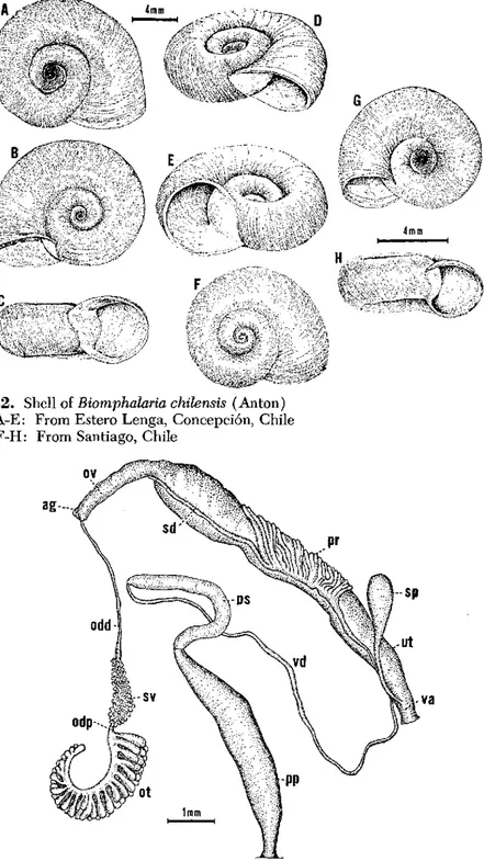
Biomphalaria chilensis
(Anton, 1839)
The Shell (Fig. 32)
Shells of average size measure 9 to 11 mm in diameter and about 3 mm in width. The largest specimen available measured 13 mm in diameter and 4 mm in width. Specimens with 4 to 41/2 whorls measure 9 to 11 mm in diameter. The specimen measuring 13 mm in diameter had 51/2 whorls. The whorls, which increase rapidly in diameter, are rounded and not carinate. The aperture is unarmed and irregularly circular, with a tendency in larger specimens to be more wide than long. In some instances the aperture is slightly deviated towards the left.
The right side is flattened along the periphery and slightly concave toward the center. The left side is more concave than the right side and deeply umbilicate. On both sides the whorls are smoothly rounded.
The Soft Parts (Fig. 33)
The body is elongate, as in most members of the family. The color is uniformly blackish.
The ovotestis consists of many club-shaped diverticula arranged in two or three rows. The diverticula, usually 20 to 27 in each row, are simple, unbranched, and seldom bifurcated. The seminal vesicle is very well developed and beset with many small-sized protuberances and a few large diverticula. A short duct links the seminal vesicle to the ovotestis. The spermatheca is elongate, sac-like, and attached to the vagina by the short spermathecal duct, which is always shorter than the spermatheca itself. The uterus enlarges gradually to join the nidamental gland, which is very swollen. The oviduct is very short. The vagina has no visible corrugation or pouch. The prostate is com-paratively short and bears from 7 to 13 diverticula. The diverticula are usually branched at the tips, though never arborescent, and closely packed at their point of origin. The two or three last distal diverticula, especially the last one, are very elongate and lie over the sperm duct. The penial complex is elongate. The preputium is nearly equal in length to the penis sac. The latter is slightly larger in diameter than the vas deferens, which is long and somewhat irregular in diameter.
e.i
-- ·
~-'s
L,
-I
ci:-:::. : ' ,- ~-:
Fig. 32. Shell of Biomphalaria chilensis (Anton)
A-E: From Estero Lenga, Concepción, Chi F-H: From Santiago, Chile
ag-* 4mm
le
ESsp
ut
In the lateral radular teeth the middle cusp is truncated and not pointed, giving it a squarish appearance (Fig. 66E).
Geographical Distribution
The species is only known with certainty from Chile.
Ecological Features
No data have been recorded.
Genetics
B. chilensis is reproductively isolated from two Brazilian populations
of B. glabrata, one from Recife (Pernambuco) and the other from
Santa Luzia (Minas Gerais). It is also isolated from B. tenagophila
from Sáo Paulo, Brazil. In three out of six reciprocal crosses attempted between this species and B. straminea from Recife, fertile hybrids were
obtained, although the number of viable eggs was small (Barbosa, Carneiro, and Barbosa, 1956).
Epidemiological Importance
This species is a potential host for a Brazilian strain of S. mansoni.
Laboratory infection was obtained in 3 out of 44 snail specimens (Bar-bosa and Bar(Bar-bosa, 1958b).
Nomenclature
Type locality: Maypo, Chile.
Described by Anton in 1839 as Planorbis chilensis.
Biomphalaria cousini
Paraense, 1966
The Shell (Fig. 34)
The shell grows to about 8 mm in diameter and 3 mm in width, with about 41/2 whorls increasing moderately in diameter.
3mm
Fig. 34. Shell of Biomphalaria cousini Paraense from Santo Domingo de los
The right side has a deeply sunken apical whorl and subsequent whorls surrounding a funnel-shaped concavity that broadens at the level of the penultimate whorl. The left side is broadly concave,
show-ing the surface of the whorls more plainly than the right side. There are no internal lamellae.
The Soft Parts (Fig. 35)
The pigmentation of the normally exposed parts is grayish brown. The mantle pigment is distributed in blackish patches.
There are up to about 30 ovotestis diverticula, arranged either in pairs or one after another. The diverticula are pear-shaped and pre-dominantly unbranched, though sometimes they are bifurcate and oc-casionally trifurcate. The seminal vesicle has conspicuous diverticula. The distal portion of the vagina is swollen, and the vaginal pouch is located to the right of the spermathecal duct, only vaguely indicated as a flattened elevation and hardly delimited from the surrounding sur-face. The spermatheca is usually club-shaped, with a duct about half as long as the spermathecal body and frequently well delimited from the latter. The prostate has from 5 to 11 diverticula, most of which are divided into two or three comparatively long secondary branches, which may give tertiary elongate or bud-like subdivisions. The distal half of the vas deferens is distinctly narrower than the penis sac. The penis sac is about half as long and half as wide as the preputium, and the preputium is moderately widened toward the distal end.
There is no renal ridge.
The lateral radular teeth have dagger-like or triangular cusps.
Geographical Distribution
The species is known only from Santo Domingo de los Colorados (Pichincha) Ecuador.
Ecological Features
It has been found in a slowly running brook among aquatic grasses.
Genetics
Nomenclature
Type locality: Santo Domingo de los Colorados (Pichincha)
Ecuador.
No synonyms have been recorded.
Biomphalaria glabrata
(Say, 1818)
The Shell (Figs. 36 and 37)
The shell may reach a maximum of about 40 mm in diameter, though
usually it ranges between 15 and 30 mm, the ordinary size of adults
varying with populations or geographic areas. The width is between
5 and 8 mm in most specimens at 20 mm diameter. The adult shell has
about 5 to 61/2 whorls, increasing slowly or sometimes more rapidly in
diameter.
The whorls are normally rounded on the sides, though sometimes
flattened, angular, or even carinate on the left and less frequently on
the right. The suture ranges from shallow to deep between flattened
and rounded or angular whorls, respectively.
Each side varies from broadly and shallowly to deeply concave,
generally in inverse proportion to the opposite side. The right side
may be flat or even a little convex in individuals of some populations.
The periphery varies from rounded to bluntly angular and is
fre-quently shifted to the right. The aperture may be narrow, egg-shaped,
semicircular, rounded, or transverse; it is usually subangular on the
lower left, directed forward or bent more or less leftward, and
gen-erally oblique to the right or to the left. The peristome is thin and
continuous. Specimens of about 2 to 9 mm from habitats subject to
seasonal drought may develop one or more sets of apertural lamellae
(usually six per set), which later are wholly or partly resorbed. In
some cases the whole set persists inside full-grown shells.
The Soft Parts (Figs. 38 and 39)
The pigmentation of the normally exposed parts ranges from pale
gray to deep black.
9:. ril:;·iii'A
1
6iiDiRii~~b..
´-
--- 6a¿a
Fig. 36. Shell of
(Say) from Lake do Norte, Brazil
Biomphalaria glabrata
Estremoz, Rio Grande
5mm
·
1
~A~~
D5mm
rey
dr
-lmm
i3 !
Fig. 38. Ceiling of mantle cavity of Biomphalaria glabrata (Say) from Brazil.
Imm
'&*
va
utFig. 39. Reproductive system of Biomphalaria glabrata (Say) from Brazil. See