Radio graphy, co m pute d to m o graphy
and m agne tic re so nance im aging
at 0.5 Te sla o f m e chanically induce d
o ste o arthritis in rabbit kne e s
1Programa de Pós-graduação em Medicina Veterinária, and Departamentos de 2Cirurgia e Anestesiologia Veterinária, and 3Reprodução Animal e Radiologia, Faculdade de Medicina Veterinária e Zootecnia, Universidade Estadual Paulista, Botucatu, SP, Brasil
Departamentos de 4Cirurgia e O rtopedia and 5Doenças Tropicais e Diagnóstico por Imagens, Faculdade de Medicina de Botucatu, Universidade Estadual Paulista, Botucatu, SP, Brasil
6Departamento de Bioestatística, Instituto de Biociências, Universidade Estadual Paulista, Botucatu, SP, Brasil
S.R. Torelli1, S.C. Rahal2, R.S. Volpi4, S. Yamashita5, M.J. Mamprim3 and A.J. Crocci6
Abstract
In the present experimental study we assessed induced osteoarthritis data in rabbits, compared three diagnostic methods, i.e., radiography (XR), computed tomography (CT) and magnetic resonance imaging (MRI), and correlated the imaging findings with those obtained by macroscopic evaluation. Ten young female rabbits of the Norfolk breed were used. Seven rabbits had the right knee immobilized in extension for a period of 12 weeks (immobilized group), and three others did not have a limb immobilized and were maintained under the same conditions (control group). Alterations observed by XR, CT and MRI after the period of immobilization were osteophytes, osteochon-dral lesions, increase and decrease of joint space, all of them present both in the immobilized and non-immobilized contralateral limbs. However, a significantly higher score was obtained for the immobi-lized limbs (XT: P = 0.016, CT: P = 0.031, MRI: P = 0.0156). All imaging methods were able to detect osteoarthritis changes after the 12 weeks of immobilization. Macroscopic evaluation identified in-creased thickening of joint capsule, proliferative and connective tissue in the femoropatellar joint, and irregularities of articular cartilage, especially in immobilized knees. The differences among XR, CT and MRI were not statistically significant for the immobilized knees. However, MRI using a 0.5 Tesla scanner was statistically different from CT and XR for the non-immobilized contralateral knees. We conclude that the three methods detected osteoarthritis lesions in rabbit knees, but MRI was less sensitive than XR and CT in detecting lesions compatible with initial osteoarthritis. Since none of the tech-niques revealed all the lesions, it is important to use all methods to establish an accurate diagnosis.
Co rre spo nde nce
S.C. Rahal
Departamento de Cirurgia e Anestesiologia Veterinária Faculdade de Medicina Veterinária e Zootecnia, UNESP
Rubião Júnior, s/n 18618-000 Botucatu, SP Brasil
E-mail: sheilacr@ fmvz.unesp.br Research supported by FAPESP.
Received February 27, 2003 Accepted January 21, 2004
Ke y words
•O steoarthritis
•Rabbit knee osteoarthritis •Imaging diagnosis •Knee immobilization •Radiography
Intro ductio n
Joint movement restriction is one of a vari-ety of procedures that can be used to experi-mentally induce changes in the knee articular cartilage (1). Immobilization results in degen-erative articular alterations in both splinted and contralateral knee joints (2), with abnor-mal proliferating cartilage observed peripher-ally toward the edges of the joint cartilage (3). Osteophyte formation is observed after 14 days of immobilization (3).
Radiography (XR) is the method most frequently used to evaluate alterations in the musculoskeletal system (4). It can be used to detect hard and soft tissue changes in early stages of osteoarthritis, but multiple radio-graphic projections are necessary (5). Com-puted tomography (CT) is an accurate ex-amination technique for osteoarthritis lesions, which most frequently involve the knee (6). It provides excellent contrast resolution and permits differentiation of soft tissue struc-tures that cannot be seen precisely by con-ventional radiography (4). Magnetic reso-nance imaging (MRI) is a sensitive imaging modality for the internal anatomy of the knee joint without the need for any type of con-trast agent or joint manipulation (7).
The aim of the present study was to com-pare the efficacy of XR, CT and MRI for the diagnosis of osteoarthritis induced by con-tinuous immobilization in rabbits, and to correlate the imaging findings with macro-scopic evaluation used as a “gold” standard.
Mate rial and Me thods
The Ethics Committee of the Faculty of Veterinary Medicine and Animal Science, UNESP, Botucatu, approved the study.
Ten female Norfolk rabbits, initial age from 2.5 to 3 months and initial weight 2.5 kg, housed in individual cages of standard size were used. The animals received com-mercial food and water ad libitum. Seven rabbits had the right knee immobilized in
extension for a period of 12 weeks to induce degenerative joint disease (immobilized group) and three others were not immobi-lized (control group). The method used to immobilize the knee was an individually shaped PVC splint applied to the caudal aspect of the right leg from the proximal end of the thigh to the distal end of the limb (2,3,8-10). Rabbits were premedicated with acepromazine (1.0 mg/kg, im) and anesthe-tized with sodium pentobarbital (15 mg/kg, iv), and both knees were evaluated by XR (TUR D800, Germany), CT (GE-SYTEC-2001, Japan) and MRI(GE-MR-MAX, 0.5 Tesla, Japan), before immobilization (TI) and 12 weeks after immobilization (TII). Radiographic examinations were performed at our Veterinary Hospital, and CT and MRI in the Imaging Diagnostic Service of the Faculty of Medicine, UNESP, Botucatu. Af-ter 12 weeks, the animals were sacrificed for macroscopic examination.
Radiographs were obtained in lateral, craniocaudal and skyline projections. XR features observed were osteophyte forma-tion, femoral condyle subchondral lesions, tibial subchondral lesions, femoral condyle irregularity, femoral osteochondral reaction, and increase or decrease of articular space.
in-crease or dein-crease of articular space.
MRI was performed on sagittal and coro-nal planes using a 0.5 Tesla scanner. Three imaging pulse sequences were used: a) T1-weighted spin echo (SE) with a repetition time (RT) of 700 ms, echo time (ET) of 25 ms (700/25), acquisition time of 2 min and 59 s, and NEX of 2; b) T2-weighted SE with an RT of 2000 ms, ET of 100 ms (2000/100), with an acquisition time of 8 min and 32 s, and NEX of 2; c) proton density-weighted SE with an RT of 2000 ms, ET of 25 ms (2000/25), with an acquisition time of 8 min and 32 s, and NEX of 2. The other param-eters used were the same for all sequences: 15-cm field of view and 3-mm thick sections with a 3-mm intersection gap and a 224 x 128 matrix. An extremity coil was used. Each MR image was examined for the pres-ence of femoral condyle osteochondral le-sion, tibial osteochondral lele-sion, fibular teochondral lesion, femoropatellar joint os-teochondral lesion, osteophyte formation in femur, patella and femoropatellar joint, and increase or decrease of synovial fluid. One reader evaluated the XR images and another one evaluated the CT and MRI images. All exams were printed on radiological films. The readers did not know which limb was immobilized. The same scoring system was used to quantify degenerative joint disease for all the diagnostic methods. This system was based on a previous score reported by Vasseur and Berry (9). Each finding was assigned a value of 0 (absence), 1 (mild), 2 (moderate) or 3 (severe) depending on the severity of the changes and the values were summed to obtain a cumulative osteoarthri-tis score.
A macroscopic study was performed to obtain reliable reference data as a “gold standard” and to determine the accuracy of the diagnostic test XR, CT and MRI. The distal end of the femur and the proximal end of the tibia were separated, and the gross appearance of the joint surfaces and the peri-articular soft tissues was evaluated. The
ar-ticular cartilage was observed for loss of brightness, irregularities or presence of ul-cers. Ligaments and the joint capsule were evaluated for rupture and thickness.
Data for immobilized and non-immobi-lized contralateral knees at TII, and between TI and TII for both knees were analyzed statistically by the nonparametric Wilcoxon rank sum test. Friedman repeated measures analysis of variance on ranks was used to compare the three methods of diagnosis (XR, CT and MRI) after 12 weeks of immobiliza-tion (TII) using the scores obtained for each one. Non-immobilized contralateral knees from the immobilized group and all knees from the control group were compared at TII by the nonparametric Mann-Whitney rank sum test. Differences were considered to be significant when the P value was <0.05.
Re sults
The osteoarthritis scores obtained by the three diagnostic methods are presented in Table 1.
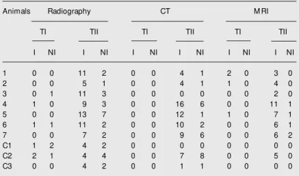
Table 1. Total scores obtained w ith the three diagnostic methods based on a previous score reported by Vasseur and Berry (9).
Animals Radiography CT M RI
TI TII TI TII TI TII
I NI I NI I NI I NI I NI I NI
1 0 0 11 2 0 0 4 1 2 0 3 0
2 0 0 5 1 0 0 4 1 1 0 4 0
3 0 1 11 3 0 0 0 0 0 0 2 0
4 1 0 9 3 0 0 16 6 0 0 11 1
5 0 0 13 7 0 0 12 1 1 0 7 1
6 1 1 11 2 0 0 10 2 0 0 6 1
7 0 0 7 2 0 0 9 6 0 0 6 2
C1 1 2 4 2 0 0 0 0 0 0 0 0
C2 2 1 4 4 0 0 7 8 0 0 5 0
C3 0 0 4 2 0 0 1 1 0 0 0 0
Figure 1. Radiography of the right knee of a rabbit from the control group (A) and of an im-mobilized knee (B) show ing pa-tellar ligament mineralization (ar-row ). A = rabbit C3; B = rabbit 1 (see Table 1).
Radiographic findings
The data in Table 1 obtained before im-mobilization (TI) differed significantly from those obtained after 12 weeks of immobili-zation (TII) for both immobilized (P = 0.016) and non-immobilized contralateral knees (P = 0.016). After 12 weeks, the immobilized
and non-immobilized contralateral knees showed different osteoarthritis scores (P = 0.016), whereas the scores for non-immobi-lized contralateral knees and control knees were not significantly different (P = 0.295). A decrease of articular space was the most common change observed, also detected in the control group. The least common changes
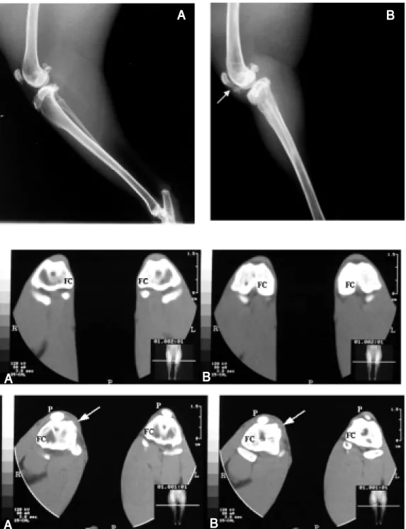
Figure 2. Top: Sequence of CT t ransverse planes (A, B) of knees from the control group (anim al num ber 3). Fem oral condyles (FC) are demonstrated.
Bottom: Sequence of CT trans-verse planes (A, B) of immobi-lized and non-immobiimmobi-lized con-tralateral knees (animal number 2). The patellas (P) and FC can be observed. An osteophyte is evident on the medial condyle of the right (R) immobilized joint (arrow s). L = left joint.
B
B
B
B
B
A
A
A
A
A
B
B
B
B
B
A
A
A
A
A
B
B
B
B
B
A
were femoral subchondral lesions and osteo-phyte formation in the tibia and trochlear groove. The control group did not show osteochondral reactions or osteophyte for-mation in the femoral condyles or patella (Figure 1A). Other changes were seen in some animals, such as femur head sublux-ation in non-immobilized contralateral limbs (four rabbits), muscle group asymmetry be-tween immobilized and non-immobilized contralateral limbs (all immobilized rabbits) and patellar ligament mineralization (one rabbit; Figure 1B).
Compute d tomography findings
Immobilized knees and non-immobilized contralateral knees were significantly differ-ent (P = 0.031) after 12 weeks of immobiliza-tion (Table 1). There was a significant differ-ence between TI and TII for both immobilized (P = 0.031) and non-immobilized contralateral knees. However, there was no difference be-tween the non-immobilized contralateral knees and the control group knees. No animal showed osteochondral lesions in the fibula and patella or osteophyte formation in the patella. Never-theless, osteochondral lesions in femur, tibia and trochlear groove were the most common changes. The control group presented few alterations by CT, like a mild decrease of articular space in the right and left limbs (Fig-ure 2, top). Osteophytes were observed espe-cially in the immobilized joint (Figure 2, bot-tom). Muscle asymmetry was visible upon CT examination in all immobilized animals.
Magne tic re sonance imaging findings
Statistical analysis confirmed the differ-ence between immobilized and non-immobi-lized contralateral limbs at TII (P = 0.0156, Table 1). The difference between TI and TII was significant for immobilized knees (P = 0.0156), but not for the non-immobilized con-tralateral knees. Comparison of the scores for the non-immobilized contralateral knees and
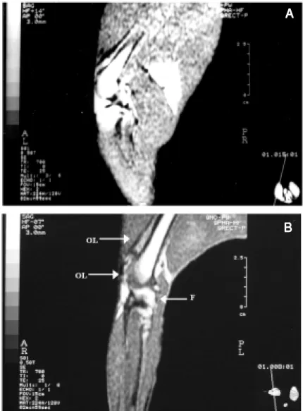
control knees did not show significant differ-ences. The animals showed some osteoarthri-tis alterations, the most common being osteo-chondral lesion (Figure 3A), but articular car-tilage injuries could not be detected. The fibula and patella did not show any osteochondral change and increase of synovial fluid was observed in only two rabbits. One animal from the control group had a lesion revealed by MRI, but no changes were detected in the other two animals (Figure 3B). Some other alterations were soft tissue asymmetry between
Figure 3. A, Sagittal plane of the right non-immobilized joint of an animal from the control group (animal number 3) in a T1-w eighted spin echo sequence. B, Sagittal plane of an immobilized joint (animal number 5). Articular fluid (F) and osteochondral lesion (OL) in the femoral condyle and in the proximal trochlear groove in a T1-w eighted spin echo sequence are observed.
A
A
A
A
A
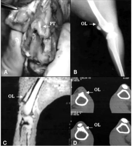
Figure 4. Proliferating tissue and osteochondral lesion in the proximal trochlear groove of an immobilized knee (animal number 5). A, Proliferating tissue (PT) observed by macro-scopic examination; B, osteochondral lesion (OL) observed by radiography; C, OL detected by magnetic resonance imaging; D, OL detected by computed tomography.
lar joint and on the femoral condyle similar to cartilaginous tissue was more evident in immobilized knees (Figure 4). Ulcers on the articular cartilage were present in only one immobilized knee. Irregularities on the ar-ticular surface were very common in both immobilized and non-immobilized contralat-eral knees, especially fine irregularities as-sociated with loss of brightness. Menisci did not show macroscopic changes and no liga-ment was ruptured or thick.
Muscle asymmetry was revealed by all imaging methods. The proliferated tissue on the proximal femoropatellar joint was dem-onstrated by the three methods (Figure 4), but only moderate to severe proliferation was visualized. Mild proliferated tissue was not detected by any method. Areas with proliferating tissue detected by macroscopic examination were associated with osteochon-dral lesion in imaging exams. Increased thick-ness of the joint capsule was not revealed by the imaging methods.
Comparison of the methods did not dem-onstrate a statistical difference among XR, CT and MRI in the immobilized knee; how-ever, in the non-immobilized contralateral knees MRI 0.5 Tesla yielded a significantly higher score than XR and CT (P = 0.027).
D iscussio n
The injuries observed in immobilized knees were compatible with osteoarthritis, as described in other studies (2). A wide variability of results was seen among ani-mals for both joints, possibly due to differ-ences in the extent of movement restriction produced by the splint (1).
Young animals have different abilities to repair joint lesions when compared to adults (11, apud 2). This is relevant information when we are comparing immobilized or non-immobilized contralateral joints to observe osteoarthritis alterations. All animals had the growth plates almost or totally closed after the 12 weeks, so this fact probably did limbs noted in all animals, and fluid collection
in soft tissue observed in two animals and better visualized in T2-weighted SE images. Meniscus and cruciate ligaments were not well defined by MRI.
Macroscopic findings
Macroscopic evaluation of the immobi-lized group showed connective tissue on the femoropatellar joint border, muscle asym-metry and increased thickness of the joint capsule, especially on immobilized limbs. The control group did not present an in-creased thickening of the knee capsule. Pro-liferated tissue on the proximal
femoropatel-B
B
B
B
B
A
A
A
A
A
D
D
D
D
D
C
not have an effect on the observed lesions. Adult rabbits show abnormalities such as proliferating cartilage or osteophyte forma-tion within 10 to 14 days of immobilizaforma-tion (3), findings that were detected here after 12 weeks or 84 days and that were not so marked. This was probably due to the fact that young rabbits were used.
Injuries present in non-immobilized con-tralateral knees were less severe than in the immobilized knees, but were clearly visible on the XR, which indicated small degenera-tive changes probably normal or due to me-chanical stress from moving on three legs or to humoral factors (8). The method was ap-propriate to diagnose osteoarthritis in rab-bits (4,8,12). The XR permitted the observa-tion of osteophytes in different parts of the knee, especially in immobilized joints (3). Osteophytes have been described as some of the most common alterations seen in dogs submitted to cranial cruciate ligament rup-ture, removal of medial meniscus or ligation of veins (5,11,13). The joint space is impor-tant to characterize osteoarthritic joints (3,4, 11,13). This space includes synovial fluid with articular cartilage, joint effusion, liga-ments, intracapsular fat, and menisci that make it difficult to assess joint space by a radiographic exam (4). However, the increase of articular space present in three animals could be associated with joint effusion.
CT is considered to be a good method for demonstrating morphological and structural changes of the knee anatomy without any kind of contrast (6). It was possible to differ-entiate the immobilized and non-immobi-lized contralateral knees by CT. The most common changes observed by CT in immo-bilized and non-immoimmo-bilized contralateral knees were the osteochondral lesions, but less severe lesions were present in the non-immobilized contralateral knees, just as ob-served on the XR. The proliferating tissue in proximal femoropatellar joint and femoral condyles was better visualized by CT than by XR because of the elimination of
super-imposed structures (4). Cruciate ligament alterations were not evident on the axial plane. Sagittal and coronal reconstructions, which are described as the better views for this ligament (6), were not performed. Dem-onstration of meniscal tears was not pos-sible, but advances in surface coils and a better resolution of imaging permit the diag-nosis of hyaline cartilage degeneration, meniscal tears and meniscal degeneration (14).
differentiation of cartilage, like the fat-sup-pressed ones, were not determined because of the limitation of the scanning technique. With MRI it was possible to differentiate immobilized and non-immobilized contralat-eral knees, as done by XR and CT. Osteo-chondral lesions were also the most common alterations observed by MRI. Joint effusion was observed on T2-weighted SE, a sequence that permitted a better differentiation of the surrounding musculature (7,12).
Osteoarthritis changes were seen in both immobilized and non-immobilized contralat-eral knee joints, in agreement with earlier studies about articular degeneration result-ing from immobilization (2,8). However, the scores obtained for non-immobilized con-tralateral knees from the immobilized group did not differ from those for the control group, probably because the contralateral knees developed normal changes for rabbits of this age kept under these conditions. De-generative injuries were observed in non-immobilized joints evaluated by XR as de-scribed in other studies (8). Nevertheless, when transection of the right cranial cruciate ligament was used to induce osteoarthritis in dogs, radiographic injuries were not detected in the contralateral knee (5).
The lesions in the immobilized knee were compatible with osteoarthritis, leading to
differentiation of the immobilized limb and confirming the efficiency of the induction method. The information obtained with each diagnostic method was complementary and helped to diagnose osteoarthritis, but no method detected all lesions present in the joints. The small number of readers must be considered to be a limitation of this study. XR was a good method for the assessment of bone changes, but did not have good contrast resolution and some structures were not well defined. CT had a better contrast resolution and could show bone lesions in more detail than XR. MRI had excellent contrast resolu-tion, especially for soft tissue; however, it did not detect more changes than XR or CT. Immobilized knees did not show differences between the three diagnostic methods, but for the non-immobilized contralateral knees only XR and CT were compatible, probably because of mild alterations observed in these joints, especially in soft tissue.
We conclude that the techniques used in the present study, XR, CT and MRI 0.5 Tesla scanner, similarly detected osteoarthritis le-sions in rabbit knees. XR and CT showed more bone osteoarthritis changes than MRI with a 0.5 Tesla scanner. None of the meth-ods revealed all the lesions detected by macroscopic examination, especially mild changes.
Re fe re nce s
1. Sood SC (1971). A study of the effects of experimental immobilisa-tion on rabbit articular cartilage. Journal of Anatomy, 108: 497-507. 2. Paukkonen K, Jurvelin J & Helminen HJ (1986). Effects of immobili-zation on the articular cartilage in young rabbits - a quantitative light microscopic stereological study. Clinical Orthopaedics and Related Research, 206: 270-280.
3. Langenskiöld A, M ichelsson JE & Videman T (1979). Osteoarthritis of the knee in the rabbit produced by immobilization. Acta Ortho-paedica Scandinavica, 50: 1-14.
4. Carrig CB (1997). Diagnostic imaging of osteoarthritis. Veterinary Clinics of North America. Small Animal Practice, 27: 777-813. 5. Widmer WR, Buckw alter KA, Braustein EM , Hill M A, O’Connor BL
& Visco DM (1994). Radiographic and magnetic resonance imaging of the stifle joint in experimental osteoarthritis of dogs. Veterinary
Radiology and Ultrasound, 35: 371-383.
6. Passariello R, Trecco F, De Paulis F & De Amicis R (1983). Com-puted tomography of the knee joint: clinical results. Journal of Computer Assisted Tomography, 7: 1043-1049.
7. Hartzman S, Reicher M A, Bassett LW, Duckw iler GR, M andelbaum B & Gold RH (1987). M R imaging of the knee. Part II. Chronic disorders. Radiology, 162: 553-557.
8. Videman T (1982). Experimental osteoarthritis in rabbit. Acta Orthopaedica Scandinavica, 53: 339-347.
9. Vasseur PB & Berry CR (1992). Progression of stifle joint osteoar-throsis follow ing reconstruction of the cranial cruciate ligament in 21 dogs. Journal of the American Animal Hospital Association, 28: 129-136.
induzida pela imobilização contínua do joelho em coelhos. M aster’s thesis, Departamento de Cirurgia e Anestesiologia Veterinária, Fa-culdade de M edicina de Botucatu, Universidade Estadual Paulista, Botucatu, SP, Brazil.
11. Shapiro F & Glimcher M J (1980). Induction of osteoarthrosis in the rabbit knee joint. Clinical Orthopaedics and Related Research, 147: 287-295.
12. Videman T, Eronen I & Friman C (1981). Glycosaminoglycan me-tabolism in experimental osteoarthritis caused by immobilization.
Acta Orthopaedica Scandinavica, 52: 11-21.
13. Tirgari M (1978). A study of the effects of various experimental surgical procedures designed to produce degenerative arthritis in dogs.
Journal of the American Animal Hospital Association, 14: 757-765.
14. Konig H, Sauter R, Deimling M & Vogt M (1987). Cartilage disorders: comparison of spin-echo, CHESS, and FLASH sequence M R im-ages. Radiology, 164: 753-758.
15. Recht M P, Kramer J, M arcelis S, Pathria M N, Trudell D, Haghighi P, Sartoris DJ & Resnick D (1993). Abnormalities of articular cartilage in the knee: analysis of available M R techniques. Radiology, 187: 473-478.