Differentiation from Human Embryonic Stem Cells
Hananeh Fonoudi1,2, Meghdad Yeganeh3, Faranak Fattahi1,2, Zaniar Ghazizadeh1, Hassan Rassouli3, Mehdi Alikhani3, Bahareh Adhami Mojarad1,2, Hossein Baharvand1,4*, Ghasem Hosseini Salekdeh3,5*, Nasser Aghdami1,6*
1Department of Stem Cells and Developmental Biology, Cell Science Research Center, Royan Institute for Stem Cell Biology and Technology, Academic Center for Education, Culture and Research, Tehran, Iran,2Department of Biotechnology, College of Science, University of Tehran, Tehran, Iran,3Department of Molecular Systems Biology Biology, Cell Science Research Center, Royan Institute for Stem Cell Biology and Technology, Academic Center for Education, Culture and Research, Tehran, Iran, 4Department of Developmental Biology, University of Science and Culture, Academic Center for Education, Culture and Research, Tehran, Iran,5Department of Systems Biology, Agricultural Biotechnology Research Institute of Iran, Karaj, Iran,6Department of Regenerative Medicine, Cell Science Research Center, Royan Institute for Stem Cell Biology and Technology, Academic Center for Education, Culture and Research, Tehran, Iran
Abstract
Background:Human embryonic stem cells (hESCs) have the potential to provide an unlimited source of cardiomyocytes, which are invaluable resources for drug or toxicology screening, medical research, and cell therapy. Currently a number of obstacles exist such as the insufficient efficiency of differentiation protocols, which should be overcome before hESC-derived cardiomyocytes can be used for clinical applications. Although the differentiation efficiency can be improved by the genetic manipulation of hESCs to over-express cardiac-specific transcription factors, these differentiated cells are not safe enough to be applied in cell therapy. Protein transduction has been demonstrated as an alternative approach for increasing the efficiency of hESCs differentiation toward cardiomyocytes.
Methods:We present an efficient protocol for the differentiation of hESCs in suspension by direct introduction of a LIM homeodomain transcription factor, Islet1 (ISL1) recombinant protein into the cells.
Results: We found that the highest beating clusters were derived by continuous treatment of hESCs with 40mg/ml
recombinant ISL1 protein during days 1–8 after the initiation of differentiation. The treatment resulted in up to a 3-fold increase in the number of beating areas. In addition, the number of cells that expressed cardiac specific markers (cTnT, CONNEXIN 43, ACTININ, and GATA4) doubled. This protocol was also reproducible for another hESC line.
Conclusions:This study has presented a new, efficient, and reproducible procedure for cardiomyocytes differentiation. Our results will pave the way for scaled up and controlled differentiation of hESCs to be used for biomedical applications in a bioreactor culture system.
Citation:Fonoudi H, Yeganeh M, Fattahi F, Ghazizadeh Z, Rassouli H, et al. (2013) ISL1 Protein Transduction Promotes Cardiomyocyte Differentiation from Human Embryonic Stem Cells. PLoS ONE 8(1): e55577. doi:10.1371/journal.pone.0055577
Editor:David S. Milstone, Brigham and Women’s Hospital, United States of America
ReceivedJuly 18, 2012;AcceptedJanuary 3, 2013;PublishedJanuary 30, 2013
Copyright:ß2013 Fonoudi et al. This is an open-access article distributed under the terms of the Creative Commons Attribution License, which permits unrestricted use, distribution, and reproduction in any medium, provided the original author and source are credited.
Funding:This study was funded by grants provided from Royan Institute and the Iranian Council of Stem Cell Research and Technology. The funders had no role in study design, data collection and analysis, decision to publish, or preparation of the manuscript.
Competing Interests:The authors have declared that no competing interests exist.
* E-mail: [email protected] (NA); [email protected] (GHS); [email protected] (HB)
Introduction
Cardiomyocytes derived from human embryonic stem cells (hESCs) potentially offer large numbers of cells for biomedical and industrial applications. Current protocols for differentiation of cardiomyocytes from hESCs are time consuming, have low yield, and lack reproducibility (for review see, ref [1]). However, for the applicability of these cells in biomedicine it is necessary to produce sufficient numbers of functional cardiomyocytes or their progen-itors. This requires the development of large-scale expansion of hESCs and their controlled differentiation protocols. In recent years technologies for the suspension expansion of hESCs and application of bioreactors have been introduced [2,3,4,5]. For
example, we recently expanded hESCs as carrier-free suspension aggregates for an extended period of time [6].
On the other hand, the differentiation of cardiomyocytes from hESCs has progressed rapidly through a growth factor-mediated approach. Although the efficiency of differentiation protocols has increased over time, a desirable efficiency has not been attained by these methods.
It has been shown that the forced expression of instructive transcription factors such as Tbx5 and Nkx2.5 successfully increased the differentiation efficiency toward cardiomyocytes [7,8].
marker of myocardial lineage during mammalian cardiogenesis and marks a common population of progenitors in the heart that can differentiate into cardiomyocytes, smooth muscle, and endothelial cells [9,10]. It has been demonstrated that approxi-mately 97% of cells within the outflow tract, 92% of cells within the right ventricle, 65% of cells within the left atria, 70% of cells within the right atria, and approximately 20% of cells within the left ventricle of a normal heart are ISL1-positive. Thus, two-thirds of the cells within the entire heart originate from ISL1-positive progenitor cells [11]. It has also been shown that ISL1 is required for survival, proliferation, and migration of progenitor cells into the cardiac tube [12]. Cells differentiated from Isl1 knockdown ESCs have shown severely reduced beating frequencies and compromised expression of cardiac sarcomeric genes (Myh6,Myh7, Mlc2a, and Mlc2v). On the other hand, over-expression of Isl1 during spontaneous differentiation of mouse ESCs into EBs resulted in a higher expression level of cardiac muscle genes compared with the control. A 2-fold over-expression ofIsl1led to a 25% increase in the number of cardiac cells [13], and the expression level of Nkx2.5 (a cardiovascular progenitor marker) increased after over-expression ofIsl1in hESCs [14]. These and other data have proven that ISL1 acts at the top of a cascade of cardiac transcription factors in the myocardial lineage [12].
Although these reports represent a critical step forward in determining the potential of ISL1 in cardiac differentiation, genetic alteration of cells continues to raise safety concerns due to transgene reactivation and insertional mutagenesis [15]. Ultimate-ly, derivation of cardiomyocytes without viral integration is essential for the generation of safe cells for therapeutic applica-tions.
Protein transduction has been shown to be an alternative approach for the over-expression of a desired gene in the absence of genetic manipulation [16]. However, because of eukaryotic cell membrane structure the directed intracellular delivery of proteins is less efficient in these cells. A significant exception to this rule is the application of protein transduction domains (PTDs), also known as cell-penetrating peptides (CPPs) that are capable of transporting cargo across the membrane and delivering biologi-cally active proteins inside the cell. The initial discovery of CPPs originated from the observation that the HIV TAT transactivator could translocate across the plasma membrane by its 11 basic amino acids (residues 47–57), the TAT PTD. It has been shown that TAT has a higher efficiency for protein delivery into the cells when compared to other PTD signals [16,17,18]. The positive charges allow the protein to interact with lipid rafts in a negatively charged membrane and overcome the cell membrane barrier by different mechanisms, including macropinocytosis [19,20,21].
Recent studies have demonstrated that protein transduction of transcription factors can stimulate over-expression of its genes and initiate the specific pathway that is needed for differentiation toward a particular cell fate. By transduction of TAT-PDX-1 protein into hESCs, insulin protein production was induced [22]. In another experiment, Stock et al. succeeded in doubling the efficiency of oligodendroglial differentiation of mouse ESC-derived neural stem cells by NKX2.2 protein transduction [23].
In this study, we successfully applied a TAT-base protein transduction system to deliver the TAT-ISL1 protein into hESCs with the intent to improve the cardiomyocyte differentiation rate under a suspension culture condition. We have demonstrated that the application of TAT-ISL1 increased the differentiation of cardiomyocytes (2–3 folds) without genetic modification.
Materials and Methods
Cloning of ISL1 cDNA
Total RNA from hESC-derived cardiac precursor cells was extracted using TRIzol reagent (Invitrogen, CA) and treated with RNase-free Dnase (Invitrogen, Carlsbad, CA, USA). Reverse transcription was performed under the conditions recommended by the manufacturer using Super ScriptIII reverse transcriptase (Invitrogen, Carlsbad, CA, USA) and Oligo dT primer. Next, the Isl1fragment was amplified by PCR using (pfx) DNA polymerase (Invitrogen, Carlsbad, CA, USA) and cloned into the pENTR-D/ TOPO Gateway entry vector according to the supplier’s instructions (Invitrogen, Carlsbad, CA, USA). PCR forward and reverse primers were 5’ CACC TGC GGA CCG GGC AGG GG 3’ and 5’ TTA GCC TCC CGA TTT GGC 3’, respectively. Forward primers included the 4 base pair sequences (CACC) necessary for directional cloning on the 59 end of the forward primer.
Construction of pDest17/ISL1 expression vector
cDNA from the pENTER D-TOPO/ISL1 entry clone was transferred into the pDest17 gateway expression vector using an LR clonase recombination according to the manufacturer’s instructions (Invitrogen, Carlsbad, CA, USA). The expression vector was transformed to E. coli strain BL21 (DE3; Novagen, Madison, WI, USA) by the heat shock method according to the supplier’s manual (User Protocol TB009 Rev. F 0104). The sequence ofIsl1was verified by DNA sequencing.
Recombinant fusion protein expression and purification
For recombinant fusion protein expression, the selected clones were grown until the OD600 reached 0.8. Recombinant fusion protein expression was then induced by the addition of isopropyl-d-thiogalactopyranoside (IPTG). The expressed His6-TAT-ISL1 fusion proteins (rISL1) were purified by immobilized metal affinity chromatography (IMAC) and eluted with 8 M urea (pH 3.5), then desalted by Tris (5 mM) that contained 50% glycerol and maintained at -20uC until use. Identical volumes of elution fractions were mixed with 1/5 volume of 5x loading buffer [1 M Tris–HCl (pH 6.8), 10% w/v SDS, 0.05% w/v bromophenol blue, 50% glycerol, and 200 mMb-mercaptoethanol], heated at 95uC for 5 min and then analyzed by SDS-PAGE on a 12% (w/v) separating gel. This was followed by staining with 0.1% Coomassie brilliant blue (CBB) R-250. CBB tainted protein bands of interest were excised from the SDS-PAGE gel and samples analyzed by a Matrix-assisted laser desorption/ionization-tandem time of flight mass spectrometry (MALDI TOF/TOF MS).
Gel shift assay
To investigate the interaction of rISL1 with DNA, we applied a gel shift assay with some modifications. Genomic DNA was extracted from the hESCs.
Suspension cell culture and differentiation protocol
Royan H5 and Royan H6 hESC lines [24] were used in this study. Suspension culture of hESCs was performed according to a recently published protocol [6]. Briefly, cells were treated with 10 mM ROCK inhibitor Y-27632 (Sigma-Aldrich, Y0503) 1 h prior to dissociation from Matrigel. Cells then were washed by Ca2+- and Mg2+-free phosphate buffered saline (PBS; Gibco, 21600-051) and incubated with 0.05% trypsin at 37uC for 4– 5 min. Dissociated cells were transferred into non-adhesive bacterial plates (60 mm; Griner, 628102) at 156104viable cells/ ml in hESC medium that had been conditioned on mouse embryonic fibroblasts (MEFs) [25], which contained 10 mM ROCK inhibitor. After 2 days, half of the medium was replaced by the hESC medium conditioned on MEFs. The medium was changed every other day.
Differentiation of the cells into cardiomyocytes in suspension was performed according to the Laflamme et al. protocol [26] with some modifications. Briefly, 6-day old spheres were treated by 100 ng/ml Activin A for 1 day in RPMI medium (Gibco, 51800-035) supplemented with 2% B27 without vitamin A, followed by 4 days of 10 ng/ml BMP4. At day 5, the spheres were plated on gelatin-coated plates in RPMI/B27 medium without cytokines. Beating clusters were observed 5 days post-plating. In the rISL1 treated group the recombinant protein was added from days 1–8 after initiation of differentiation induction. All experiments with hESCs were performed under supervision of the Institutional Review Board and Institutional Ethical Committee of Royan Institute.
Stability and penetration of the rISL1 protein
To analyze the stability and penetration of recombinant proteins, 40mg/ml rISL1 and elution buffer (as control) were added to the aggregated hESC differentiation media one day after differentiation initiation. Cells and culture media were collected after 2, 6, 12, 24, 36, and 48 h. The quantity of rISL1 in the cell extracts and media were analyzed by Western blot and qRT-PCR as described below.
Penetration was further confirmed by immunostaining analysis of adherent and aggregated hESC colonies treated with 40mg/ml rISL1 protein and elution buffer (as control) for 2 h. Cells were washed 3 times by PBS/tween to ensure the removal of all rISL1 proteins that were loosely bound to the cell surfaces. The penetration of rISL1 was then investigated using anti-ISL1 and TAT antibodies.
ISL1-GFP reporter assay
A 4kb fragment that contained theIsl1promoter was isolated from human genomic DNA extracted from Royan H5 using the Expand Long Template PCR System (Roche, 10201179). The PCR forward primer was 5’ CATGCAAGATC-TAATCGTCTGTTCCTGGTAC 3’ and the reverse was 5’ CTCGATCTTAAGGGGCTGTTCTGGCTCTGG 3’. The iso-lated fragment was then cloned into the pIRES2-EGFP vector. For transfection hESCs, cells were plated at 200,000 cells/6 cm diameter dish, and transfected 24 h later. 3mg of plasmid DNA was mixed with 4ml of X-tremeGENE 9 DNA Transfection Reagent (Roche, 06 365 787 001) and 300ml of DMEM/F12 for 15–30 minutes and then applied to cells for a total volume of 1 ml of hESCs culture medium. After 48 h, the medium was replaced by fresh media that contained 100mg/ml G418 (active concen-tration). After 1–2 weeks, transfected colonies taken up and cultured. In order to examine the ability of rISL1 to induce its own gene expression, we treated undifferentiated aggregated ISL1-GFP
cells with 40mg/ml rISL1 and elution buffer (as a control); cells were examined after 5 days for GFP expression by flow cytometry.
Western blotting
Proteins were separated by 12% SDS-PAGE electrophoresis at 100 V for 2 h using a Mini-PROTEAN 3 electrophoresis cell (Bio-Rad, Hercules, CA, USA) then transferred to a PVDF membrane by wet blotting (Bio-Rad, Hercules, CA, USA). Membranes were blocked for 1 h with 5% BSA and incubated for 1.5 h at room temperature (RT) with the respective primary antibodies [anti-ISL1 (Abcam, ab86472, 1:5000) and anti-b-TUBULIN 49 (Millipore, P07437, 1:5000)]. At the end of the incubation period membranes were rinsed 3 times (15 min each) with PBS-tween-20 (0.05%) and incubated with the peroxidase-conjugated secondary antibody [anti-mouse (Millipore, 1:6000)], as appropriate for 1 h at RT. The blots were visualized with Sigma detection reagents (Sigma, C9107) and films were scanned with a densitometer (GS-800, Bio-Rad, Hercules, CA, USA). Quantification of immuno-logical signals was performed by Image Master software. The volume of each band was analyzed by dividing the volume percent of ISL1 by the housekeeping gene (ISL1/b-TUBULIN49) in order to assure uniformity of the protein amounts loaded on the gels.
Immunofluorescence staining
For immunofluorescence staining, differentiated cells were washed twice in PBS and fixed in 4% paraformaldehyde for 20 min at RT. For permeabilization, 0.2% (v/v) Triton X-100 was used for 20 min. Nonspecific antibody binding was blocked for 30 min at 37uC with 10% heat-inactivated serum. Cells were incubated overnight at 4uC with the appropriate primary antibodies: GATA4 (1:200, Santa Cruz, SC-1237); MHC (1:200, Abcam, Ab15); ISL1 (1:200, Abcam, Ab86472); NKX2.5 (1:200, R&D Systems, AF2444); DESMIN (1:200, Abcam, Ab8470); ACTININ (1:200, Abcam, Ab78505); and TAT (1:200, Cell Application Inc., CB0888). Cells were then washed twice with PBS/0.1% Tween-20 for 5 min and incubated with the appro-priate secondary antibody in PBS. Fluorescence-conjugated secondary antibodies, goat anti-mouse IgG FITC (1:200, Aldrich, F9006), goat anti-rabbit IgG FITC (1:200, Sigma-Aldrich, F1262), goat anti-mouse IgG Qdot 585 (1:100, Invitro-gen, Q11011MP), and rabbit anti-goat IgG FITC (1:200, Sigma-Aldrich, F7367) were used as appropriate for 1 h at 37uC. After two washes with PBS+0.1% Tween-20 for 5 min, cells were counterstained with DAPI (Sigma-Aldrich, D8417) and analyzed with a fluorescent microscope (Olympus, IX71).
Flow cytometric analysis
at least 3 times. Acquired data were analyzed with WinMDI software (The Scripps Research Institute, La, Jolla, CA, USA).
Quantitative reverse transcriptase PCR (qRT-PCR)
Gene expression was assessed by qRT-PCR for genes of interest. The PCR mix in each well included 10ml of SYBRHPremix Ex TaqTM II (RR081Q, Takara Bio, Inc.), 6ml dH2O, 1ml each of the forward and reverse primers (5 pmol/ml),
and 2ml of single strand cDNA (16 ng/ml) in a final reaction volume of 20ml. Primer sequences are given in Table S1. PCR was performed on a Rotor-GeneTM6000 Real-Time PCR System (Corbett Life Science) using the following program of 95uC for 10 min (stage 1) and 95uC for 10 s, 60uC for 20 s, and 72uC for 20 s (stage 2), for 40 cycles.
qRT-PCR was conducted using 3 biologically independent replicates. Thermal conditions for all genes were the same. The annealing temperature was 60uC. Amplification specificity was verified by the melting curve method. Relative gene expression was calculated by the DDCT method [27]. Target genes normalized by the reference geneGapdh; CT did not vary under
different experimental conditions when equal amounts of RNA were used. PCR efficiencies for different primer pairs were,1 as determined by a standard curve method on serially diluted templates. All data are represented as log2-linear plots.
Statistical analysis
All quantitative experiments including Western blot, qRT-PCR, and flow cytometry were performed using 3 biologically indepen-dent replicates. Significant differences between groups were examined by the student’s t-test. P,0.05 was considered statistically significant. Data were presented as mean6SD.
Results and Discussion
Direct protein transduction into the cells both in vitro and in vivo is an efficient alternative to genetic manipulation, which leads to the production of safe cells required for cell therapy. Using this method, the concentration and duration of proteins in the cells can be easily controlled [28,29].
Figure 1. Daily qRT-PCR analysis in aggregate differentiation of hESCs.Undifferentiated aggregates of hESCs were treated by Activin A for 1 day and then for 4 days by BMP4. At day 5, the aggregates were plated without cytokines. The data show the maximum expression of the mesoendodermal marker,Brachyury, one day after Activin A treatment (day 2 after differentiation initiation). By continuing differentiation with BMP4 for the next 4 daysIsl1, a marker of precardiac mesoderm, andActininwere reached to their highest expression level.Isl1expression was remained at high level for the next 3 days and by decreasing its expression,Mef2c, a cardiac progenitor marker showed its maximum expression and after that, other cardiac progenitor genes,Gata4 Nkx2.5andTbx5reached to their highest expression level respectively. Finally, the expression ofMHCandcTnT, which are structural cardiomyocytes markers, got to maximum level (Fig. 1). These data shows that 3-dimentional structures of the cells are very important for cardiac differentiation and aggregated differentiation method enhances cardiac differentiation and functionality.Target genes were normalized by the reference geneGapdh. The relative expression was calculated by dividing the normalized target gene expression of the treated sample with that of the undifferentiated state (day 0). All data represented as log2-linear plots. All data are statistically significant otherwise marked with ‘‘ns’’ (P.0.05).
We have demonstrated that the transduction of rISL1 protein enhanced hESCs differentiation into the beating cardiomyocytes phenotype.
Establishment of a cardiac differentiation protocol
hESCs were expanded in feeder and serum-free conditions on Matrigel-coated plates. hESCs (Royan H5) were induced to differentiate under adherent conditions and as aggregates in the absence of bFGF, with the addition of Activin A for one day and BMP4 for four days, and subsequently in growth factor-free medium in the presence of 2% B27, as described previously [26]. In adherent conditions none of the colonies were able to beat during 40 days of culture (Fig. S1A). RT-PCR analysis of differentiated cells during days 0–14 showed the onset of the expression of cardiac markers,Brachyury, Isl1, Mef2c, Tbx5, Gata4, Nkx2.5,aMHC,bMHC, andMLC2v(Fig. S1B). This showed that despite morphological changes and expression of some cardiac genes in the differentiated cells they are not functional, therefore they were unable to beat. In aggregate conditions, hESCs initially expanded in suspension aggregates as previously described [6]. Next, 6-day aggregates were induced to differentiate as described above and plated on gelatin-coated plates. In this way, 2062.5% of the aggregates showed evidence of beating 5 days post-plating. qRT-PCR analysis of aggregate differentiation of hESCs showed the maximum expression of the mesoendodermal marker, Brachyury, one day after Activin A treatment (day 2 after differentiation initiation) (Fig. 1). By continuing differentiation with BMP4 for the next 4 days Isl1, a marker of precardiac mesoderm, and Actinin were reached to their highest expression level.Isl1expression was remained at high level for the next 3 days and by decreasing its expression, Mef2c, a cardiac progenitor marker showed its maximum expression level. After that, other cardiac progenitor genes,Gata4, Nkx2.5andTbx5reached to their highest expression level respectively. Finally, the expression of MHCandcTnT, which are structural cardiomyocytes markers, got to maximum level (Fig. 1). These data showed that in aggregate differentiation, the 3-D structure of cells enhanced cardiac differentiation and functionality.
Expression pattern of ISL1 during differentiation
ISL1 is necessary for the proliferation and survival of ISL1+ progenitor cells and the inhibition of further cardiac differentia-tion, therefore a decrease in its expression level is necessary before ISL1+
progenitor cells can differentiate into cardiac cells [11,12]. It is important to define the optimum time for addition and
removal of the rISL1 protein in order to achieve the highest number of ISL1+progenitor and cardiac cells, respectively. To address this, we have analyzed the expression pattern of Isl1 during the first 15 days of differentiation using qRT-PCR. Isl1 expression was detected after Activin A treatment and reached a maximum level at days 7–8 (Fig. 1). Therefore, we decided to add rISL1 protein to the differentiation medium from days 1–8 after the initiation of differentiation. In this condition, rISL1 treatment was started from the first day ofIsl1gene expression and none of theIsl1expressing cells were missed. We did not continue rISL1 treatment after day 8, because it inhibited further differentiation.
Generation of cell-permeable rISL1 protein
A 522 bp of theIsl1native sequence was amplified from human cardiac precursor cell mRNA with specific primers and its DNA sequence was confirmed by sequencing. The right orientation of the primary cloning of Isl1 was demonstrated by a PCR that utilized T7 forward andIsl1reverse primers for the pENTER D-TOPO/ISL1 entry clone. A fusion protein (rISL1 protein) that consisted of the TAT transduction domain for protein transduc-tion, a N-terminus histidine tag for protein purificatransduc-tion, and the ISL1 protein was then generated using a bacterial pDest17/ISL1 expression vector (Fig. 2A). Since we used theIsl1native sequence for protein expression, there was no exogenous nuclear localiza-tion signal included. To produce rISL1 protein in the bacterial host, we used the pDest17 expression vector system which is one of the most frequently employed ways to efficiently and effectively synthesize heterologous proteins in prokaryotic cells. This system possesses the characteristics of having an exceptionally strong promoter allowing high-level production of recombinant proteins. The rISL1 protein (42 kDa) was successfully purified from the bacterial expression system as demonstrated by SDS-PAGE and Western blot analysis using anti-ISL1antibody (Figs. 2B and C). The identity of the expressed protein was also confirmed by mass spectrometry (data not shown). We confirmed that the rISL1 protein was bound to digested genomic DNA by using a gel shift assay (Fig. 2D) which showed that not only the purified protein was rISL1, but that it was functional and had the ability to bind with DNA in vitro.
Penetration and stability of the rISL1 protein
Cellular uptake and stability of the rISL1 protein was confirmed by Western blot analysis of cell lysates from control or rISL1-treated hESCs (Fig. 2E). Our results showed a higher abundance of ISL1 in the rISL1-treated group compared to the control, which
Figure 2. Generation, purification, and penetration of rISL1 protein.(A) Construction of the ISL1 protein expression vector in fusion with the TAT transduction domain for penetration into the cells and N-terminus histidine tag for purification of recombinant protein. (B) Over-expressing rISL1fusion protein (42 kDa) was purified from the bacterial expression system as demonstrated by Coomassie blue staining by SDS-PAGE (M: Size marker; T: Total protein; E: Elution ). (C) Western blot analysis using anti-ISL1 antibody. (D) Gel shift assay confirmed that native rISL1 could bind to digested genomics DNA. The first lane is the ladder. The second lane shows DNA. Lanes 3 and 4 are rISL1 proteins at 2 different concentrations (5 and 10mg). The other lines are DNA treated by different amounts of protein. The band of rISL1 is detectable in protein-loaded lines, which disappeared in
the DNA-treated group although the other non-specific proteins were still visible. This indicated that only the ISL1 protein had the ability to interact with DNA. (E) The stability of the rISL1 protein was evaluated under cell culture conditions in the presence and absence of hESCs for 2 days by Western blot analysis. Temporal analysis showed that the rISL1 protein was detectable in medium up to 48 h in the presence or absence of cells; no significant decrease in the amount of protein was observed. Cellular uptake of the recombinant protein was also analyzed by Western blot analysis of cell lysates from vehicle (glycerol)- or rISL1-treated hESCs. (F) Quantification of Western blot analysis of experiments in Fig. 2E. The percentage volume of ISL1 protein was calculated by dividing the percentage volume of ISL1 protein at a defined time by the percentage volume of b-TUBULIN. Expression of the ISL1 protein was significantly higher when hESCs were treated with rISL1 in comparison with vehicle-treated cells. (G) Quantitative RT-PCR ofISL1gene expression at different time points after addition of the rISL1 protein. Data is represented as log2-linear plot. These data show significant increase in the level ofIsl1expression in comparison to untreated group in all the time points after addition of the rISL1 protein (P,0.05). (H) ISL1-GFP reporter assay. Histograms show the percentages of GFP+cells in rISL1 treated and control groups and indicate that after addition of
rISL1 protein on undifferentiated cells, the percentages of ISL1 expressing cells are increased (P,0.05). This result suggests that rISL1 enhances its own gene expression. The experiment was performed for at least 3 independent biological replicates. (I) Cellular uptake of rISL1 protein into the adherent and sphere hESCs, visualized by immunostaining with both anti-TAT and ISL1 antibodies. *: P,0.05.
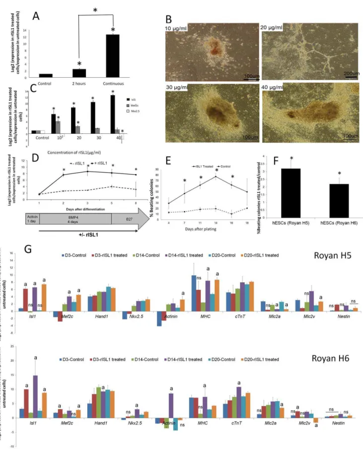
Figure 3. Optimization of the effect of rISL1 protein on hESCs.(A) To evaluate the effect of discontinuous (2 h/day) or continuous rISL1 protein addition on hESCs (Royan H5) differentiation, cells were treated continuously or discontinuously from days 1–8 post initiation of differentiation.Isl1qRT-PCR analysis of differentiated cells at day 8 showed higher significant endogenousIsl1expression in hESCs in the continuous protocol.Thus continuous treatment was applied in the next steps. )* : P,0.05( (B) To determine the best concentration of rISL1 protein for cardiac differentiation, cells were treated with four different concentrations of recombinant protein: 10, 20, 30, and 40mg/ml in continuous treatment of
hESCs during days 1–8 after initiation of differentiation. During differentiation, cells that were treated by 10 and 20mg/ml rISL1 protein were
suggested efficient penetration of rISL1protein into the cells (Fig. 2F). Temporal analysis showed that the rISL1 protein was detectable in medium up to 48 h in the presence or absence of cells; no significant decrease in the amount of protein was observed (Fig. 2E). rISL1 protein stability was further confirmed by monitoringIsl1gene expression at different time points after the addition of the protein. Quantitative RT-PCR results indicated that during 48 h after addition of the protein expression of theIsl1 gene was 16- to 32-fold higher than the control group (Fig. 2G). To examine the ability of rISL1 to regulate its own expression, we used an ISL1 reporter assay and added rISL1 protein on the ISL1-GFP reporter cell line. Our data showed that after rISL1 treatment, undifferentiated cells expressed 20.5963.67% GFP vs. 4.8261.25% GFP in the control group (Fig. 2H ). This result has suggested that rISL1 acts in a positive feedback loop, enhancing its gene expression.
Based on these results, we decided to add rISL1 protein into the differentiation media every other day during medium replace-ment. Cell penetration of the rISL1 protein was further confirmed by immunostaining analysis of adherent and aggregated cells using both anti-TAT and ISL1 antibodies. Two hours following transduction of hESCs, most cells were positive for the labeled TAT or ISL1 protein (Fig. 2I). rISL1 proteins were detected around the nucleus in adherent and aggregated cells, which suggested the ability of the recombinant protein to penetrate deep inside aggregated cells (Fig. 2I). This finding was consistent with the prevailing view that TAT can promote cellular uptake via endocytosis [30,31,32]
Defining rISL1 treatment conditions
Previous studies have demonstrated that discontinuous or continuous addition of recombinant protein into cell culture media is also an important factor that should be considered [23,33]. In order to optimize protein transduction, the cells were either discontinuously (2 h/day) or continuously (from days 1–8) treated by rISL1. qRT-PCR analysis of differentiated cells at day 8 showed higher endogenous Isl1 expression in hESCs in the continuous protocol (P,0.05, Fig. 3A). The following experiments were performed by continuous protein treatment.
To study the dose dependency of rISL1 transduction, hESCs were exposed to different concentrations of the purified protein (10, 20, 30, and 40mg/ml) in the continuous treatment of hESCs during days 1–8 after differentiation initiation. We observed that the concentration greater than 40mg/ml was lethal (data not shown). The differentiating cells at concentrations of 10 and 20mg/ml of the rISL1 protein were morphologically similar to hematopoietic and endothelial progenitors, while 30 and 40mg/ ml rISL1 protein showed cardiomyocytes and muscular appear-ances (Fig. 3B). According to qRT-PCR analysis, 40mg/ml rISL1
protein induced more endogenousIsl1and less expression ofMef2c and Nkx2.5 (Fig. 3C). These data were consistent with previous data which has shown that ISL1 marks a common population of progenitors in the heart that can differentiate into cardiomyocytes, smooth muscle, and endothelial cells [9,10]. It seems that different levels of ISL1 protein direct cells towards specific lineages. However, more experiments are needed to find the exact amount of ISL1 expression required for each lineage differentiation.
Increasing cardiac differentiation using rISL1 protein
Based on the above mentioned experiments, we continuously added 40mg/ml of rISL1 protein into differentiation medium from days 1–8 after differentiation initiation. The effect of rISL1 protein on the expression of endogenousIsl1was analyzed using qRT-PCR at 1, 2, 3, 5, and 8 days after differentiation initiation. Our results showed that treated cells expressed higher endogenous Isl1 than the untreated control (P,0.05, Fig. 3D). We further continued differentiation to obtain beating clusters. The beating areas appeared at day 5 post-plating of aggregates and the percent of beating areas were significantly higher in rISL1-treated cells compared to the control (Fig. 3E). The difference was more pronounced at 14 days after plating when the percent of beating areas reached 75610% in rISL1-treated cells compared to 2062.5% in the control when more than 1000 embryoid bodies were assessed in each group (Fig. 3E). Therefore, rISL1 treatment resulted in a 3.260.05 fold increase in the number of beating areas (Fig. 3F). In order to check reproducibility of this protocol the same experiments were performed using another hESC line, Royan H6. Our data indicated that rISL1 treatment could also cause a 2.260.4 fold increase in the number of beating areas in Royan H6. Temporal expression of cardiac genes showed the highest levels ofIsl1, Mef2c, Hand1, Nkx2.5, Actinin, MHC, cTnT, Mlc2aandMLC2vat day 14 in both hESC lines, Royan H5 and Royan H6 (Fig. 3G). Our data showed thatMLC2v expression increased in the rISL1-treated groups while MLC2a decreased. These results suggested that differentiated cells were directed toward ventricular cardiomyocytes. This observation was consis-tent with previous reports in which approximately 92% of cells within the right ventricle and about 20% of cells within the left ventricle of a normal heart were ISL1-positive [11].
Immunofluorescence staining of differentiated cells showed expressions of NKX2.5, GATA4, ISL1, ACTININ, DESMIN, and MHC (Fig. 4A). By flow cytometric analysis, higher expressions of cTnT, CONNEXIN 43, ACTININ, and GATA4 were detected (P,0.05, Fig. 4B).
Based on theIsl1gene expression profile, rISL1 was added to the cell culture media from days 1–8 of differentiation, when the expression level of endogenousIsl1was first detected (day 1) and reached its maximum level (day 8). The addition of rISL1 with
muscular appearances. It seems that 30 and 40mg/ml rISL1 protein are better concentrations for cardiac differentiation. )* : P,0.05( (C) qRT-PCR
analysis of differentiated cells at day 8 by different concentrations of rISL1 also showed that 40mg/ml of the rISL1 protein induced more endogenous
Isl1, but lessMef2candNkx2.5expressions. )* : P,0.05( (D) Schematic diagram of the differentiation protocol by the addition of rISL1 protein (40mg/
ml), which was added after induction with Activin A (days 1–8). qRT-PCR analysis of endogenousIsl1expression in hESCs demonstrated that treated cells expressed higher significant endogenousIsl1than the untreated control. )* : P,0.05( (E) The percentage of beating clusters in continuous treatment of hESCs by 40mg/ml rISL1 protein during days 1–8 after differentiation initiation in comparison with the control (vehicle-treated) group.
The percentage of beating clusters in the rISL1-treated group was significantly higher than the untreated group at day 14 after plating (75610% vs. 2062.5%). )* : P,0.05( (F) rISL1 treatment resulted in a 3.260.5 fold increase in the number of beating areas in comparison with untreated control group. rISL1 also caused a 2.260.4 fold increase in the other hESC line, Royan H6, which shows the reproducibility of this protocol for another hESC line. )* : P,0.05( (G) In order to assess the expression of cardiac-specific genes, we collected samples at 3 stages: day 3 after plating (the day of rISL1 removal); day 14 after plating (day of maximum beating); and day 20 after plating (day that beating decreased and cells were mature) by qRT-PCR in two hESC lines. Target genes were normalized by the reference geneGapdh. The relative expression was calculated by dividing the normalized target gene expression of treated hESCs with rISL1 protein and elution buffer (as control) with that of the undifferentiated state (day 0). All data are statistically significant in comparison with undifferentiated state (day 0) otherwise marked with ‘‘ns’’ (ns: P.0.05). a: P,0.05 in comparison with control group (elution buffer treated group). All data were represented as log2-linear plots.
Figure 4. Marker analysis of hESC-derived cardiomyocytes.Cardiomyocytes were dissociated at differentiation day 14 post plating, cultured for an additional 2 days, and assessed by (A) immunofluorescence staining and (B) flow cytometry for cardiac-specific markers. Flow cytometric analysis showed significant increases in the levels of cTnT, CONNEXIN43, ACTININ, and GATA4 protein expression (P,0.05). ). The experiment was performed for at least 3 independent biological replicates. *: P,0.05.
BMP4 to the cell culture media may also enhance its effect. It has been demonstrated that ISL1 promoted BMP expression (such as BMP4 and BMP7), and the expression level of BMP4 was reduced in ISL1 mutant cells [11]. It is likely that rISL1 increases the number of beating cells through enhancing the expression of the gap junction protein Connexin40 (a major protein in the conduction system) by BMP signaling. It has been shown that BMP signaling is necessary for the expression of T-box transcrip-tion factors [34]. Connexin40 is one of the direct downstream targets of the T-box transcription factors, which play an important role in the conduction system [35].
Taken together, these data indicate that direct delivery of the transcription factor ISL1 by protein transduction enhanced the cardiomyocyte differentiation in hESCs in vitro.
Conclusions
In this study we showed that under cell culture conditions purified rISL1 protein was stable for at least 48 h. When the protein was added to hESCs cultures, it efficiently penetrated into the cells and enhanced the differentiation of the two hESC lines into cardiac cells up to 3-fold.
This approach may pave the way for the scaled up expansion of hESCs as carrier-free suspension aggregates for an extended period of time. It provides a controlled environment for a homogeneous culture and simplifies the handling and controlling of the differentiation of hESCs, which are required for their applications in bioreactor culture systems and cell therapy.
Another advantage of this method is the lack of genetic manipulation of hESCs which may decrease the risk of their application in cell therapy. In conclusion, our data indicate that by addition of rISL1 protein into the differentiation medium we have successfully produced large numbers of functional cardiomyocytes that can be easily applied in drug discovery or cell therapy. However, further research is necessary to additionally increase the efficiency of differentiation using this method.
Supporting Information
Figure S1 Adherent differentiation and gene expression of hESCs (Royan H5) by the Laflamme et al. protocol [26]. (A) D0: Undifferentiated colony of hESCs. D1: One day after addition of Activin A. D5: Four days after treatment by BMP4. D15: Fifteen days after differentiation initiation. (B) RT-PCR data of daily expression of cardiac genes after differentiation induction.
(TIF)
Table S1 Primers and reaction conditions used in real time RT-PCR analysis.
(DOC)
Author Contributions
Conceived and designed the experiments: GHS NA HB. Performed the experiments: HF MY. Analyzed the data: GHS NA HB. Contributed reagents/materials/analysis tools: HR BAM FF ZG MA. Wrote the paper: HB GHS NA.
References
1. Zhu WZ, Hauch KD, Xu C, Laflamme MA (2009) Human embryonic stem cells and cardiac repair. Transplant Rev (Orlando) 23: 53–68.
2. Amit M, Laevsky I, Miropolsky Y, Shariki K, Peri M, et al. (2011) Dynamic suspension culture for scalable expansion of undifferentiated human pluripotent stem cells. Nature Protocols 6: 572–579.
3. Krawetz R, Taiani JT, Liu S, Meng G, Li X, et al. (2010) Large-scale expansion of pluripotent human embryonic stem cells in stirred-suspension bioreactors. Tissue Eng Part C Methods 16: 573–582.
4. Zweigerdt R, Olmer R, Singh H, Haverich A, Martin U (2011) Scalable expansion of human pluripotent stem cells in suspension culture. Nature Protocols 6: 689–700.
5. Serra M, Brito C, Leite SB, Gorjup E, von Briesen H, et al. (2009) Stirred bioreactors for the expansion of adult pancreatic stem cells. Ann Anat 191: 104– 115.
6. Rezaei Larijani M, Seifinejad A, Pournasr B, Hajihoseini V, Hasani N, et al. (2011) Long-term Maintenance of Undifferentiated Human Embryonic and Induced Pluripotent Stem Cells in Suspension. Stem Cells Dev: [Epub ahead of print].
7. Fijnvandraat AC, Lekanne Deprez RH, Christoffels VM, Ruijter JM, Moorman AF (2003) TBX5 overexpression stimulates differentiation of chamber myocardium in P19C16 embryonic carcinoma cells. J Muscle Res Cell Motil 24: 211–218.
8. Hiroi Y, Kudoh S, Monzen K, Ikeda Y, Yazaki Y, et al. (2001) Tbx5 associates with Nkx2-5 and synergistically promotes cardiomyocyte differentiation. Nature genetics 28: 276–280.
9. Kattman SJ, Huber TL, Keller GM (2006) Multipotent flk-1+cardiovascular progenitor cells give rise to the cardiomyocyte, endothelial, and vascular smooth muscle lineages. Developmental cell 11: 723–732.
10. Moretti A, Caron L, Nakano A, Lam JT, Bernshausen A, et al. (2006) Multipotent embryonic isl1+progenitor cells lead to cardiac, smooth muscle, and endothelial cell diversification. Cell 127: 1151–1165.
11. Cai CL, Liang X, Shi Y, Chu PH, Pfaff SL, et al. (2003) Isl1 identifies a cardiac progenitor population that proliferates prior to differentiation and contributes a majority of cells to the heart. Developmental cell 5: 877–889.
12. Laugwitz KL, Moretti A, Caron L, Nakano A, Chien KR (2008) Islet1 cardiovascular progenitors: a single source for heart lineages? Development 135: 193–205.
13. Kwon C, Qian L, Cheng P, Nigam V, Arnold J, et al. (2009) A regulatory pathway involving Notch1/b-catenin/Isl1 determines cardiac progenitor cell fate. Nature cell biology 11: 951–957.
14. Bu L, Jiang X, Martin-Puig S, Caron L, Zhu S, et al. (2009) Human ISL1 heart progenitors generate diverse multipotent cardiovascular cell lineages. Nature 460: 113–117.
15. Moore JC, Van Laake LW, Braam SR, Xue T, Tsang SY, et al. (2005) Human embryonic stem cells: genetic manipulation on the way to cardiac cell therapies. Reproductive toxicology 20: 377–391.
16. Noguchi H, Matsushita M, Okitsu T, Moriwaki A, Tomizawa K, et al. (2004) A new cell-permeable peptide allows successful allogeneic islet transplantation in mice. Nature medicine 10: 305–309.
17. Derossi D, Joliot AH, Chassaing G, Prochiantz A (1994) The third helix of the Antennapedia homeodomain translocates through biological membranes. Journal of Biological Chemistry 269: 10444.
18. Matsushita M, Tomizawa K, Moriwaki A, Li ST, Terada H, et al. (2001) A high-efficiency protein transduction system demonstrating the role of PKA in long-lasting long-term potentiation. The Journal of Neuroscience 21: 6000– 6007.
19. Wadia JS, Stan RV, Dowdy SF (2004) Transducible TAT-HA fusogenic peptide enhances escape of TAT-fusion proteins after lipid raft macropinocytosis. Nature medicine 10: 310–315.
20. El-Sayed A, Futaki S, Harashima H (2009) Delivery of macromolecules using arginine-rich cell-penetrating peptides: ways to overcome endosomal entrap-ment. The AAPS journal 11: 13–22.
21. Ziegler A, Nervi P, Du¨rrenberger M, Seelig J (2005) The cationic cell-penetrating peptide CPPTAT derived from the HIV-1 protein TAT is rapidly transported into living fibroblasts: optical, biophysical, and metabolic evidence. Biochemistry 44: 138–148.
22. Do Kwon Y, Oh SK, Kim HS, Ku SY, Kim SH, et al. (2005) Cellular manipulation of human embryonic stem cells by TAT-PDX1 protein transduction. Molecular Therapy 12: 28–32.
23. Stock K, Nolden L, Edenhofer F, Quandel T, Bru¨stle O (2010) Transcription factor-based modulation of neural stem cell differentiation using direct protein transduction. Cellular and Molecular Life Sciences 67: 2439–2449.
24. Baharvand H, Ashtiani SK, Taee A, Massumi M, Valojerdi MR, et al. (2006) Generation of new human embryonic stem cell lines with diploid and triploid karyotypes. Dev Growth Differ 48: 117–128.
25. Baharvand H, Matthaei KI (2004) Culture condition difference for establish-ment of new embryonic stem cell lines from the C57BL/6 and BALB/c mouse strains. In Vitro Cell Dev Biol Anim 40: 76–81.
26. Laflamme MA, Chen KY, Naumova AV, Muskheli V, Fugate JA, et al. (2007) Cardiomyocytes derived from human embryonic stem cells in pro-survival factors enhance function of infarcted rat hearts. Nat Biotechnol 25: 1015–1024. 27. Livak KJ, Schmittgen TD (2001) Analysis of relative gene expression data using real-time quantitative PCR and the 2(-Delta Delta C(T)) Method. Methods 25: 402–408.
29. Nagahara H, Vocero-Akbani AM, Snyder EL, Ho A, Latham DG, et al. (1998) Transduction of full-length TAT fusion proteins into mammalian cells: TAT-p27ˆ Kˆ iˆ pˆ 1 induces cell migration. Nature medicine 4: 1998–1912. 30. Fittipaldi A, Ferrari A, Zoppe´ M, Arcangeli C, Pellegrini V, et al. (2003) Cell
membrane lipid rafts mediate caveolar endocytosis of HIV-1 Tat fusion proteins. Journal of Biological Chemistry 278: 34141.
31. Console S, Marty C, Garcı´a-Echeverrı´a C, Schwendener R, Ballmer-Hofer K (2003) Antennapedia and HIV transactivator of transcription (TAT)‘‘protein transduction domains’’ promote endocytosis of high molecular weight cargo upon binding to cell surface glycosaminoglycans. Journal of Biological Chemistry 278: 35109.
32. Sandgren S, Cheng F, Belting M (2002) Nuclear targeting of macromolecular polyanions by an HIV-Tat derived peptide. Journal of Biological Chemistry 277: 38877–38883.
33. Kim D, Kim CH, Moon JI, Chung YG, Chang MY, et al. (2009) Generation of human induced pluripotent stem cells by direct delivery of reprogramming proteins. Cell Stem Cell 4: 472–476.
34. Yang L, Cai CL, Lin L, Qyang Y, Chung C, et al. (2006) Isl1Cre reveals a common Bmp pathway in heart and limb development. Development 133: 1575–1585.