at the bedside.(1-5) During prolonged transport, the patient does not receive the same level of intensive care, and this can result in complica-tions.(1,3-18)
When deciding to transport a critically ill patient, the potential benefits should be
Introduction
Technological advances have led to conside-rable improvement in intensive care medicine, in terms of treatment aspects as well as diagnostic techniques. Despite the current sophistication of intensive care units (ICUs), neither all necessary care nor all appropriate exams can be offered
Intrahospital transport of patients on invasive ventilation:
cardiorespiratory repercussions and adverse events*
Transporte intra-hospitalar de pacientes sob ventilação invasiva: repercussões cardiorrespiratórias e eventos adversos
Lea Tami Suzuki Zuchelo, Paulo Antônio Chiavone
Abstract
Objective: To determine the occurrence of cardiorespiratory alterations and to identify adverse events during the intrahospital transport of patients on invasive ventilation. Methods: A prospective observational non-randomized study was conducted at two tertiary hospitals between April of 2005 and December of 2006. We included patients on invasive ventilation who required intrahospital transport during the study period. Exclusion criteria were as follows: being under suspicion of brain death; being submitted to alternate periods of mechanical ventilation/nebulization via a T-piece; and being transported to the operating room. Prior to and after transport, we evaluated blood gas analysis results, vital signs, use of medications by means of a continuous infusion pump, parameters regarding the mechanical ventilator, duration of transport, transport distance and number of professionals involved. Results: We included 48 patients in a total of 58 intrahospital transports. Relevant cardiorespiratory alterations were identified in 39 transports, totaling 86 episodes, as well as 16 adverse events related to equipment or personnel failure, such as problems related to batteries and to miscommunication. Conclusions: During the intrahospital transport of patients on invasive ventilation, cardiorespiratory alterations were common (67.2%), and adverse events occurred in 75.7% of the transports.
Keywords: Patient transfer; Intensive care; Respiration, artificial; Ventilators, mechanical.
Resumo
Objetivo: Verificar a ocorrência de alterações cardiorrespiratórias e identificar eventos adversos durante o transporte intra-hospitalar de pacientes sob ventilação invasiva. Métodos: Estudo observacional prospectivo não-randomi-zado, conduzido em dois hospitais terciários, entre abril de 2005 e dezembro de 2006. Foram incluídos pacientes sob ventilação invasiva que necessitaram de transporte intra-hospitalar durante o período do estudo. Os critérios de exclusão foram: estar sob suspeita de morte encefálica; ter sido submetido a períodos de ventilação mecânica e de nebulização em tubo T; e ter sido transportado para o centro cirúrgico. Antes e após o transporte, os seguintes parâmetros foram avaliados: gasometria arterial, sinais vitais, uso de medicamentos através de uma bomba de infusão contínua, parâmetros do ventilador mecânico, duração do transporte, distância percorrida e número de profissionais envolvidos. Resultados: Foram incluídos 48 pacientes, num total de 58 transportes. Observou-se alte-ração cardiorrespiratória importante em 39 transportes, totalizando 86 episódios, assim como 16 eventos adversos relacionados à falha de equipamento e falha da equipe, dentre eles problemas com baterias e falhas de comuni-cação. Conclusões: Durante o transporte intra-hospitalar de pacientes submetidos à ventilação invasiva, alterações cardiorrespiratórias foram frequentes (67,2%), e eventos adversos ocorreram em 75,7% dos transportes realizados
Descritores: Transferência de pacientes; Cuidados intensivos; Respiração artificial; Respiradores mecânicos.
* Study carried out at the Santa Casa School of Medical Sciences in São Paulo, São Paulo, Brazil.
Correspondence to: Léa Tami Suzuki Zuchelo. Rua Coronel Joaquim Ferreira de Souza, 212, Alto do Mandaqui, CEP 02419-070, São Paulo, SP, Brasil.
Tel 55 11 2236-4811. E-mail: [email protected] Financial support: None.
submitted to alternating periods of mechan-ical ventilation/nebulization via a T-piece; and having been transported to the operating room due to access-related difficulties in collecting the necessary data within the ICU.
After the medical team had made the deci-sion to transport a patient, the following data were collected: identification; diagnosis; Glasgow coma scale or Ramsay sedation scale score; Acute Physiologic and Chronic Health Evaluation (APACHE) II score; lung injury score; duration of intubation or tracheotomy; medica-tion delivered via a continuous infusion pump (CIP); and transport destination.
The pre-transport period was defined as the moment before initiating the preparation of the patient for transport (prior to the disconnection of the mechanical ventilator and the discontinu-ation of medicdiscontinu-ation delivered via the CIP).
The post-transport period was defined as the moment after the return of the patient to the ICU and admission by the nursing team (resumption of monitoring, medication delivery and mechanical ventilation).
In the pre-transport and the post-transport periods, the maximum tolerance period for data collection was 15 min.
For patients transported in order to undergo tests, post-transport data were evaluated upon their return to the original units. In the case of one-way transports (transfers between sectors), the post-transport evaluation was conducted upon the arrival of the patient at the destination.
In the pre-transport and post-transport periods, the following parameters were evalu-ated: blood gas; heart rate (HR); respiratory rate (RR); systolic blood pressure (SBP); diastolic blood pressure (DBP); mean arterial pressure; SpO2; weighed against the risks to which the patient
will be exposed.(5,19-21) Due to these factors, appropriate planning, a well-trained team and the use of reliable equipment are indispensible during the intrahospital transport of critically ill patients, since this is a population at high risk for complications and instability inherent to the underlying disease.
The objective of the present study was to identify cardiorespiratory alterations occurring in patients transported to diagnostic units or during transfers between sectors, as well as to identify the adverse events that occur during intrahospital transport, with the aim of assisting the patient during this phase necessary to the treatment.
Methods
This was an operational study, with a prospective non-randomized design, carried out in the ICUs and in the Semi-intensive Care Units (SICUs) of the Santa Casa Central Hospital in São Paulo, in the period between July and December of 2006, as well as in the ICU and in the SICUs (general and emergency) of the Ipiranga Hospital between April and September of 2005 and between September and December of 2006. Both hospitals are located in the city of São Paulo.
Written informed consent was obtained from the patients, their guardians or family members. The research project and the informed consent form were both approved by the research ethics committees of the institutions involved.
We included all patients on invasive ventila-tion who required intrahospital transport during the study period.
The exclusion criteria were as follows: being under investigation of brain death; being
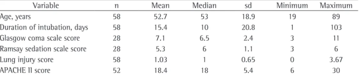
Table 1 - Descriptive analysis of the variables age, duration of intubation, Glasgow coma scale score, Ramsay sedation scale score, lung injury score and Acute Physiology and Chronic Health Evaluation II score for the patients on mechanical ventilation submitted to intrahospital transport (Santa Casa Central Hospital in São Paulo and Ipiranga Hospital, between April of 2005 and December of 2006).
Variable n Mean Median sd Minimum Maximum
Age, years 58 52.7 53 18.9 19 89
Duration of intubation, days 58 15.4 10 20.8 1 103
Glasgow coma scale score 28 7.1 6.5 2.4 3 11
Ramsay sedation scale score 28 5.3 6 1.1 3 6
Lung injury score 58 1.03 1 0.65 0 3.67
APACHE II score 52 18.4 18 5.4 6 30
medications delivered via the CIP; mechanical ventilator parameters; the PaO2/FiO2 ratio; trans-port duration; distance covered; professionals involved (intensivists, residents, physical thera-pists, nurses and nursing assistants/ technicians); and complications.
All units involved in this study presented a similar pattern of transport, and the patients were submitted to intrahospital transport according to the routine of the sector. The type of ventilation used was decided by the team itself, according to the clinical status of the patient and the availability of the equipment in the sector.
Adverse events were defined as any event, expected or not, that affected patient stability. The following criteria were used in order to define cardiorespiratory alterations: ≥ 20 bpm change in HR; ≥ 10 breaths/min change in RR; ≥ 20 mmHg change in SBP; ≥ 20 mmHg change in DBP; ≥ 5% decrease in SpO2; ≥ 0.07 change in pH; ≥ 10 mmHg change in PaCO2; ≥ 10 mmHg decrease in PaO2; ≥ 5% decrease in SaO2; and ≥ 20% decrease in PaO2/FiO2 ratio.
The statistical analysis was conducted using the Mann-Whitney and Kruskal-Wallis tests for comparisons among two and three groups, respectively.
In all tests, the level of significance was set at 5% (p < 0.05).
Results
We evaluated 48 patients, in a total of 58 transports, of which 30 occurred at the Santa Casa Central Hospital in São Paulo and 28 occurred at the Ipiranga Hospital. However, not all variables were considered in the 58 transports. In some cases, these data were not included on the data collection form. In other cases, the data had to be excluded from the analysis. For example, since venous blood gas analysis findings were considered inappro-priate and only arterial blood gas analysis results were included, these data were evaluated in only 46 cases.
The descriptive analyses of the demographic and clinical variables are shown in Table 1.
The APACHE II score was used for patient stratification in the characterization of the sample regarding the severity of the patients, without the objective of evaluating pre- and post-transport alterations.(22) Patient scores
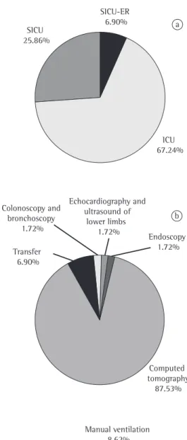
SICU-ER 6.90% SICU
25.86%
ICU 67.24%
a
c Manual ventilation
8.62%
Mechanical ventilation 39.66% Transport
ventilation 51.72%
b
Transfer 6.90% Colonoscopy and
bronchoscopy 1.72%
Echocardiography and ultrasound of
lower limbs 1.72%
Endoscopy 1.72%
Computed tomography
87.53%
The patients were also divided in subgroups and compared regarding the following cardi-orespiratory variables: HR, RR, SBP, DBP, SpO2, pH, PaCO2, PaO2, SaO2 and PaO2/FiO2. The subgroups analyzed and the differences found are described in Table 3.
All of the transport teams included at least one physician, and there were two physicians present in one of the transports. In addition, there were nursing assistants/technicians present in all of the transports. In 11 cases and in 1 case, the patients were transported by two assistants/ technicians and three assistants/technicians, respectively. A physiotherapist was present in 22 transports, and a nurse was present in 8.
The mean transport duration was 52 min, and the mean distance covered was 325 m.
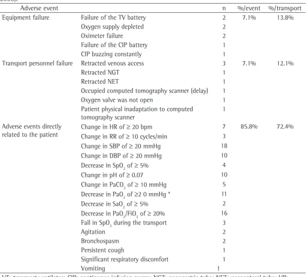
In 44 of the transports, adverse events (n = 112) were reported. The divisions regarding the nature of the complications were classified as follows: equipment failure; human error; and adverse effects directly related to the patient status (Table 4).
ranged from 6 to 30, with a mean value of 18.4, which characterizes a varied sample in terms of patient severity.
Neurology patients accounted for 38 (65.5%) of the 58 transports, followed by pulmonology patients (n = 6, 10.3%), gastroenterology patients (n = 4, 6.9%), vascular patients (n = 3, 5.2%) and other patients (n = 7, 12.1%).
Figure 1 shows the origin and destination sectors, as well as the type of ventilation used during transport.
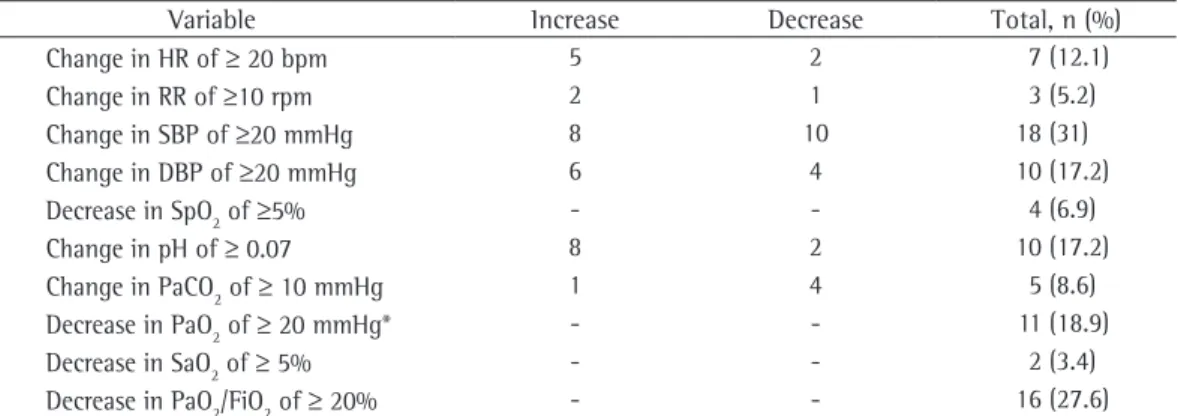
Among the 58 transports, there were 39 cases (67.2%) in which the patient presented at least one episode of relevant cardiorespiratory altera-tion. A detailed description is shown in Table 2. Among the 58 transports, there were 13 cases (22.4%) in which the patient was using vasoactive drugs (noradrenaline, dobutamine, dopamine or sodium nitroprusside) delivered via the CIP, which was turned off in only 1 case. In that case, the patient presented a decrease in HR of 25 bpm, an increase in SBP of 80 mmHg and an increase in DBP of 30 mmHg.
Table 2 - Number of patients submitted to intrahospital transport and presenting major cardiorespiratory alterations (Santa Casa Central Hospital in São Paulo and Ipiranga Hospital, between April of 2005 and December of 2006).
Variable Increase Decrease Total, n (%)
Change in HR of ≥ 20 bpm 5 2 7 (12.1)
Change in RR of ≥10 rpm 2 1 3 (5.2)
Change in SBP of ≥20 mmHg 8 10 18 (31)
Change in DBP of ≥20 mmHg 6 4 10 (17.2)
Decrease in SpO2 of ≥5% - - 4 (6.9)
Change in pH of ≥ 0.07 8 2 10 (17.2)
Change in PaCO2 of ≥ 10 mmHg 1 4 5 (8.6) Decrease in PaO2 of ≥ 20 mmHg* - - 11 (18.9)
Decrease in SaO2 of ≥ 5% - - 2 (3.4)
Decrease in PaO2/FiO2 of ≥ 20% - - 16 (27.6)
HR: heart rate; RR: respiratory rate; SBP: systolic blood pressure; and DBP: diastolic blood pressure; *The patients in which FiO2 was decreased were excluded.
Table 3 - Comparison among subgroups of patients submitted to intrahospital transport in relation to cardiorespiratory variations and differences found (Santa Casa Central Hospital in São Paulo and Ipiranga Hospital, between April of 2005 and December of 2006).
Groups compared Differences found p
Under sedation vs. not under sedation In the group under sedation, both RR and HR tended to increase during transport, whereas in the group
with no sedation, the opposite occurred.
RR: p < 0.01* HR: p = 0.04*
Under sedation vs. sedation discontinued - p > 0.05*
With VADs vs. without VADs - p > 0.05*
Manual ventilation vs. MV vs. TV - p > 0.05**
presented—but also due to the poor monitoring of the patients. During most of the transports, we used a pulse oximeter to monitor the patient. However, the oximeter often became impre-cise due to interference, temperature drops or decreased perfusion, making its reading impos-sible or unreliable. In such cases, there was a gap in the monitoring, since there was no other device available to register SpO2.
Continuous and systematized monitoring throughout the transport should be mandatory, so that the magnitude and duration of these alterations, as well as the occurrence of arrhyth-mias and other electrocardiographic alterations, can be registered with greater precision. Thus, the appropriate measures could be taken as soon as possible, as they are in the ICUs.
Discussion
The intrahospital transport of critically ill patients for complimentary tests is currently indispensible, and this process must be well planned and executed, in order to minimize the risks to which the patients are exposed. However, in the present study, we found that the appro-priate precautions were not always taken.
One of the most alarming results found in the present study was the high incidence of cardiorespiratory alterations—relevant alterations occurred in more than 67% of the transports (total, 86 episodes). This is a worrisome result, not only due to the fact per se—since the popu-lation was composed of critically ill patients with instability inherent to the pathology
Table 4 - Adverse events observed during intrahospital transport of patients on invasive mechanical ventilation (Santa Casa Central Hospital in São Paulo and Ipiranga Hospital, between April of 2005 and December of 2006).
Adverse event n %/event %/transport
Equipment failure Failure of the TV battery 2 7.1% 13.8% Oxygen supply depleted 2
Oximeter failure 2
Failure of the CIP battery 1 CIP buzzing constantly 1
Transport personnel failure Retracted venous access 3 7.1% 12.1%
Retracted NGT 1
Retracted NET 1
Occupied computed tomography scanner (delay) 1 Oxygen valve was not open 1 Patient physical inadaptation to computed
tomography scanner
1
Adverse events directly related to the patient
Change in HR of ≥ 20 bpm 7 85.8% 72.4% Change in RR of ≥ 10 cycles/min 3
Change in SBP of ≥ 20 mmHg 18 Change in DBP of ≥ 20 mmHg 10 Decrease in SpO2 of ≥ 5% 4 Change in pH of ≥ 0.07 10 Change in PaCO2 of ≥ 10 mmHg 5 Decrease in PaO2 of ≥2 0 mmHg * 11 Decrease in SaO2 of ≥ 5% 2 Decrease in PaO2/FiO2 of ≥ 20% 16 Fall in SpO2 during the transport 3
Agitation 2
Bronchospasm 2
Persistent cough 1
Significant respiratory discomfort 1
Vomiting 1
ical ventilator itself was employed. Therefore, there were few transports involving manual ventilation, which was primarily used only during transport within the unit.
One group of authors observed a high inci-dence of complications during patient transports to the tomography sector, which, according to the authors, might be attributed to the physical isolation of the patient during the procedure.(11)
The type of ventilation used seems to affect patient stability during transport.(10,23-26) However, in the present study, no statistically significant differences were found among the types of venti-lation used, according to the variables studied. However, the number of transports involving manual ventilation was very small (n = 5).
We found no differences between the use of the transport ventilator and that of the mechan-ical ventilator itself. It is of note that, in our study, most of the patients presented neuro-logical rather than respiratory complications. The latter group of patients would probably benefit from transport with the mechanical ventilator itself, which would favor the pulmo-nary mechanics, since it would not be necessary to disconnect/reconnect the patient from the mechanical ventilator/to the transport ventilator, and depressurization of the respiratory system would therefore be avoided.
In one case, the sodium nitroprusside CIP was shut off, due to the technical unfeasi-bility of transporting the patient together with the CIP. This patient presented major altera-tions in HR, SBP and DBP. The SBP increased by 80 mmHg, from 140 mmHg to 220 mmHg. This shows that, when technical conditions or the equipment used do not offer total safety to the patients, it is probably better to wait for a more appropriate time to transport the patient. It is important to give the safety and stability of the patient priority over hospital routines and bureaucratic procedures. The patients whose CIPs remained activated did not present relevant variations and, when compared with a group of patients who made no use of vasoactive drugs, no statistical differences were found regarding blood gas or hemodynamic alterations, showing that the patients transported while receiving vasoactive drugs presented the same alterations as did those not receiving such drugs. Therefore, the use of vasoactive drugs, if maintained, is not a contraindication to transport.
In a study evaluating a total of 103 transports,(9) 113 episodes of significant intratransport alterations (requiring intervention) were reported, which underscores the importance of maintaining the level of care and monitoring offered in the ICUs.
In most studies evaluating blood gas altera-tions, a change in pH, tending toward alkalosis, and a decrease in PaCO2
(10,23,24) have been reported. This can be explained by the anxiety or pain of the patient, which increases sponta-neous ventilation, or even due to the inability of some transport ventilators to maintain the established tidal volume.
In our study, there were 10 transports (17.2%) in which the patients presented variations in pH of ≥ 0.07, presenting a tendency toward alka-losis in 8 of those 10 cases, and 5 transports (8.6%) in which the patients presented varia-tions in PaCO2 of ≥ 10 mmHg, also presenting hypocapnia in 4 of those 5 cases.
Since, on most occasions, blood gases were collected by the medical team and the nurse, delays in their collection might have occurred in the post-transport period. Such delays could have influenced the post-transport PaCO2, giving the impression that it had been stable.
Some authors reported a tendency toward a decrease in oxygenation during transport.(3,4,13) However, we observed a tendency on the part of the transport team to increase the FiO2 prior to transport, as was done in 20 of the transports. This could explain why we found no statistically relevant alterations in PaO2 or SaO2.
Despite the fact that FiO2 was increased in more than 34% of the transports, we observed a decrease in PaO2 in 56.5% of the patients. Even if we consider that blood gases were not collected immediately after transport, the aim of the team as a whole should be to optimize ventilation and, especially, oxygenation. Therefore, this value might have been even higher than 56.5%.
Nearly half of the patients (44.8%) presented a decrease in the PaO2/FiO2 ratio; in 27.6% of the cases, this decrease represented a change of > 20% in relation to the baseline value.
mechan-advanced life support and capable of promptly establishing an artificial airway,(22,23,28-30) which was not always the case in the transports we evaluated. In addition, although all of the trans-ports were also conducted in the presence of a resident, this was often a first-year resident without sufficient experience to deal with criti-cally ill patients.
We conclude that, during intrahospital transport of patients on invasive ventilation, cardiorespiratory alterations often occur. Adverse events occurred in 75.7% of the transports evaluated.
Ideally, all transports would be carried out by trained professionals, principally physicians specialized in intensive care, and the appropriate monitoring of patient vital signs, through the use of an electrocardiograph, pressure monitor, pulse oximeter, etc., would be uninterrupted. In addition, equipment to deal with complications, such as a defibrillator, should routinely be trans-ported together with the patient.
We recommend that additional studies be conducted, and that such studies involve continuous intratransport monitoring of patient vital signs, as well as evaluation and quantifica-tion of alteraquantifica-tions occurring during transport, so that a protocol for intrahospital transport can be developed.
References
1. Weg JG, Haas CF. Safe intrahospital transport of critically ill ventilator-dependent patients. Chest. 1989;96(3):631-5.
2. Kalisch BJ, Kalisch PA, Burns SM, Kocan MJ, Prendergast V. Intrahospital transport of neuro ICU patients. J Neurosci Nurs. 1995;27(2):69-77.
3. Brokalaki HJ, Brokalakis JD, Digenis GE, Baltopoulos G, Anthopoulos L, Karvountzis G. Intrahospital transportation: monitoring and risks. Intensive Crit Care Nurs. 1996;12(3):183-6.
4. Waydhas C. Intrahospital transport of critically ill patients. Crit Care. 1999;3(5):R83-9.
5. Chang DW; American Association for Respiratory Care (AARC). AARC Clinical Practice Guideline: in-hospital transport of the mechanically ventilated patient--2002 revision & update. Respir Care. 2002;47(6):721-3. 6. Taylor JO, Chulay, Landers CF, Hood W Jr,
Abelman WH. Monitoring high-risk cardiac patients during transportation in hospital. Lancet. 1970;2(7685):1205-8.
7. Hanning CD, Gilmour DG, Hothersal AP, Aitkenhead AR, Venner RM, Ledingham IM. Movement of the critically ill within hospital. Intensive Care Med. 1978;4(3):137-43. 8. Insel J, Weissman C, Kemper M, Askanazi J, Hyman
AI. Cardiovascular changes during transport of Another interesting fact is that, although the
CIPs of some patients under sedation had to be switched off during the transport, there was no statistically significant difference, in terms of blood gas and hemodynamic alterations, between those in whom the sedation was main-tained and those in whom it was not. This shows that CIP-delivered sedation can safely be discon-tinued during transport, thereby decreasing the amount of equipment and facilitating the management of the patient.
In the present study, we identified 112 adverse events, of which 16 were related to equipment failure or human error (problems with batteries or communication). These episodes could have been avoided by better planning, especially regarding the batteries for the equipment used during the transport, as well as by better guide-lines regarding communication among the teams involved.
One group of authors found that complica-tions were reported in 176 transport reports; there were 191 episodes, 61% of which were attributed to human error; the authors suggested better training of the team.(21)
In another study,(18) intrahospital transport was described as a high-risk procedure associ-ated with possible complications, since, in 33% of the 64 transports evaluated, the patients presented major alterations, evolving to cardi-orespiratory arrest in 2 cases. The authors of that study discussed the need for standardizing the transport and the precautions taken during this procedure.
Another group of authors stated that patient transport is safe, and that, in fact, the patients who require transport are those who are more critically ill and therefore evolve to death more rapidly, regardless of whether they undergo intrahospital transport.(27)
In our study, it would have been of interest to include a control group composed of non-transported patients with APACHE II scores equivalent to those of the transported patients, so that we could have determined whether the alterations found in the transported patients were related to patient severity or to the trans-port itself.
Nurses Transfer Guidelines Task Force. Crit Care Med. 1993;21(6):931-7.
20. Ferdinande P. Recommendations for intra-hospital transport of the severely head injured patient. Working Group on Neurosurgical Intensive Care of the European Society of Intensive Care Medicine. Intensive Care Med. 1999;25(12):1441-3.
21. Beckmann U, Gillies DM, Berenholtz SM, Wu AW, Pronovost P. Incidents relating to the intra-hospital transfer of critically ill patients. An analysis of the reports submitted to the Australian Incident Monitoring Study in Intensive Care. Intensive Care Med. 2004;30(8):1579-85.
22. Chiavone PA, Sens YA. Evaluation of APACHE II system among intensive care patients at a teaching hospital. Sao Paulo Med J. 2003;121(2):53-7.
23. Gervais HW, Eberle B, Konietzke D, Hennes HJ, Dick W. Comparison of blood gases of ventilated patients during transport. Crit Care Med. 1987;15(8):761-3.
24. Tobias JD, Lynch A, Garrett J. Alterations of end-tidal carbon dioxide during the intrahospital transport of children. Pediatr Emerg Care. 1996;12(4):249-51. 25. Dockery WK, Futterman C, Keller SR, Sheridan MJ,
Akl BF. A comparison of manual and mechanical ventilation during pediatric transport. Crit Care Med. 1999;27(4):802-6.
26. Nakamura T, Fujino Y, Uchiyama A, Mashimo T, Nishimura M. Intrahospital transport of critically ill patients using ventilator with patient-triggering function. Chest. 2003;123(1):159-64.
27. Szem JW, Hydo LJ, Fischer E, Kapur S, Klemperer J, Barie PS. High-risk intrahospital transport of critically ill patients: safety and outcome of the necessary “road trip”. Crit Care Med. 1995;23(10):1660-6.
28. Manji M, Bion JF. Transporting critically ill patients. Intensive Care Med. 1995;21(10):781-3.
29. Warren J, Fromm RE Jr, Orr RA, Rotello LC, Horst HM; American College of Critical Care Medicine. Guidelines for the inter- and intrahospital transport of critically ill patients. Crit Care Med. 2004;32(1):256-62.
30. Lamblet LC, Teixeira AP, Corrêa AG. Transporte intra-hospitalar de pacientes graves. In: Knobel E, Laselva CR, Moura Jr DF, editors. Terapia intensiva: enfermagem. São Paulo: Atheneu; 2006. p. 85-92.
critically ill and postoperative patients. Crit Care Med. 1986;14(6):539-42.
9. Indeck M, Peterson S, Smith J, Brotman S. Risk, cost, and benefit of transporting ICU patients for special studies. J Trauma. 1988;28(7):1020-5.
10. Hurst JM, Davis K Jr, Branson RD, Johannigman JA. Comparison of blood gases during transport using two methods of ventilatory support. J Trauma. 1989;29(12):1637-40.
11. Smith I, Fleming S, Cernaianu A. Mishaps during transport from the intensive care unit. Crit Care Med. 1990;18(3):278-81.
12. Pereira Jr GA, Nunes TL, Basile-Filho A. Transporte do paciente crítico. Medicina (Ribeirão Preto). 2001;34(2):143-53.
13. Waydhas C, Schneck G, Duswald KH. Deterioration of respiratory function after intra-hospital transport of critically ill surgical patients. Intensive Care Med. 1995;21(10):784-9.
14. Romano M, Raabe OG, Walby W, Albertson TE. The stability of arterial blood gases during transportation of patients using the RespirTech PRO. Am J Emerg Med. 2000;18(3):273-7.
15. Shirley PJ, Stott SA. Clinical and organizational problems in patients transferred from the intensive care unit to other areas within the hospital for diagnostic procedure [abstract]. Proceedings of the Intensive Care Society and Riverside Group ‘State of Art’ Meeting; 2000 Dec 7-8; London, UK. Br J Anaesth. 2001;87(2):346-7.
16. Zanetta G, Robert D, Guérin C. Evaluation of ventilators used during transport of ICU patients -- a bench study. Intensive Care Med. 2002;28(4):443-51.
17. Shirley PJ, Bion JF. Intra-hospital transport of critically ill patients: minimising risk. Intensive Care Med. 2004;30(8):1508-10.
18. Damm C, Vandelet P, Petit J, Richard JC, Veber B, Bonmarchand G, et al. Complications during the intrahospital transport in critically ill patients [Article in French]. Ann Fr Anesth Reanim. 2005;24(1):24-30. 19. Guidelines for the transfer of critically ill patients.
Guidelines Committee of the American College of Critical Care Medicine; Society of Critical Care Medicine and American Association of Critical-Care
About the authors
Lea Tami Suzuki Zuchelo
Professor. Centro Universitário da Capital – UNICAPITAL, Capital University Center – São Paulo, Brazil.
Paulo Antônio Chiavone