PICTURES IN DIGESTIVE PATHOLOGY
1130-0108/2015/107/12/759-760 REVISTA ESPAÑOLA DE ENFERMEDADES DIGESTIVAS COPYRIGHT © 2015 ARÁN EDICIONES, S. L.
REV ESP ENFERM DIG (Madrid) Vol. 107, N.º 12, pp. 759-760, 2015
Duodenal gastrointestinal stromal tumor and endoscopic ultrasound
Fernando M. Castro-Poças1,2, Tarcísio P. Araújo3, Jorge D. Silva2,4, Carlos A. Lopes2,5 and Miguel M. Saraiva1,2
1Department of Gastroenterology. Institute of CUF-ManoPH. Porto, Portugal. 2Institute of Biomedical Sciences Abel Salazar. University of Porto. Portugal. 3Department of Gastroenterology. Santo António Hospital. Porto Hospital Center. Porto, Portugal. 4Department of Surgery. Santa Maria Hospital. Porto,
Portugal. 5Department of Pathology. Institute of CUF-ManoPH. Porto, Portugal
CASE REPORTS
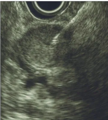
Case report 1. 73-year-old patient performed an upper endoscopy (UE) (Fig. 1), revealing a subepithelial lesion (20 mm) in the second part of the duodenum. Endoscopic ultrasound (EUS) showed a homogeneous lesion, 25x13.3 mm, hypoechoic, regular margins, originated in muscularis propria, two centimeters distal to papilla of Vater (PV) (Fig. 2). We suspected of a gastrointestinal stromal tumor (GIST). Surgery was advised. Laparoscopic excision con-firmed the diagnosis.
Case report 2. 51-year-old patient underwent UE, revealing a bulge in the second part of the duodenum. EUS showed a heterogeneous lesion, 27.5x18.5 mm, hypoechoic, irregular margins, originated in muscularis propria, three centimeters distal to PV (Fig. 3). Diagnostic hypothesis was GIST, surgery being advised. The patient demanded clinical test with higher diagnostic accuracy. EUS guided fine-needle-aspiration (FNA) was performed (Fig. 4). Cytology (Fig. 5) showed spindle cells aggregates; malignant signs were not seen. This pattern suggested a mesenchymal tumor without malignancy. Low risk GIST
was probable, although leiomyoma could be considered. For final diagnosis, an immunocytochemical study for CD117 and CD34 was done in a cell-block specimen that showed positivity in some cells for both. The final diagno-sis was low risk GIST. Surgery was performed. Histology confirmed the diagnosis.
DISCUSSION
GISTs in the duodenum are very rare (1). The treatment of choice is surgical resection of the tumor (2,3). Optimal surgical procedure has not yet been established (1,3,4). Sur-gery implies morbidity, so a definitive pre-operative diag-nosis, when there is doubt in resection, is essential (4,5).
Fig. 1. Subepithelial lesion in the second part of the duodenum.
760 F. M. CASTRO-POÇAS ET AL. REV ESP ENFERM DIG (MADRID)
REV ESP ENFERM DIG 2015; 107 (12): 759-760
The proximity to PV seems to be an important factor affecting surgical approach (3). The precise location is imperative. We used EUS to characterize the lesions and their anatomical relations to PV, allowing a more guided surgery. In one of the cases, there was the need to perform FNA supporting the diagnostic hypothesis.
REFERENCES
1. Buchs NC, Bucher P, Gervaz P, et al. Segmental duodenectomy for gastrointestinal stromal tumor of the duodenum. World J Gastroenterol 2010;16:2788-92. DOI: 10.3748/wjg.v16.i22.2788
2. Bodega-Quiroga I, Tejedor-Togores P, Sáez-García MA, et al. Gas-trointestinal stromal tumors (GIST): New treatment options expecta-tions. Rev Esp Enferm Dig 2013;105:506-7. DOI: 10.4321/S1130-01082013000800016
3. Kamath AS, Sarr MG, Nagorney DM, et al. Gastrointestinal stromal tumour of the duodenum: Single institution experience. World J Gas-troenterol 2013;19:6000-10.
4. Beham A, Schaefer IM, Cameron S, et al. Duodenal GIST: A single center experience. Int J Colorectal Dis 2013;28:581-90. DOI: 10.1007/ s00384-012-1432-8
5. Goh BK, Chow PK, Kesavan S, et al. Outcome after surgical treatment of suspected gastrointestinal stromal tumors involving the duodenum: Is limited resection appropriate? J Surg Oncol 2008;97:388-91.
Fig. 3. Heterogeneous lesion, mainly hypoechoic, with irregular margins, originated in muscularis propria.
Fig. 4. Fine needle aspiration guided by endoscopic ultrasound.
Fig. 5. Cytology. A. Spindle cells with abundant cytoplasm. No atypia neither malignancy signs are present (DiffQuick staining, x400). B. CD117 staining in cell-block material.