1 Department of Physical Therapy, Universidade Federal de São Carlos (UFSCar), São Carlos, SP, Brazil 2 Department of Physical Therapy, Centro Universitário Central Paulista (UNICEP), São Carlos, SP, Brazil 3 Department of Statistics, UFSCar, São Carlos, SP, Brazil
Received: 02/28/2013 Revised: 07/21/2013 Accepted: 09/19/2013 a r t i c l e
Determination of exacerbation predictors in patients
with COPD in physical therapy – a longitudinal study
Diego M. Marino1, Kamilla T. Marrara1,2, Juliano F. Arcuri1,
Cecília Candolo3, Maurício Jamami1, Valéria A. Pires Di Lorenzo1
ABSTRACT | Background: Chronic obstructive pulmonary disease (COPD) typically presents the characteristic clinical condition of exacerbation, with more intense symptoms associated with greater functional loss and consequently lower chances of patient survival. Objectives: This study sought to determine the predictors of exacerbation, alone or in combination, in patients with chronic obstructive pulmonary disease (COPD) who received physical therapeutic treatment over 6 months. Method: This was an observational, longitudinal and prospective study in which 63 COPD patients residing within the municipality of São Carlos, SP, Brazil were evaluated. These patients had COPD stages II and III and were entered into a physical therapy program, consisting of 3 periods of assessment over 6 months. We evaluated the occurrence of acute exacerbation as well as the patients’ body mass index (BMI), fat-free mass (FFM), fat-free mass index, forced expiratory volume in 1 second (FEV1), dyspnea, distance walked (DW) in the 6-minute walk test (6MWT) and handgrip strength. Results: When applying Cox settings with each covariate separately, the results revealed 5% significance only for the DW in the 6MWT, which demonstrated an interaction between BMI and FFM. Comparison of the 3 periods of assessment across the covariates measured showed a significant difference only for the DW between evaluations in the 3rd and 6th months. Conclusion: Upon analyzing the predictors of risk over 6 months of follow-up in
patients with COPD, we found that the DW in the 6MWT was associated with the risk of exacerbation, although this risk also depended on the covariates BMI and FFM.
Keywords: COPD; physical exertion; physical therapy; survival; rehabilitation. Clinical Trials.gov Identifier: NCT01464736
HOW TO CITE THIS ARTICLE
Marino DM, Marrara KT, Arcuri JF, Candolo C, Jamami M, Pires Di Lorenzo VA. Determination of exacerbation predictors in patients with COPD in physical therapy – a longitudinal study. Braz J Phys Ther. 2014 Mar-Apr; 18(2):127-136. http://dx.doi. org/10.1590/S1413-35552012005000146
Introduction
Chronic obstructive pulmonary disease (COPD) typically presents the clinical characteristic of exacerbation, although patients demonstrate different clinical forms and levels of severity1,2.
Exacerbation is defined as an acute increase in respiratory symptoms that exceeds normal daily variation and that leads to the need for changes in the medications regularly used by the patient, and this definition accurately describes the exacerbations related to COPD3 and/or the use of healthcare resources4. The most frequent characteristic of exacerbation is an increase in dyspnea, which requires the patient to seek medical attention1,2.
However, there is also evidence of reduced tolerance to physical efforts, and physical inactivity is more accentuated in patients with frequent exacerbations,
which also makes them more susceptible to new exacerbations5.
According to Donaldson et al.6, greater numbers of exacerbations are associated with greater functional loss and, consequently, according to Cardoso1, a reduced likelihood of COPD patient survival. According to Beghé et al.7, exacerbation of COPD refers to the exacerbation of respiratory symptoms in these patients, which may or may not be related to respiratory mechanisms.
In this study, an interaction between tolerance to physical efforts and the occurrence of exacerbations in COPD patients was expected. Our objective was to determine the predictors of exacerbation, in isolation or in combination, in patients with COPD who participated in moderate-intensity physical therapy treatment over 6 months.
Method
This was an observational, longitudinal and prospective study in which patients from the municipality of São Carlos, SP, Brazil, were evaluated from January 2010 to September 2011. The participants were of both genders, with a clinical diagnosis of stage II or III COPD3 and were monitored for 6 months, with evaluations at the initial time of contact, after 3 months and after 6 months.
In accordance with Resolution 196/96 of the National Health Council (Conselho Nacional de Saúde (CNS)), all of the patients signed the informed consent form after receiving guidance. The study was approved by the Research Ethics Committee of the Federal University of São Carlos (Universidade Federal de São Carlos - UFSCar), São Carlos, SP, Brazil (Opinion no 432/2008).
Inclusion and exclusion criteria
The inclusion criteria consisted of patients with a clinical diagnosis of COPD, from both genders, who demonstrated a forced expiratory volume in 1 second (FEV1)/forced vital capacity (FVC)<70%, with moderate to severe obstruction3, and who were ex-smokers and clinically stable at the time they were contacted.
The exclusion criteria included patients with pulmonary infections at the time of initial contact, diseases associated with cardiopathies and rheumatic and orthopedic diseases that made it impossible to perform any of the tests due to exercise limitations.
Experimental procedure
All of the patients who participated in the study were evaluated at 3 times points (every 3 months) for 6 months. First, an initial contact was scheduled with the patient, during which they were invited to participate in the study after the research objectives were explained.
The patients were placed in a physical therapy program, consisting of 2 to 3 sessions per week, for a period of 3 months. The program consisted of aerobic physical training on a rolling treadmill or bicycle ergometer for at least 20 minutes with an intensity
of 80% of the velocity and the slope obtained in the incremental, symptom-limited cardiopulmonary exercise test (CPT). The intensity of the training was constantly adjusted8 to maintain the sensation of dyspnea between 4-6 on the BORG-CR10 scale, always respecting a heart rate that was 85% of the maximum heart rate.
The physical therapy program also involved guidelines for respiratory exercises8, free exercises with the upper limbs (UULL) and lower limbs (LLLL) and stretching the muscles of the neck, trunk, UULL and LLLL3; bronchial hygiene maneuvers were administered only when necessary.
In all of the evaluations, the patients were subjected to a general and specific evaluation consisting of the following: identification of the occurrence of exacerbation; anthropometric evaluation; verification of body composition according to the body mass index (BMI), fat free mass (FFM) and fat free mass index (FFMI); obstruction of the airways, as evaluated by spirometry through the FEV1, dyspnea by the Modified Medical Research Council (mMRC) scale, tolerance to physical efforts by the distance walked (DW) in the 6-minute walk test (6MWT) and evaluation of hand grip strength.
Evaluation of exacerbation
The exacerbation of COPD was carefully evaluated in this study according to the appearance or worsening of more than 2 signs and/or symptoms (dyspnea, sputum purulence or sputum volume, coughing or wheezing) for more than 2 consecutive days.
The occurrence as well as the number of exacerbations after the first evaluation was questioned at the second evaluation period (3rd month) as well as the third period (6th month). In addition, when present, exacerbation of the clinical status during the corresponding period was evaluated, which was characterized by sustained worsening of the health condition of patients who required an unscheduled visit to a healthcare facility, such as the emergency room, a basic healthcare unit or the responsible pulmonologist. The number of patients who quit the study was also recorded over the 6-month period.
Anthropometrics
The BMI was calculated according to the method of Willett9 using the following classification system: BMI<20 Kg/m2 was considered low weight; BMI between 20 and 24.9 Kg/m2 as normal weight; BMI between 25 and 29.9 Kg/m2 as overweight and BMI ≥30 Kg/m2 as obese10.
Body composition
Additionally, the FFM was verified by bioelectrical impedance analysis (BIA) at 50 kHz on a digital scale
(Ironman, Tanita model BC-553, Illinois, USA),
according to the manufacturer’s recommendations. The patients were directed to fast for 3 hours prior to testing.
For the FFMI analysis, the formula described by Steiner et al.11 was used, and FFMI = fat free mass/ height2.
Spirometry
Spirometry was performed by the responsible pulmonologist before and after the use of a bronchodilator to verify the degree of obstruction, considering the ratio FEV1/FVC<70% and FEV1. The patients were then classified as having stage II obstruction (50%≤FEV1<80% of the predicted) or stage III obstruction (30%≤FEV1<50% of the predicted)3.
Dyspnea
Evaluation of the degree of dyspnea was performed using the mMRC scale12. This scale is based on the degrees of different activities that lead to dyspnea, varying between “0” (individual is not bothered by a lack of air except when subjected to vigorous exercise) to “4” (individual presents with a significant lack of air from leaving their home or even from changing clothes).
The patients determined the number on the scale that best related to their dyspnea during an interview applied by the same examiner.
Tolerance to physical effort
Tolerance to physical effort was evaluated according to the DW in the 6MWT. This test was performed in a flat corridor, 30 meters in length, according to the standardized method of the American Thoracic Society (ATS)13. The DW was recorded at the end in meters14.
For the calculation of the predicted distance, the formula (predicted distancem) = 622.461 - (1.846 * ageyears) + (61.503 * 1)15 was used, with knowledge of the age of the evaluated patients.
Handgrip strength
Handgrip strength evaluations were performed on the dominant upper limb using a Jamar
dynamometer (Jackson, MI 49203 USA), which provided measurements in kilograms-force (kgf)16 and was adjusted to the second position. The patients remained seated, with their elbow flexed at 90° and their forearm and wrist in a neutral position, according to the standardized method proposed by the American Society of Hand Therapists (ASHT).
Three maximum voluntary contractions were requested with a rest of 30 seconds between contractions, and the average of the values obtained was considered for statistical analysis. These measurements did not vary by more than 5%.
Data analysis
The Shapiro-Wilk test was performed to verify the normality of the data, with verification of a non-parametric distribution. Descriptive statistics were performed for sample characterization, and the data were expressed as the median and interquartile interval and presented in tables and figures.
Regarding the initial analysis of the risk of exacerbation, non-parametric analysis of time until exacerbation was performed using the construction of a survival curve to exacerbation. The Kaplan-Meier17 estimator and 95% confidence intervals were also used.
The influence of BMI, FFM, FFMI, FEV1, dyspnea, DW in the 6MWT and handgrip strength as predictors of exacerbation was verified through the adjustment of the Cox model, and patients who experienced exacerbation were excluded from the sample for the statistical analysis.
With the objective of comparing the 3 periods of evaluation for each covariate, the Friedman test followed by the post-hoc Dunn test was selected.
For comparisons between the 3 evaluation periods, the GraphPad InStat for Windows application was used. The analyses and figures presented were obtained using the R statistical software (free software) and the SAS 9.3 software (SAS/STAT, Version 9.3 of the SAS System for Windows). The hazard ratio (HR) was also calculated, and the 95% confidence interval (CI) was used. The significance level adopted was p<0.05.
Results
Considering the entire study period, 63 patients were initially recruited. However, 12 patients participated only in the first evaluation, making it impossible to monitor them over the 6-month period due to personal problems (4), abandonment (6), fall (1) and surgery (1). Of the 51 patients who remained in the study, 20 showed stage II obstruction and 31 stage III obstruction. Four presented exacerbation in the first 3 months of monitoring and, of the 47 patients who continued, 13 showed exacerbation between the 3rd and 6th months of monitoring, and 5 abandoned the study for personal problems (4) or surgery (1), as shown in Figure 1. There was no occurrence of death over the 6-month monitoring period.
The anthropometric, spirometry, dyspnea, DW in the 6MWT, predicted distance in the 6MWT and handgrip strength characteristics for the patients included in this study are shown in Table 1.
Upon applying adjustments to the Cox model for each of the covariates separately, a significance of 5% was observed for only the DW in the 6MWT (p=0.02), which demonstrated an interaction between the DW in the 6MWT with BMI (p=0.01) and FFM (p=0.01).
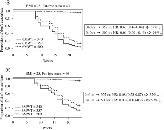
Based on information in the literature18 and the values obtained for the first and third quartiles of the sample, respectively, the following median values were measured for the DW in the 6MWT: 357, 340 and 500 m. These results highlight the large variation observed in this study for this variable.
Values of 23 and 25 kg/m2 were considered for BMI and values of 43 and 46 kg were considered for FFM, taking into consideration the greater frequency of these values in the sample included in this study. Considering a BMI of 23 kg/m2 and an FFM of 43 kg, the risk of exacerbation in patients with a DW of 357 m in the 6MWT was approximately 0.3 times less than the risk of exacerbation for patients with a DW of 340 m. Moreover, upon increasing the DW to 500 m, the risk of exacerbation was 0.95 times less (Figure 2A).
After evaluating the FFM at 46 kg, and maintaining the same BMI value, the risk of exacerbation among
patients who walked 357 m in the 6MWT was approximately 0.21 times lower than the risk of exacerbation for patients with a DW of 340 m. In addition, by increasing the DW to 500 m, this risk was approximately 0.89 times lower; in other words, this risk was reduced by approximately 90% (Figure 2B).
With a BMI of 25 kg/m2 and FFM of 43 kg, the risk of exacerbation for patients who walked 357 m in the 6MWT was 0.37 times less compared to patients who walked 340 m. In comparison, after walking 500 m, this risk was approximately 0.99 times lower; in other words, this risk was reduced by 99%, as illustrated in Figure 3A. With maintenance of the BMI and a FFM of 46 kg, the risk of exacerbation was approximately 0.32 times less in patients who walked 357 m in the 6MWT compared to patients who walked 340 m; with the increase of the DW to 500 m, this risk was 0.97 times lower (Figure 3B).
Table 2 shows the values for the covariates of body composition, airway obstruction, dyspnea, DW in the 6MWT and handgrip strength in the 3 described evaluations, demonstrating significant differences only for the DW between the 3- and 6-month evaluations.
Discussion
Principal findings
The principal finding of this study was that the DW in the 6MWT, in association with BMI and FFM, predicted the risk of exacerbation among COPD patients monitored over a 6-month period. Additionally, a reduction was observed in the tolerance to physical efforts over 6 months of monitoring relative to the 3rd month.
Importance of this study
Because few data have been reported in the literature concerning the evaluation of DW in the 6MWT as a predictor of exacerbation18 in COPD patients enrolled in an exercise program, this study contributes to the field by showing that the evaluation of DW in the 6MWT could be used to predict the risk of exacerbation, while also taking into consideration BMI and FFM.
During exacerbation of COPD and even after its occurrence, there is a decrease in tolerance to physical efforts, and a certain percentage of patients with COPD do not recuperate even 3 months after the occurrence of exacerbation19. This finding most likely justifies the results observed in this study with regard to the reduction of DW in the 6MWT, even among patients undergoing physical therapy.
Because of the interaction between DW and body composition variables, it is important to include a range of variables in the evaluation and monitoring of these patients in an attempt to heed guidelines and referrals considering the occurrence of exacerbations.
DW, BMI and FFM as determinants of exacerbation in COPD
This study demonstrated that the DW in the 6MWT, in association with BMI and FFM, could predict the risk of exacerbation in patients with COPD. In addition, there is evidence of the association between body mass and mortality in patients with COPD, which highlights this variable as an important extrapulmonary factor that may signal the need for disease intervention20. Additionally, a relationship was also previously observed between body mass and acute exacerbation in COPD, accompanied with the imbalance generated by a decrease in food ingestion and an increase in the energy used by the patient21.
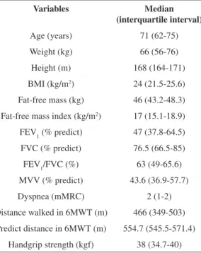
Slinde et al.22 and Vestbo et al.23 reported that free fat mass was related to mortality in COPD; however, its impact on functional limitation remains unknown. According to some authors23, BMI is an Table 1. Anthropometric characteristics, spirometry, dyspnea,
distance walked and predicted distance in the 6MWT and handgrip strength of the studied patients (n=63).
Variables Median
(interquartile interval)
Age (years) 71 (62-75)
Weight (kg) 66 (56-76)
Height (m) 168 (164-171)
BMI (kg/m2) 24 (21.5-25.6)
Fat-free mass (kg) 46 (43.2-48.3)
Fat-free mass index (kg/m2) 17 (15.1-18.9)
FEV1(% predict) 47 (37.8-64.5)
FVC (% predict) 76.5 (66.5-85)
FEV1/FVC (%) 63 (49-65.6) MVV (% predict) 43.6 (36.9-57.7)
Dyspnea (mMRC) 2 (1-2)
Distance walked in 6MWT (m) 466 (349-503)
Predict distance in 6MWT (m) 554.7 (545.5-571.4)
Handgrip strength (kgf) 38 (34.7-40)
Data reported for the median (interquartile interval). BMI: body
mass index. FEV1: forced expiratory volume in one second. FVC:
independent factor for prognosis in this disease, with reduced body mass associated with an increase in mortality. According to Lan et al.24, low BMI exerts an influence on COPD, independent of the degree of airway obstruction. Furthermore, it is worth reporting that age, physical inactivity, hypoxia and the use of
medications, primarily corticoids, was also shown to influence weight loss and the decrease in FFM25.
According to Schols et al.26, in addition to FFM serving as a predictor of mortality, this variable can be used for the staging of COPD because it acts as a systemic marker of disease severity.
Figure 2. Proportion of exacerbations considering variations in the DW in the 6MWT and the interaction with the fixed value of BMI (23 kg/m2) and FFMof 43 kg (A) and 46 kg (B). Data are expressed as the HR: hazard ratio and CI: confidence interval (95%).
Table 2. Corporal composition, air way obstruction, dyspnea, distance walked in 6MWT and handgrip strength of patients over the six months of follow-up.
Variables Initial assessment (n= 63) 3º month (n= 47) 6º month (n= 29)
BMI (kg/m2) 24 (21.5-25.6) 22.5 (20.7-25.5) 22.3 (20.8-26.1)
Fat-free mass (kg) 46 (43.2-48.3) 45.4 (43.5-48.6) 45.4 (43.6-48.1)
Fat-free mass index (kg/m2) 17 (15.1-18.9) 16.7 (15.8-17.2) 16.3 (16.1-17.3)
FEV1(% predict) 47 (37.8-64.5) 46.8 (37.2-58.4) 47.8 (37.8-66.1)
Dyspnea (mMRC) 2 (1-2) 2 (1.5-2) 2 (1-2)
Distance walked in 6MWT (m) 466 (349-503) 470 (349-537) 402 (252-486)*
Handgrip strength (kgf) 38 (34.7-40) 37.3 (34.8-39) 37.3 (33.4-40)
This study found that tolerance to exercise, as evaluated by the DW in the 6MWT, was shown to predict the risk of exacerbation in patients with COPD, provided that there was an association with BMI and FFM. According to Celli27, the capacity to perform exercise is one of the most important variables to be considered in the prognosis of COPD, as tolerance to physical effort shows a progressive decline and functionality is generally decreased with disease progression.
Anzueto28 reported that the reduction in physical activity could be demonstrated by the decrease in DW in the 6MWT, resulting in a higher probability of new exacerbations and hospitalizations3,29. Similarly, according to Rondelli et al.30, intolerance to physical effort is one characteristic of individuals with COPD, which is related to progressive incapacity, with a consequent reduction in survival.
According to Enright et al.31 and Pinto-Plata et al.32, DW appears to be a good predictor of mortality because it reflects not only respiratory function but also cardiopulmonary and peripheral muscular system function, in addition to relating to other
important outcomes in patients with COPD, such as dyspnea31.
Kessler et al.33 and Carr et al.34 observed that patients with COPD after exacerbation show a reduction in DW in the 6MWT. The former study showed that the increased risk of hospitalization among these patients occurred in association with DW values less than 367 m, while in this study, it was observed that patients with episodes of exacerbation showed an average DW of 372 m.
It is also worth reporting that, as in the present study, Carr et al.34 monitored approximately 60 patients with COPD for 6 months to verify the occurrence of exacerbation and demonstrated that the average DW in the 6MWT of patients who exacerbated was 350 m, compared to 416 m for those who did not exacerbate. In the present study, the patients who did not exacerbate walked 459.9 m on average.
data shown in the present study, as older patients with COPD showed lower tolerance to physical efforts.
Upon monitoring the performance of patients with COPD according to the DW in the 6MWT for 2 years, Pinto-Plata et al.32 observed a progressive increase in survival with an increase in DW. Additionally, there is evidence that a reduction of 30 meters or more in DW in the 6MWT increases the risk of death, and this therefore represents a clinically significant minimum difference in patients with COPD36.
In the present study, the risk of exacerbation, which is considered to be one of the primary causes of morbidity/mortality37, was shown to be reduced with an increase in DW. Therefore, this variable may predict the risk of exacerbation for these patients, provided there is an association with BMI that is between the reference values used in this study.
Previously, FEV1 was regarded as the variable that best represented COPD with respect to the severity of the situation and the response of the patient considering the treatment modalities38. However, O’Donnell et al.39 demonstrated that FEV
1 is limited when the intent is to predict the capacity for physical efforts because it does not evaluate this condition in a systemic manner, and this finding has since been confirmed by the results of other studies. Moreover, because a decline in the DW in the 6MWT is observed among more serious patients, the DW has been shown to be a more sensitive variable for the detection of clinical changes associated with COPD as compared to the pulmonary function test, thus highlighting its importance for monitoring these patients40,41.
Fletcher and Peto42 observed a relationship between airway obstruction and survival while monitoring patients with COPD. However, FEV1 does not fully express the disease complexity and manifestations, considering that other clinical variables better predict mortality in these individuals27.
According to Vestbo et al.23 and Rabe et al.37, exacerbations in COPD are important events that influence disease severity and progression. Moreover, episodes of exacerbation result in an increase in the provision of medical care, implying a negative impact, as it is one of the primary causes of morbidity/ mortality, on patient quality of life.
The limitations of this study were that we evaluated only one convenience sample treated at only a single study center. However, the results obtained demonstrate the importance of physical exercise, specifically aerobic physical training associated with a physical therapy treatment program, for patients with COPD, as previously shown in the systematic review by Puhan et al.43. Thus, the
goal for these patients is to obtain an increase and/ or maintenance in tolerance to physical efforts and a consequent reduction in the risk of exacerbation, provided that patients performed monitoring and drug treatment with the responsible pulmonologist.
Conclusion
In summary, over 6 months of monitoring of COPD patients undergoing physical therapy, we identified the DW in the 6MWT as the determinant predictor of the risk of exacerbation, although this association also depended on the covariates of BMI and FFM.
References
1. Cardoso AP. DPOC - agudização aguda – diagnosticar, prevenir e tratar. Pulmão RJ - Atualizações Temáticas. 2009;1(1):68-70.
2. Figueiredo AB, Silva SRB Fº, Lôbo RR, Moriguti JC. Exacerbação da doença pulmonar obstrutiva crônica. Medicina (Ribeirão Preto). 2010;43(3):223-30.
3. Global Initiative for Chronic Obstructive Lung Disease - GOLD. Global strategy for the diagnosis, management, and prevention of chronic obstructive pulmonary disease. Bethesda: Global Initiative for Chronic Obstructive Lung Disease; 2011.
4. Marchiori RC, Susin CF, Dal Lago L, Felice CD, Brandão da Silva D, Severo MD. Diagnóstico e tratamento da DPOC exacerbada na emergência. Rev AMRIGS. 2010;54(2):214-23.
5. Donaldson GC, Wilkinson TMA, Hurst JR, Perera WR, Wedzicha JA. Exacerbations and time spent outdoors in chronic obstructive pulmonary disease. Am J Respir Crit Care Med. 2005;171(5):446-52. PMid:15579723. http:// dx.doi.org/10.1164/rccm.200408-1054OC
6. Donaldson GC, Seemungal TA, Bhowmik A, Wedzicha JA. Relationship between exacerbation frequency and lung function decline in chronic obstructive pulmonary disease. Thorax. 2002;57(10):847-52. PMid:12324669 PMCid:PMC1746193. http://dx.doi.org/10.1136/ thorax.57.10.847
7. Beghé B, Verduri A, Roca M, Fabbri LM. Exacerbation of respiratory symptoms in COPD patients may not be exacerbations of COPD. Eur Respir J. 2013;41(4):993-5. PMid:23543648. http://dx.doi. org/10.1183/09031936.00180812
8. Langer D, Probst VS, Pitta F, Burtin C, Hendriks E, Schans CPVD, et al. Clinical Practice Guideline for physical therapy in patients with Chronic Obstructive Pulmonary Disease (COPD) - Portuguese version. Rev Bras Fisioter. 2009;13(3):183-204. http://dx.doi.org/10.1590/ S1413-35552009005000034
10. Prescott E, Almdal T, Mikkelsenz KL, Tofteng CL, Vestbo J, Lange P. Prognostic value of weight change in chronic obstructive pulmonary disease: results from the Copenhagen City Heart Study. Eur Respir J. 2002;20(3):539-44. PMid:12358326. http://dx.doi.org/1 0.1183/09031936.02.00532002
11. Steiner MC, Barton RL, Singh SJ, Morgan MDL. Bedside methods versus dual energy X-ray absorptiometry for body composition measurement in COPD. Eur Respir J. 2002;19(4):626-31. PMid:11998990. http://dx.doi.org/10 .1183/09031936.02.00279602
12. Mahler D, Wells C. Evaluation of clinical methods for rating dyspnea. Chest. 1988;93(3):580-6. http://dx.doi. org/10.1378/chest.93.3.580
13. ATS Statement: guidelines for the six-minute walk test. Am J Respir Crit Care Med. 2002;166(1):111-7. PMid:12091180. http://dx.doi.org/10.1164/ ajrccm.166.1.at1102
14. Homann D, Stefanello JMF, Góes SM, Leite N. Impaired functional capacity and exacerbation of pain and exertion during the 6-minute walk test in women with fibromyalgia. Rev Bras Fisioter. 2011;15(6):474-80. PMid:22218712. http://dx.doi.org/10.1590/S1413-35552011000600008 15. Iwama AM, Andrade GN, Shima P, Tanni SE, Godoy I,
Dourado VZ. The six-minute walk test and body weight-walk distance product in healthy Brazilian subjects. Braz J Med Biol Res. 2009;42(11):1080-5. PMid:19802464. http://dx.doi.org/10.1590/S0100-879X2009005000032 16. Figueiredo IM, Sampaio RF, Mancini MC, Silva FCM,
Souza MAP. Teste de força de preensão utilizando dinamômetro Jamar. Acta Fisiatr. 2007;14(2):104-10. 17. Colosimo EA, Giolo SR. Análise de Sobrevivência
Aplicada. São Paulo: Edgard Blucher; 2006.
18. Spruit MA, Polkey MI, Celli B, Edwards LD, Watkins ML, Pinto-Plata V, et al. Predicting outcomes from 6-minute walk distance in chronic obstructive pulmonary disease. J Am Med Dir Assoc. 2012;13(3):291-7. PMid:21778120. http://dx.doi.org/10.1016/j.jamda.2011.06.009
19. Pamplona P, Morais L. Treino de exercício na doença pulmonar crónica. Rev Port Pneumol. 2007;XIII(1):101-28. 20. Eisner MD, Blanc PD, Sidney S, Yelin EH, Lathon
PV, Katz PP, et al. Body composition and functional limitation in COPD. Respir Res. 2007;8(27):7. PMid:17261190 PMCid:PMC1797017. http://dx.doi. org/10.1186/1465-9921-8-7
21. Vermeeren MA, Schols AM, Wouters EF. Effects of an acute exacerbation on nutritional and metabolic profile of patients with COPD. Eur Respir J. 1997;10:2264-9. PMid:9387951. ttp://dx.doi.org/10.1183/09031936.97. 10102264
22. Slinde F, Gronberg A, Engstrom CP, Rossander-Hulthen L, Larsson S. Body composition by bioelectrical impedance predicts mortality in chronic obstructive pulmonary disease patients. Respir Med. 2005;99(8):1004-9. http:// dx.doi.org/10.1016/j.rmed.2004.09.024
23. Vestbo J, Prescott E, Almdal T, Dahl M, Nordestgaard BG, Andersen T, et al. Body mass, fat-free body mass, and prognosis in patients with chronic obstructive pulmonary disease from a random population sample: findings from
the copenhagen city heart study. Am J Respir Crit Care Med. 2006;173(1):79-83. PMid:16368793.
24. Lan CC, Su CP, Chou LL, Yang MC, Lim CS, Wu YK. Association of body mass index with exercise cardiopulmonary responses in lung function-matched patients with chronic obstructive pulmonary disease. Heart Lung. 2012;41(4):374-81. PMid:22421506. http://dx.doi. org/10.1016/j.hrtlng.2012.02.010
25. Marquis K, Debigare R, Lacasse Y, LeBlanc P, Jobin J, Carrier G, et al. Midthigh muscle cross-sectional area is a better predictor of mortality than body mass index in patients with chronic obstructive pulmonary disease. Am J Respir Crit Care Med. 2002;166:809-13. PMid:12231489. http://dx.doi.org/10.1164/rccm.2107031
26. Schols AMWJ, Broekhuizen R, Weling-Scheepers CA, Wouters EF. Body composition and mortality in chronic obstructive pulmonary disease. Am J Clin Nutr. 2005;82(1):53-9. PMid:16002800.
27. Celli BR. Predictors of mortality in COPD. Respir Med. 2010;104(6):773-9. PMid:20417082. http://dx.doi. org/10.1016/j.rmed.2009.12.017
28. Anzueto A. Impact of exacerbations on COPD. Eur Respir Rev. 2010;19(116):113-8. PMid:20956179. http://dx.doi. org/10.1183/09059180.00002610
29. Pitta F, Troosters T, Spruit MA, Probst VS, Decramer M, Gosselink R. Characteristics of physical activities in daily life in chronic obstructive pulmonary disease. Am J Respir Crit Care Med. 2005;171(9):972-7. PMid:15665324. http://dx.doi.org/10.1164/rccm.200407-855OC
30. Rondelli RR, Dal Corso S, Simões A, Malaguti C. Métodos de avaliação da fadigabilidade muscular periférica e seus determinantes energético-metabólicos na DPOC. J Bras Pneumol. 2009;35(11):1125-35. PMid:20011849. http:// dx.doi.org/10.1590/S1806-37132009001100011 31. Enright PL, McBurnie MA, Bittner V, Tracy RP,
McNamara R, Arnold A, et al. The 6-min walk test: a quick measure of functional status in elderly adults. Chest. 2003;123(2):387-98. PMid:12576356. http://dx.doi. org/10.1378/chest.123.2.387
32. Pinto-Plata VM, Cote C, Cabral H, Taylor J, Celli BR. The 6-min walk distance: change over time and value as a predictor of survival in severe COPD. Eur Respir J. 2004;23(1):28-33. PMid:14738227. http://dx.doi.org/10. 1183/09031936.03.00034603
33. Kessler R, Faller M, Fourgaut G, Weitzenblum E. Predictive factors of hospitalization for acute exacerbation in a series of 64 patients with chronic obstructive pulmonary disease. Am J Respir Crit Care Med. 1999;159(1):158-64. PMid:9872834. http://dx.doi. org/10.1164/ajrccm.159.1.9803117
34. Carr SJ, Goldstein RS, Brooks D. Acute exacerbations of COPD in subjects completing pulmonary rehabilitation. Chest. 2007;132(1):127-34. PMid:17475631. http:// dx.doi.org/10.1378/chest.07-0269
36. Polkey MI, Spruit MA, Edwards LD, Watkins ML, Pinto-Plata V, Vestbo J, et al. Six-minute-walk test in chronic obstructive pulmonary disease minimal clinically important difference for death or hospitalization. Am J Respir Crit Care Med. 2013;187(4):382-6. PMid:23262518. http:// dx.doi.org/10.1164/rccm.201209-1596OC
37. Rabe KF, Hurd S, Anzueto A, Barnes PJ, Buist SA, Calverley P, et al. Global strategy for the diagnosis, management, and prevention of chronic obstructive pulmonary disease: GOLD executive summary. Am J Respir Crit Care Med. 2007;176(6):532-55. PMid:17507545. http://dx.doi. org/10.1164/rccm.200703-456SO
38. Celli BR, Cote CG, Lareau SC, Meek PM. Predictors of Survival in COPD: more than just the FEV1. Respir Med. 2008;102(1):S27-35. http://dx.doi.org/10.1016/ S0954-6111(08)70005-2
39. O’Donnell DE, Lam M, Webb KA. Measurement of symptoms, lung hyperinflation, and endurance during exercise in chronic obstructive pulmonary disease. Am J Respir Crit Care Med. 1998;158(5):1557-65. PMid:9817708. http://dx.doi.org/10.1164/ ajrccm.158.5.9804004
40. Kuzma AM, Meli Y, Meldrum C, Jellen P, Butler-Lebair M, Koczen-Doyle D, et al. Multidisciplinary care of the patient with chronic obstructive pulmonary disease. Proc
Am Thorac Soc. 2008;5(4):567-71. PMid:18453373 PMCid:PMC2645337. http://dx.doi.org/10.1513/ pats.200708-125ET
41. Rodrigues SL, Silva CAM, Lima T, Viegas CAA, Rodrigues MP, Ribeiro FA. Influência da função pulmonar e da força muscular na capacidade funcional de portadores de doença pulmonar obstrutiva crônica. Rev Port Pneumol. 2009;15(2):199-214.
42. Fletcher C, Peto R. The natural history of chronic airflow obstruction. Br Med J. 1977;1(6077):1645-8. http://dx.doi. org/10.1136/bmj.1.6077.1645
43. Puhan MA, Gimeno-Santos E, Scharplatz M, Troosters T, Walters EH, Steurer J. Pulmonary rehabilitation following exacerbations of chronic obstructive pulmonary disease. Cochrane Database Syst Rev. 2011;(10):CD005305. PMid:21975749.
Correspondence
Diego Marmorato Marino
Rua Miguel Petroni, 4900, Condomínio Eldorado, casa 296 CEP 13563-470, São Carlos, SP, Brasil