http://dx.doi.org/10.1590/0037-8682-0064-2014
Major Article
INTRODUCTION
Address to: Dr. Edelberto Santos Dias. Laboratório de Leishmanioses/CPqRR/ FIOCRUZ. Av. Augusto de Lima 1715, Barro Preto, 30190-002 Belo Horizonte, MG, Brasil.
Phone: 55 31 3349-7758 e-mail: edel@cpqrr.fi ocruz.br Received 19 March 2014 Accepted 1 August 2014
Evaluation of parasitological examination,
kDNA polymerase chain reaction and rK39-based
immunochromatography for the diagnosis
of visceral leishmaniasis in seropositive dogs from the
screening-culling program in Brazil
Shara Regina-Silva
[1], Consuelo Latorre Fortes-Dias
[2], Érika Monteiro Michalsky
[1],
João Carlos França-Silva
[3], Patrícia Flávia Quaresma
[1], Ana Cristina Vianna Mariano da Rocha Lima
[1],
Rafael Gonçalves Teixeira-Neto
[1]and Edelberto Santos Dias
[1][1]. Laboratório de Leishmanioses, Centro de Pesquisas René Rachou, Fundação Oswaldo Cruz, Belo Horizonte, MG. [2]. Diretoria de Pesquisa e Desenvolvimento, Fundação Ezequiel Dias, Belo Horizonte, MG. [3]. Departamento de Parasitologia, Universidade Federal de Minas Gerais, Belo Horizonte, MG.
ABSTRACT
Introduction:
Dogs play a primary role in the zoonotic cycle of visceral leishmaniasis (VL). Therefore, the accurate diagnosis of
infected dogs, primarily asymptomatic dogs, is crucial to the effi ciency of VL control programs.
Methods:
We investigated the
agreement of four diagnostic tests for canine visceral leishmaniasis (CVL): parasite detection, either after myeloculture or by direct
microscopic examination of tissue imprints; kinetoplast-deoxyribonucleic acid-polymerase chain reaction (kDNA-PCR); and an
immunochromatographic test (ICT). An enzyme-linked immunosorbent assay (ELISA) and an indirect immunofl uorescence test
(IFAT), both of which were adopted as part of the screening-culling program in Brazil, were used as reference tests. Our sample
set consisted of 44 seropositive dogs, 25 of which were clinically asymptomatic and 19 were symptomatic for CVL according
to ELISA-IFAT.
Results:
The highest and lowest test co-positivities were observed for ICT (77.3%) and myeloculture (58.1%),
respectively. When analyzed together, the overall percentage of co-positive tests was signifi cantly higher for the symptomatic
group compared to the asymptomatic group. However, only ICT was signifi cantly different based on the results of a separate
analysis per test for each group of dogs. The majority (93.8%) of animals exhibited at least one positive test result, with an
average of 2.66 positive tests per dog. Half of the symptomatic dogs tested positive for all four tests administered.
Conclusions:
The variability between test results reinforces the need for more effi cient and reliable methods to accurately diagnose canine VL,
particularly in asymptomatic animals.
Keywords:
Visceral leishmaniasis. Canine visceral leishmaniasis. Diagnosis.
Leishmania
.
Visceral leishmaniasis (VL) is a chronic parasitic disease
caused by the
Leishmania donovani
complex in East Africa
and the Indian subcontinent and by
Leishmania infantum
(syn.
Leishmania chagasi
) in Europe, North Africa and Latin
America. VL is fatal if left untreated and is transmitted in one
of two ways. Zoonotic VL is transmitted from animal to vector
to human, and anthroponotic VL is transmitted from human to
vector to human. In the former, humans are occasional hosts
and dogs are the main reservoir for
Leishmania
, particularly in
rural and urban domestic environments
1.
Canine visceral leishmaniasis (CVL) is an important public
health problem and is a disease of great interest to private
veterinary medicine due to its high prevalence in urban areas of
Brazil. Infected dogs may present intense cutaneous parasitism
2,3and play a primary role in the maintenance of vector infection
4.
Along with measures to control the vector population and early
treatment of human cases, euthanasia of infected dogs is part of
the VL control policy adopted by the Brazilian Ministry of Health
5.
Early and accurate diagnosis of CVL is of major importance
to the effectiveness of control programs. Although several
techniques have been proposed to serologically diagnose CVL,
a fast and safe diagnostic method with optimal sensitivity
and specifi city is not currently available
6. Until very recently,
standard serology in canine surveys of the government VL
control program used enzyme-linked immunosorbent assay
(ELISA) as a screening method followed by an indirect
immunofl uorescence test (IFAT) to confi rm ELISA-positive
samples. Currently, this methodology is being replaced by
immunochromatographic screening using the Dual Path
Platform (DPP
CVL,
Biomanguinhos
, Rio de Janeiro, Brazil)
METHODS
The main limitation of most diagnostic methods is the
detection of asymptomatic but infected dogs. In this context, the
present study evaluated the results of four different diagnostic
tests for CVL in symptomatic and asymptomatic seropositive
dogs from control program surveys in endemic areas of Brazil.
Two tests are based on
Leishmania
visualization, either after
culturing or by direct microscopic examination; one test,
kinetoplast-deoxyribonucleic acid-polymerase chain reaction,
kDNA-PCR, is a molecular biology-based method, and one test
is antibody-based but uses a recombinant protein rather than
natural
Leishmania
antigens.
Clinical samples
Forty-four dogs that tested seropositive for CVL according
to the standard methodology adopted by the Brazilian
Ministry of Health (ELISA and IFAT kits, both produced by
the
Biomanguinhos
Institute, Rio de Janeiro, Brazil) were
used in this study. These animals were collected by Zoonosis
Control Centers located in three endemic cities for VL in the
Brazilian State of Minas Gerais, Montes Claros (16°44’02”S,
43°51’23”W), Janaúba (15º47’50”S, 43º18’31”W) and Paracatu
(17°13’01”S, 46°52’17”W), all of which are classified as
intense transmission areas for the disease (Brazil 2006). The
dogs were examined by veterinary physicians and classifi ed
as asymptomatic or symptomatic according to the absence or
presence of at least one clinical sign suggestive of canine VL.
Serum samples were collected, and bone marrow aspirates
were harvested by sterilely puncturing the tibial crest for the
preparation of slide smears and parasite cultures. Ear skin,
spleen and mesenteric lymph nodes were biopsied, and the tissue
fragments were used to prepare slide imprints and to extract
total deoxyribonucleic acid (DNA). Euthanasia followed the
technical norms defi ned by Resolution no 714 from the Federal
Council of Veterinary Medicine (dated 06/02/2002) and by the
screening-culling procedure adopted by the Brazilian Ministry
of Health in its Program for Visceral Leishmaniasis Control.
Parasite culture
Novy-MacNeal-Nicolle/liver infusion tryptose (NNN/LIT)
culture medium was inoculated with bone marrow aspirates and
incubated at 25°C. The cultures were examined weekly for the
presence of
Leishmania
promastigotes. After the culture was
expanded to approximately 100 million cells, the cells were
washed with phosphate-buffered saline (PBS), and positive
samples were frozen at -20°C until use. Negative samples were
discarded after fi ve weeks of monitoring.
Polymerase chain reaction
Total DNA was organically extracted
7from
Leishmania
cultures
and biopsied tissues. DNA amplifi cation was performed with A
[5’(C/G)(C/G)(G/C)CC(C/A)CTAT(T/A)TTACACCAACCCC3’]
and B (5’GGGGAGGGGCGTTCTGCGAA3’) primers previously
designed to amplify a 120bp segment from the conserved
region of the kinetoplast DNA minicircle (kDNA) from the
Leishmania
genus
8. The amplifi ed products were visualized by
electrophoresis on a 6% polyacrylamide gel after silver staining.
The presence of a 120bp band from any of the amplifi ed tissue
samples (ear skin, spleen or mesenteric lymph nodes) was
considered a positive result. The quality of DNA extraction was
verifi ed by amplifying the IV S6 region from a constitutive gene
(cacophony) from
Lutzomyia
9.
Immunochromatographic test
Canine sera were tested using the Kalazar Detect™ test
(InBios International Inc., Seatle, Washington, USA), which is
a rapid immunochromatographic test (ICT) that qualitatively
detects antibodies specific for a recombinant
Leishmania
antigen, rK39, formed by repeats of 39 amino acids. This peptide
is a portion of a 230kDa antigen homologous to the kinesin
superfamily of motor proteins and encodes a repetitive, highly
conserved epitope in
L. infantum
and
L. donovani
10.
Direct parasitological examination
After Giemsa or panoptic staining, tissue imprints and bone
marrow smears were examined for the presence of
Leishmania
amastigotes using optical microscopy. The observation of
parasites in the imprints from any biopsied tissue (ear skin, spleen
or mesenteric lymph nodes) was considered a positive test result.
Statistical analysis
The standard serological methodology for canine visceral
leishmaniasis (ELISA-IFAT) was used as a reference, and the
performance of the tests evaluated in this study was calculated
compared to this standard method. Because all of the dogs were
positive according to the standard serological method, diagnostic
test performance was measured based on precision or accuracy
and expressed as the total number of correct classifi cations.
In this case, the test
precision
is equivalent to
sensitivity
. In
addition, given that the reference did not identify certainty of the
absence of the disease but instead a serological result, the term
sensitivity
was replaced with
co-positivity
(which is equivalent
to
relative sensitivity
).
The co-positivity of the four tests as well as the respective
positivity percentages for the two clinical groups of dogs -
symptomatic and asymptomatic - were compared using the
chi-square test
11. When necessary, Fisher’s exact test
11was
employed. The odds ratios and the 95% confi dence intervals
were calculated and adjusted for small samples
12when
applicable.
The Mann-Whitney test
13was employed to compare the
number of positive tests among the four tests, in the two clinical
groups of dogs (symptomatic and asymptomatic). Agreement
between the results of the four tests was verifi ed by calculating
the Kappa coeffi cient
14.
All statistical analyses were performed with R software
(version 3.0.1) with a 5% signifi cance level.
Ethical considerations
RESULTS
DISCUSSION
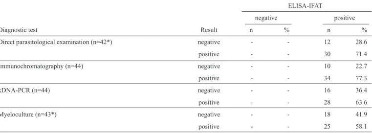
TABLE 1 -Contingency table of the co-positivities (relative sensitivity) of diagnostic tests for canine visceral leishmaniasis. The reference
test was ELISA-IFAT, which was adopted by the Brazilian Ministry of Health in the screening-culling procedure for the Surveillance and Control Program of Visceral Leishmaniasis at the time of our study. Forty-four seropositive dogs (n=44) were sampled from endemic areas of visceral leishmaniasis in the State of Minas Gerais, Brazil.
ELISA-IFAT
negative positive
Diagnostic test Result n % n %
Direct parasitological examination (n=42*) negative - - 12 28.6
positive - - 30 71.4
Immunochromatography (n=44) negative - - 10 22.7
positive - - 34 77.3
kDNA-PCR (n=44) negative - - 16 36.4
positive - - 28 63.6
Myeloculture (n=43*) negative - - 18 41.9
positive - - 25 58.1
ELISA-IFAT: enzyme-linked immunosorbent assay-indirect immunofl uorescence test; kDNA-PCR: kinetoplast-deoxyribonucleic
acid-polymerase chain reaction. *n<44 due to sample loss.
the Ethics Committee on Animal Experimentation (CETEA/
Universidade Federal de Minas Gerais
under license no.
35/2007) and the Federal Board of Veterinary Medicine (CFMV,
Resolution no. 714/2002).
Forty-four dogs seropositive for CVL by ELISA-IFAT were
assayed using direct parasitological examination, myeloculture,
ICT and kDNA-PCR, and the degree of co-positive tests were
evaluated. The highest and lowest co-positivities were obtained
for ICT (77.3%) and myeloculture (58.1%), respectively
(Table 1)
. However, differences in co-positivity were not
statistically signifi cant for any of the tests (p-value = 0.237).
kDNA-PCR showed variable co-positivity percentages based on
the tissue used for DNA extraction: 27.3% for the mesenteric lymph
nodes, 36.4% for the spleen and 50% for the skin (data not shown).
These differences in percentages were not analyzed because our
positivity criterion was the presence of a positive result in any of
the three tissues tested. The DNA extracted from the
Leishmania
biomass served as an internal control for the extraction process,
with positive results in 100% of the samples (data not shown).
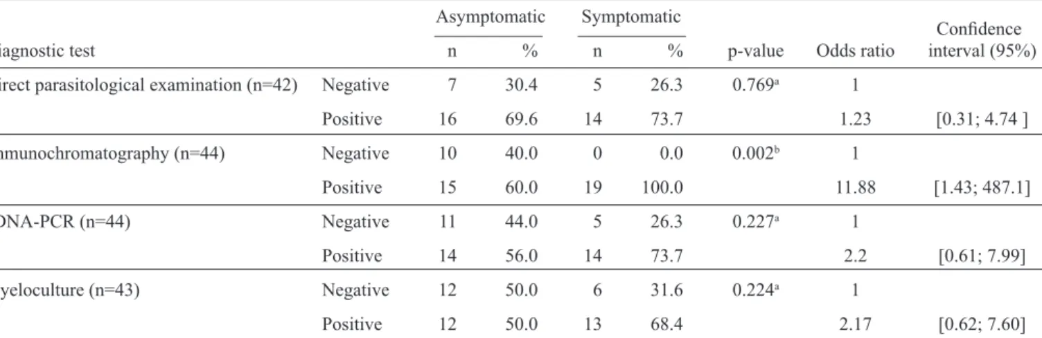
Of the 44 dogs in our sample, 25 were clinically symptomatic
and 19 were asymptomatic.
Table 2
depicts the test results
for each clinical condition. Although we observed a tendency
towards a higher percentage of co-positivity in the symptomatic
group compared to the asymptomatic group, the differences for
each test were not signifi cantly different except for ICT. This
particular assay exhibited 100% co-positivity for symptomatic
dogs compared to 60% in asymptomatic dogs, a statistically
signifi cant difference
(Figure 1)
. According to the odds ratio, the
chance of a positive ICT result in the asymptomatic group was
11.88 [1.43; 487.0] times greater than in the symptomatic group.
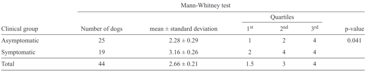
The majority (93.8%) of dogs exhibited at least one positive
CVL result for the diagnostic tests used in this study, with an
average of 2.66 positive test results per dog (
Table 3
). When
the clinical group of dogs was analyzed separately, the average
number of positive tests decreased to 2.28 in the asymptomatic
group and increased to 3.16 in the symptomatic group
(Table 3)
. Further analysis calculating the median value per
clinical group indicated that at least 50% of the asymptomatic
animals tested positive for two of the diagnostic tests for CVL.
In the symptomatic group, half of the animals showed positive
results for all four tests. Comparing the canine clinical groups,
the total number of positive tests was significantly different
(p-value = 0.041)
(Figure 2)
. Three of the asymptomatic dogs were
not co-positive for any of the diagnostic tests (data not shown).
According to the calculated κ value, there was little agreement
between the results from the four diagnostic tests for CVL
(κ = 0.348). However, the corresponding p-value (p-value = 0.000)
indicates that the tests exhibited signifi cant agreement.
In Brazil, despite efforts from the Ministry of Health to
control CVL vectors and reservoirs, VL is undergoing rapid
territorial expansion
15. The recent adoption of DPP
CVL as
the screening step for canine surveys is advantageous because
the delay between diagnosis and culling/treatment is decreased.
Currently, the long duration between sample collection and
culling, as well as the complexity of performing the procedure,
When diagnosing human patients with the goal of detecting
the clinical disease, other rapid tests have proven to be sensitive
and specifi c
18-21. However, when the goal is detecting canine
infection for control purposes, these tests demonstrated low
sensitivity and were useful only for the confi rmation of infection
in suspicious clinical cases
22-27. Therefore, it follows that ICT
identifi ed 100% of the symptomatic dogs in our hands but only
identifi ed 60% in the asymptomatic group.
PCR is considered highly sensitive and specifi c, can be
automated, and is applicable to different types of biological
samples
28-33. As previously noted
4, in contrast to
antibody-based assays, antigen-antibody-based methods such as PCR might
become more relevant indicators of infection in the future, as
these tests can still be used in vaccinated dogs that will test as
antibody-positive. In our hands, kDNA PCR identifi ed only
64% of the samples as positive. Two reasons could justify this
unexpectedly low percentage: the methodology we employed
for DNA extraction and/or a limitation of the technique itself. In
the fi rst case, residue of PCR inhibitors from the organic DNA
extraction could lead to false negative results. However, this is
unlikely because the internal control (DNA extracted from the
Leishmania
biomass) was assayed with the same methodology
and tested 100% positive. Concerning the technique itself
34,
a previous study reported a decrease in PCR sensitivity over
the course of CVL infection, from 78-88% at the 135th day
post-infection to approximately 50% after 300 days. Thus,
the sensitivity of PCR for CVL diagnosis might depend on
the sampling time during infection. For our samples, this time
point is unknown.
The tendency towards higher co-positivity percentages in
symptomatic dogs compared with asymptomatic dogs, which
was consistently observed in this study, agrees with the literature
for several diagnostic tests
2,3,34-39. Depending on the assay, this
may be related to higher antibody titers or to higher parasite
loads in clinically ill dogs
2. However, a statistically signifi cant
p-value = 0.002100
80
60
40
20
0
Direct parasitological examination
Immuno-chromatography
kDNA-PCR Myeloculture
Positive dogs (%)
Diagnostic test
Asymptomatic Symptomatic
FIGURE 1 - Co-positivity of diagnostic tests for canine visceral leishmaniasis for each clinical group of dogs. The reference test was ELISA-IFAT, which was adopted by the Brazilian Ministry of Health in the screening-culling procedure for the Surveillance and Control Program of Visceral Leishmaniasis at the time of our study. The animals were sampled from endemic areas of visceral leishmaniasis in the State of Minas Gerais, Brazil.
Statistically signifi cant differences are indicated with a p-value.
kDNA-PCR: kinetoplast-deoxyribonucleic acid-polymerase chain
reaction; ELISA-IFAT: enzyme-linked immunosorbent
assay-indirect immunofl uorescence test.
TABLE 2 - Contingency table of the co-positivities (relative sensitivity) of diagnostic tests for canine visceral leishmaniasis for each clinical group. The reference test was ELISA-IFAT, which was adopted by the Brazilian Ministry of Health in the screening-culling procedure for the Surveillance and Control Program of Visceral Leishmaniasis at the time of our study. The dogs were sampled from endemic areas of visceral leishmaniasis in the State of Minas Gerais, Brazil.
Asymptomatic Symptomatic
Confi dence
Diagnostic test n % n % p-value Odds ratio interval (95%)
Direct parasitological examination (n=42) Negative 7 30.4 5 26.3 0.769a 1
Positive 16 69.6 14 73.7 1.23 [0.31; 4.74 ]
Immunochromatography (n=44) Negative 10 40.0 0 0.0 0.002b 1
Positive 15 60.0 19 100.0 11.88 [1.43; 487.1]
kDNA-PCR (n=44) Negative 11 44.0 5 26.3 0.227a 1
Positive 14 56.0 14 73.7 2.2 [0.61; 7.99]
Myeloculture (n=43) Negative 12 50.0 6 31.6 0.224a 1
Positive 12 50.0 13 68.4 2.17 [0.62; 7.60]
ELISA-IFAT: enzyme-linked immunosorbent assay-indirect immunofl uorescence test; kDNA-PCR: kinetoplast-deoxyribonucleic
asymptomatic symptomatic
0 1 2 3 4
Number of positive diagnostic tests
p-value = 0.041
TABLE 3 - Number of positive diagnostic tests for canine visceral leishmaniasis for each clinical group. The dogs were sampled from endemic areas of visceral leishmaniasis in the State of Minas Gerais, Brazil.
Mann-Whitney test
Quartiles
Clinical group Number of dogs mean ± standard deviation 1st 2nd 3rd p-value
Asymptomatic 25 2.28 ± 0.29 1 2 4 0.041
Symptomatic 19 3.16 ± 0.26 2 4 4
Total 44 2.66 ± 0.21 1.5 3 4
FIGURE 2 - Distribution of positive results for the entire set of diagnostic tests for canine visceral leishmaniasis for each clinical group. All of the dogs were sampled from endemic areas of visceral leishmaniasis in the State of Minas Gerais, Brazil, and were
seropositive based on ELISA-IFAT. ELISA-IFAT: enzyme-linked
immunosorbent assay-indirect immunofl uorescence test.
difference in test co-positivities between the two clinical groups
of dogs was only observed for ICT. Unfortunately, a fi nal
conclusion determining the agreement of the results from the
four tests employed in this study was not possible. The
κ
value
and the p-value suggest different conclusions, possibly due to
our sample size, which precluded a more thorough analysis.
Despite testing positive serologically, three dogs from the
asymptomatic group, including a three-to-four-month-old
puppy, tested negatively for all of the administered tests. Possible
explanations include false seropositivity
40, cross-reactivity with
other diseases
41or the presence of a pre-patent infection
42,43.
In the case of the puppy, the positive serology might also be
attributable to the presence of maternal antibodies
44.
Published estimates for antibody-based immunofl uorescence
assays range from 72 to 100% sensitivity and 52 to 100%
specifi city
4. One reason for the limited specifi city of
antibody-based tests is that they are generally manufactured with antigens
from
Leishmania major
45-47instead of
Leishmania infantum
because the latter is diffi cult to culture and mass produce in
artifi cial media
47,48. Differences between these
Leishmania
species likely compromise the fi nal test results. In our hands,
even parasitological diagnosis, which is considered the gold
standard for CVL diagnosis, did not reach 100% co-positivity,
even in the symptomatic group.
Ideally, a test for CVL diagnosis should detect asymptomatic
infection, have high sensitivity, specifi city and reproducibility,
and be simple, easy to perform, inexpensive, and viable to
use in regional laboratories or adaptable to fi eld conditions.
This test should detect all dogs infected with
Leishmania,
and
use samples that can be collected non-invasively
43. Despite
the technological advances made in the development of CVL
diagnostic tests, the primary limitation for most tests is the
diagnosis of asymptomatic dogs. A previous study
16suggests
that the effectiveness of control programs could be higher if
euthanasia was directed towards infective dogs not infected,
dogs, which would require a specifi c test to predict infectivity.
Unfortunately, the development of such a test is a hurdle to
improving the success of CVL control programs. This study
reinforces the idea that more sensitive and specifi c tests need
to be developed before the effi cient diagnosis of canine VL
can be realized.
REFERENCES
The authors declare that there is no confl ict of interest.
CONFLICT OF INTEREST
1. Chappuis F, Sundar S, Hailu A, Ghalib H, Rijal S, Peeling RW, et al. Visceral leishmaniasis: what are the needs for diagnosis, treatment and control? Nat Rev Microbiol 2007; 5:873-882.
2. Reis AB, Martins-Filho OA, Teixeira-Carvalho A, Carvalho MG, Mayrink W, França-Silva JC, et al. Parasite density and impaired biochemical/hematological status are associated with severe clinical aspects of canine visceral leishmaniasis. Res Vet Sci 2006; 81:68-75. 3. Quaresma PF, Murta SM, Ferreira EC, Rocha-Lima AC, Xavier AA,
Gontijo CM. Molecular diagnosis of canine visceral leishmaniasis: identifi cation of Leishmania species by PCR-RFLP and quantifi cation
of parasite DNA by real-time PCR. Acta Trop 2009; 111:289-294. 4. Romero GA, Boelaert M. Control of visceral leishmaniasis in Latin
America: a systematic review. PLoS Negl Trop Dis 2010; 4:e584. 5. Ministério da Saúde. Brasil. Manual de Vigilância e Controle da
6. Faria AR, Andrade HM. Diagnosis of canine visceral leishmaniasis: major technological advances and few practical applications. Rev Pan-Amaz Saude 2012; 3:47-57.
7. Sambrook J, Fritsch EF, Maniatis T. Molecular cloning: a laboratory manual. New York: Cold Spring Harbor Laboratory Press; 1989. 8. Degrave W, Fernandez O, Campbell D, Bozza M, Lopez UG. Use of
molecular probes and PCR for detection and typing of Leishmania: a mini review. Mem Inst Oswaldo Cruz 1994; 89:463-469.
9. Lins RMMA, Oliveira SG, Souza NA, Queiroz RG, Justiniano SCB, Ward RD, et al. Molecular evolution of the cacophony IVS6 region in sandfl ies. Insect Mol Biol 2002; 11:117-122.
10. Burns Jr JM, Shreffl er WG, Benson DR, Ghalib HW, Badaro R, Reed SG. Molecular characterization of a kinesin-related antigen of Leishmania chagasi that detects specifi c antibody in African and American visceral
leishmaniasis. Proc Natl Acad Sci USA 1993; 90:775-779.
11. Agresti A. Categorical data analysis. 2nd edition. Wiley Series in Probability and Statistics. New York: Wiley Interscience; 2002. 12. Jewell NP. Statistics for Epidemiology. Texts in Statistical Science
1st edition. v. 58. London: Chapman & Hall; 2003.
13. Hollander M, Douglas AW. Nonparametric Statistical Methods, Solutions Manual. 2nd edition. v. 348. New York: Wiley Series in Probability and Statistics John Wiley & Sons; 1999.
14. Fleiss JL. Statistical methods for rates and proportions. 2nd edition. New York: Wiley, John and Sons Inc; 1981.
15. Werneck GL. Geographic spread of visceral leishmaniasis in Brazil. Cad Saude Publica 2010; 26:644-645.
16. Courtenay O, Quinnell RJ, Garcez LM, Shaw JJ, Dye C. Infectiousness in a cohort of Brazilian dogs: why culling fails to control visceral leishmaniasis in areas of high transmission. J Infect Dis 2002; 186: 1314-1320.
17. Reithinger R, Quinnell RJ, Alexander B, Davies CR. Rapid detection of Leishmania infantum infection in dogs: comparative study using an immunochromatographic dipstick test, enzyme-linked immunosorbent assay and PCR. J Clin Microbiol 2002; 40:2352-2356.
18. Sundar S, Reed SG, Singh VP, Kumar PC, Murray HW. Rapid accurate fi eld diagnosis of Indian visceral leishmaniasis. Lancet 1998; 351: 563-565.
19. Zijlstra E, Nur Y, Desjeux P, Khalil E, El-Hassan A, Groen J. Diagnosing visceral leishmaniasis with the recombinant K39 strip test: experience from Sudan. Trop Med Int Health 2001; 6:108-113.
20. Carvalho SF, Lemos EM, Corey R, Dietze R. Performance of recombinant K39 antigen in the diagnosis of Brazilian visceral leishmaniasis. Am J Trop Med Hyg 2003; 68:321-324.
21. Assis TS, Braga AS, Pedras MJ, Oliveira E, Barral A, Siqueira IC, et al. Multi-centric prospective evaluation of rk39 rapid test and direct agglutination test for the diagnosis of visceral leishmaniasis in Brazil. Trans R Soc Trop Med Hyg 2011; 105:81-85.
22. Reithinger R, Davies CR. Canine leishmaniasis: novel strategies for control. Trends Parasitol 2002; 18:289-290.
23. Mohebali M, Taran M, Zarei Z. Rapid detection of Leishmania infantum
infection in dogs: comparative study using an immunochromatographic dipstick rK39 test and direct agglutination. Vet Parasitol 2004; 121:239-245. 24. Otranto D, Paradies P, Sasanelli M, Spinelli R, Brandonisio O.
Rapid immuno-chromatographic test for serodiagnosis of canine leishmaniasis. J Clin Microbiol 2004; 42:2769-2770.
25. Toz SO, Chang KP, Ozbel Y, Alkan MZ. Diagnostic value of rK39 dipstick in zoonotic visceral leishmaniasis in Turkey. J Parasitol 2004; 90:1484-1486.
26. Mettler M, Grimm F, Capelli G, Camp H, Deplazes P. Evaluation of enzyme linked immunosorbent assays, an immunofl uorescent-antibody test, and two rapid tests (immunochromatographic-dipstick and gel tests) for serological diagnosis of symptomatic and asymptomatic
Leishmania infections in dogs. J Clin Microbiol 2005; 43:5515-5519. 27. Quinnell RJ, Carson C, Reithinger R, Garcez LM, Courtenay
O. Evaluation of rK39 rapid diagnostic tests for canine visceral
leishmaniasis: longitudinal study and meta-analysis. PLoS Negl Trop Dis 2013; 7:e1992.
28. Barrouin-Melo SM, Laranjeira DF, Andrade Filho FA, Trigo J, Julião FS, Franke CR, et al. Can spleen aspirations be safely used for the parasitological diagnosis of canine visceral leishmaniosis? A study on asymptomatic and polysymptomatic animals. Vet J 2005; 171:331-339. 29. Diniz SA, Melo MS, Borges AM, Bueno R, Reis BP, Tafuri WL, et al.
Genital lesions associated with visceral leishmaniosis and shedding of
Leishmania sp. in semen of naturally infected dogs. Vet Pathol 2005; 42:650-658.
30. Franceschi A, Merildi V, Guidi G, Mancianti F. Occurrence of
Leishmania DNA in urines of dogs naturally infected with leishmaniasis. Vet Res Commun 2006; 31:335-341.
31. Ferreira SA, Ituassu T, Melo MN, Andrade AS. Evaluation of the conjunctival swab for canine visceral leishmaniasis diagnosis by PCR-hibridization in Minas Gerais state, Brazil. Vet Parasitol 2008; 152: 257-263.
32. Coura-Vital W, Marques MJ, Velos VM, Roatt BM, Aguiar-Soares RD, Reis LE, et al. Prevalence and factors associated with Leishmania infantum infection of dogs from an urban area of Brazil as identifi ed by
molecular methods. PLoS Negl Trop Dis 2011; 5:e1291.
33. Ferreira SA, Almeida GG, Silva SO, Vogas GP, Fujiwara RT, Andrade AS, et al. Nasal, oral and ear swabs for canine visceral leishmaniasis diagnosis: new practical approaches for detection of Leishmania infantum DNA. PLoS Negl Trop Dis 2013; 7:e2150.
34. Quinnell RJ, Courtenay O, Davidson S, Garcez L, Lambson B, Ramos P, et al. Detection of Leishmania infantum by PCR, serology and cellular immune response in a cohort study of Brazilian dogs. Parasitology 2001; 122:253-261.
35. Killick-Kendrick R, Killick-Kendrick M, Pinelli E, Del Real G, Molina R, Vitutia MM, et al. A laboratory model of canine leishmaniasis: the inoculation of dogs with Leishmania infantum promastigotes from midguts of experimentally infected phlebotomine sandfl ies. Parasite 1994; 1:311-318.
36. Saridomichelakis MN, Mylonakis ME, Leontides LS, Koutinas AF, Billinis C, Kontos VI. Evaluation of lymph node and bone marrow cytology in the diagnosis of canine leishmaniasis (Leishmania infantum) in symptomatic and asymptomatic dogs. Am J Trop Med Hyg 2005; 73:82-86.
37. Costa-Val AP, Cavalcanti RR, Gontijo NF, Michalick MS, Alexander B, Williams P, et al. Canine visceral leishmaniasis: Relationships between clinical status, humoral immune response, haematology and Lutzomyia (Lutzomyia) longipalpis infectivity. Vet J 2007; 174:636-643.
38. Teixeira Neto RG, Giunchetti RC, Carneiro CM, Vitor RW, Coura-Vital W, Quaresma PF, et al. Relationship of Leishmania-specifi c IgG
levels and IgG avidity with parasite density and clinical signs in canine leishmaniasis. Vet Parasitol 2010; 169:248-257.
39. Almeida AB, Sousa VR, Gasparetto ND, Silva GF, Figueiredo FB, Dutra V, et al. Canine visceral leishmaniasis: diagnostic approaches based on polymerase chain reaction employing different biological samples. Diagn Microbiol Infect Dis 2013; 76:321-324.
40. Alves WA, Bevilacqua PD. Refl exões sobre a qualidade do diagnóstico da leishmaniose visceral canina em inquéritos epidemiológicos: o caso da epidemia de Belo Horizonte, Minas Gerais, Brasil, 1993-1997. Cad Saude Publica 2004; 20:259-265.
41. Vale AM, Fujiwara RT, Silva Neto AF, Miret JA, Alvarez DC, Silva JC, et al. Identifi cation of highly specifi c and cross-reactive antigens of
Leishmania species by antibodies from Leishmania (Leishmania) chagasi naturally infected dogs. Zoonoses Public Health 2009; 56:41-58. 42. Quinnell RJ, Courtenay O, Garcez L, Dye C. The epidemiology of
canine leishmaniasis: transmission rates estimated from a cohort study in Amazonian Brazil. Parasitology 1997; 115:143-156.
43. Maia C, Campino L. Methods for diagnosis of canine leishmaniasis and immune response to infection. Vet Parasitol 2008; 158:274-287. 44. Laurenti MD. Correlação entre o diagnóstico parasitológico e sorológico
45. Costa CA, Genaro O, Lana M, Magalhães PA, Dias M, Michalick MS, et al. Canine visceral leishmaniasis: evaluation of the serologic method used in epidemiologic studies. Rev Soc Bras Med Trop 1991; 24:21-25. 46. Rondon FC, Bevilaqua CM, Franke CR, Barros RS, Oliveira FR,
Alcântara AC, et al. Cross-sectional serological study of canine
Leishmania infection in Fortaleza, Ceará state, Brazil. Vet Parasitol 2008; 155:24-31.
47. Silva DA, Madeira MF, Abrantes TR, Filho CJ, Figueiredo FB. Assessment of serological tests for the diagnosis of canine visceral leishmaniasis. Vet J 2013; 195:252-253.
48. Santiago MA, Ribeiro FC, Mouta-Confort E, Nascimento LD, Schubach AO, Madeira MF, et al. Differentiation between canine cutaneous and visceral leishmaniasis by the detection of immunoglobulin G specifi c for Leishmania (Viannia) braziliensis and Leishmania (Leishmania) chagasi antigens using fl ow cytometry. Vet Parasitol 2008; 154:
341-346.