Arq Neuropsiquiatr 2008;66(1):114-116
114
TREATMENT OF CEREBRAL CYSTICERCOSIS
WITH ALBENDAZOLE IN ELEVATED DOSAGES
Marco Antônio Rocha Jr
1, Juliana Machado Santiago dos Santos
2,
Elisa Conci de Souza Gomes
2, Marco Antônio Rocha
3, Cristiane Franklin Rocha
4,
Gervásio Teles Cardoso de Carvalho
5, Bruno Silva Costa
6TRATAMENTO DE CISTICERCOSE CEREBRAL COM ALBENDAZOL EM DOSES ELEVADAS
1Professor of Neuroanatomy and Neurology at Faculdade de Ciências Médicas de Minas Gerais, Belo Horizonte MG, Brazil (FCMMG), Professor of
Neurology at PUC/MG, Neurosurgeon at SEMPER and Vera Cruz Hospitals, Belo Horizonte MG, Brazil; 2Undergraduating in Medicine, Neuroanatomy
monitor at FCMMG; 3Professor of Neuroanatomy and Neurology at FCMMG, Chief of the Neurosurgery Service at SEMPER Hospital, Belo Horizonte
MG, Brazil; 4Professor of Neurology at FCMMG, Neurologist at SEMPER Hospital, Belo Horizonte MG, Brazil; 5Professor of Neurosurgery at FCMMG,
Neurosurgeon at Santa Casa, Belo Horizonte MG, Brazil; 6Neurosurgeon at Santa Casa, Belo Horizonte MG, Brazil.
Received 11 May 2007, received in inal form 22 November 2007. Accepted 4 January 2008.
Dr. Marco Antônio Rocha Jr – Rua Helena Abdalla 25 / 703 - 30380-550 Belo Horizonte MG - Brasil. E-mail: [email protected]
Neurocysticercosis is the most common parasitic in-fection of the central nervous system (CNS)1-3. It has been
estimated that 50 million people in the underdeveloped countries are infected and the disease is endemic to Lat-in America (Equatorial Zone and Brazil), Mexico, Central America, Asia and Africa1,4. Because it is a transmissible
disease its high incidence relects the bad conditions of the local basic sanitation2. Neurocysticercosis treatment
is still surrounded by doubts. The decision about wheth-er to have a clinical or a surgical treatment is, most of the times, very dificult, especially when the location is ven-tricular or subarachnoidal5.
Whenever the clinical treatment is chosen the doubts still remain, regarding the cysticide dosage and the time of the treatment.
The case we report is illustrative in this way.
CASE
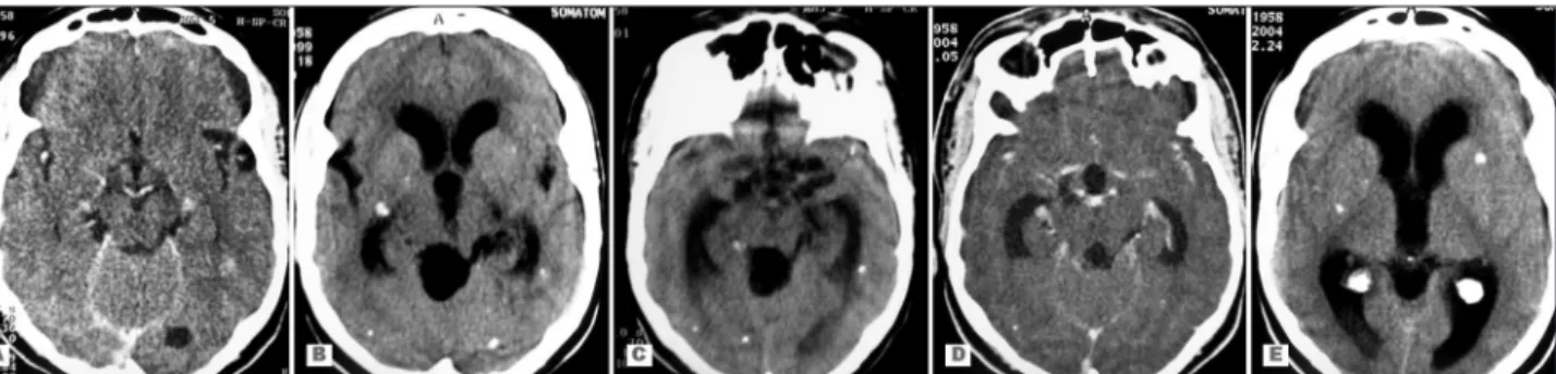
A 38 years old woman was carrier of racemose cysticercosis with cysticercus in different stages, located in the cisterna and in the parenchyma, since 1996 (Fig 1A). History of seizures, which have been controlled by antiepileptic drugs.
In 1999, the patient developed cysts in the quadrigeminal cisterna, with supratentorial dilatation of the ventricular system, without symptoms of intracranial hypertension (Fig 1B). The pa-tient evolved in an asymptomatic way, despite the growing cysts inside the quadrigeminal cisterna, which were evident in a cra-nial computed tomography (CT) in 2001 (Fig 1C). In September 2004, the lesion in the quadrigemial cisterna disappeared (Figs 1D and 1E). There are no known documents or reliable reports about the use of cysticides during this time.
In October 2005, the patient developed right eyelid pto-sis. The magnetic resonance imaging (MRI) revealed cistyc le-sions, which suggested cisternal cysticercosis in the suprasellar and perimesencephalic region on the right side. The third
cra-nial nerve was compromised, in association with communicant hydrocephaly. In December 2005, the patient was admitted at SEMPER hospital prostrated, with headache and vomiting. CT re-vealed an increase in the cistyc lesion of the prepontin cisterna and a discret increase of the hydrocephaly regarding previous studies (Figs 2A, 2B). The patient was submitted to ventriculo-peritoneal shunt and the symptoms were solved (Fig 2C). How-ever, the oculomotor paralysis persisted on the right side. During this hospitalization, the patient was also submitted to a treat-ment with albendazole 15 mg/Kg for 8 days.
In January 30th 2006, the patient was readmitted with
paral-ysis of the right third nerve and left hemiparesis. MRI revealed a resolution of the hydrochepaly and an increase on the size of the lesion in the prepontine cisterna, with a signiicant compression of the mesencephalous, which justiied the alternate syndrome (Fig 2D and 2E). Also, there is still presence of bilateral chronic subdural hematoma, bigger at right, which was probably a con-sequence of the shunt (Fig 2F). The patient went through surgical drainage of the hematoma but there was no improvement of the hemiparesis and ptosis. The ventricular cateter was not removed.
A new therapeutic treatment was accomplished using alben-dazole 30 mg/Kg dose, for 30 days, along with dexamethasone IV during the irst week of treatment. The patient was discharged and completed the treatment at home, with oral corticoids in a decreasing way.
Control CT after 15 days of treatment showed a resolution of the subdural hematoma, but also the persistence of the le-sion in the cistern (Figs 3-A and 3-B). The patient’s neurological deicits did not change.
Arq Neuropsiquiatr 2008;66(1)
115 Cerebral cysticercosis treatment: albendazole Rocha Jr et al.
Fig 1. (A) Cranial CT showing lesions of cerebral cysticercosis; (B and C) Cranial CT showing cyst in the quadrigeminal cistern and dilatation of the ventricular system; (D and E) Cranial CT showing the disappearing of the cyst in the quadrigeminal cistern.
Within 6 months of follow-up, the patient presented no neu-rological deicits, and the CT showed almost completely resolu-tion of the lesion caused by neurocysticercosis (Fig 3D).
DISCUSSION
Most people with cyticercosis are assymptomatic.
Their clinical manifestation depends on the location, size and number of cysts and on the host’s immune response to the parasite1. Seizures appear in 70% to 90% of the
patients1. Intracranial hypertension, headache,
meninge-al syndrome and psychiatric disturbs are other recurrent manifestations6.
Fig 2. (A) MR of the encephalous revealing cis-tyc lesions in the suprasellar and perimesence-phalic region on the right side; (B) Cranial CT revealed an increase in the cistyc lesion of the prepontin cistern; (C) Cranial CT with intraven-tricular cateter; (D and E) MR of the encepha-lous revealing a resolution of the hydrocephaly and an increase on the size of the lesion in the prepontin cistern; (F) MR of the encephalous revealing bilateral chronic subdural hematoma.
Arq Neuropsiquiatr 2008;66(1)
116
Cerebral cysticercosis treatment: albendazole Rocha Jr et al.
The hexacanth embryo reaches the CNS, via the vas-cular system. It passes through the capillar wall in the en-cephalic parenchyma, develops into the cystic form and then turns into Cisticercus celullosae, a benign form of the disease2,5. In this period, it shows a thin membrane with a
colorless liquid in the inside associated with the scolex. The cysticercus which develop inside the ventricle and subarachnoidal space assumes the Cisticercus racemosus form, a malign form of the disease, wich is characterized by an irregular enhance membrane, absence of scolex and grouped in multiples vesicles2. Hydrocephaly is the most
common manifestation. The prognosis is bad, and the therapeutic response is low, poor and the life span is short.
In the CNS, the cysticercus shows a natural evolutive process that culminates in its degeneration in an approxi-mated time of 2 to 5 years2. During this period, they go
through 4 stages7: (1) vesicular: active form, with an
albes-cent membrane, translualbes-cent, with colorless liquid and the scolex in the inside; (2) colloidal: there is an enhancing membrane e substitution from the lipid liquid by an albes-cent gel; (3) granular: calcium is deposited in the gel; (4) nodular: there is a completely calciication of the cyst.
Treatment of neurocysticercosis may be clinical or sur-gical. Surgical treatment is traditionally recommended for the intracranial forms which improve with local compres-sion of the encephalous and cranial nerves or intracranial hypertension.
The effectiveness of the surgical procedure to remove the cistycercus from the cisterna of the base is doubt-ful2. Ordinarily, cistycercus are multiple and show a partial
degeneration as well as adherence to the cranial nerves, vessels and encephalic parenchyma because of arach-noiditis, meaning that an attempt to completed lysis can be disastrous2. When a choice must be made between a
surgical or a pharmacological conduct, there is still much controversy, especially if the location of the cyst is at the ventricular or subarachnoidal space. The conduct must be individualized for each case5.
Because these considerations, we choose to try the clinical treatment of the reported patient. Albendazole has been used in a 15 mg/Kg dose for 15 days and more recently for 8 days2.
Singhi3, in a prospective, randomized, double blind
study with 122 children with neurocysticercosis, treated half of the patients with albendazole (15 mg/Kg) for 7 days and the other group was treated with the same drug and the same dosage for 4 weeks. He came to the con-clusion that the 1 week treatment was as effective as the treatment for 28 days. The exactly time of the terapy with
albendazole still remains unclear3. Other authors
recom-mended the treatment with albendazole for 28 to 30 days and that can reach even 60 days of treatment1,4,5.
Contro-versy arises also over the dose of treatment. Recently stud-ies have been made regarding the treatment of racemose neurocysticercosis with elevated dosages of albendazole.
Comparative studies between the treatment of sub-arachnoidal and intraventricular forms of the parasite with a 15 and a 30 mg dose of albendazole, had shown an involution of the cysticercus much more signiicant in the groups treated with higher doses of the cysticid5,8,9. In our
patient we used 30 mg/Kg dose, for 30 days.
It seems to be consensual as well as in our reported case, the treatment among with corticoid therapy (dexa-methasone)1,2,4,8. Predisone may be employed when a
long-term treatment is needed, especially in cases of chronic arachnoiditis. For the intracranial hypertension, manitol would be indicated as well. Some authors recommended antihistaminics instead of dexametasone, and this is also useful in diabetes and blood hypertension5,9.
Elevated dosage of albendazole does not seem to be associated to the increase of side effects when compared with the usual doses5,8,9.
In conclusion, administration of elevated dosages of albendazole has been shown to be more effective and free from the side effects when compared to the treatment with conventional dosages. Clinical follow-up for longer periods seems to be necessary. The need of repeated cy-cles of the treatment is still questioned by some authors.
REFERENCES
1. Cerdas C, Retana M, Ramírez G, Valenciano A. Neurocisticercosis parenquimatosa activa: reporte de um caso y revisión de la literatura. Rev Costorricense 2004;25:41-47.
2. Colli BO, Carlotti CGJr. Fisiopatologia, diagnóstico e tratamento da ticercose do sistema nervoso central. Temas atuais de neurocirurgia: cis-ticercose do SNC, Sociedade de Neurocirurgia do Estado de São Paulo 2003;4-19.
3. Singhi P, Dayal D. One week versus four weeks of albendazole thera-py for neurocysticercosis in children: a randomized, placebo-controlled double blind tiral. Pediatr Infect J 2003;22:268-272.
4. Kalra V, Dua T, Kumar V. Eficacy of albendazole and short-course dexa -methasone treatment in children with 1 or 2 ring-enhancing lesions of neu-rocysticercosis: a randomized controlled trial. J Pediatr 2003;143:111-114. 5. Agapejev S, Silva MD, Ueda AK. Severe forms of neurocysticercosis:
treatment with albendazole. Arq Neuropsiquiatr 1996;54:82-93. 6. Souza SEM. Neurocisticercose. In: Souza SEM (Ed). Tratamento das
doenças neurológicas. Rio de Janeiro: Guanabara Koogan,2000:13-15. 7. Machado LR, Nobrega JP, Barros NG, Livramento JA, Bacheschi LA,
Spina-França A. Computed tomography in neurocysticercosis: a 10-year long evolution analysis of 100 patients with an appraisal of a new classiication. Arq Neuropsiquiatr 1990;48:414-418.
8. Rivera G, Hernández S, González ED, et al. Albendazole trial at 15 or 30 mg/Kg/day for subarachnoid and intraventricular cysticercosis. Neu-rology 2006;66:436-438.