897
https://doi.org/10.1590/0004-282X20170163
IMAGES IN NEUROLOGY
Central nervous system vasculitis in a patient
with HIV infection: a diagnostic challenge
Vasculite do sistema nervoso central em um paciente com infecção por HIV: um
desafio diagnóstico
Nícolas de O. Amui
1*, Roberta G. F. Silva
2*, Danilo M. C. Costa
3, Orlando G. P. Barsottini
1, José Luiz Pedroso
1,
Paulo Roberto Abrão Ferreira
2A 37-year-old woman with HIV infection (CD4:
5 cells/µl) presented with acute dysarthria and right
hemi-paresis. A brain CT showed nodular hyperdense lesions; MRI
depicted cortico-subcortical hyperintense signals (Figure 1);
MRI and CT angiography disclosed arterial stenosis
suggest-ing vasculitis (Figure 2). he cerebrospinal luid PCR con
-irmed cytomegalovirus infection. Highly-active antiretroviral
therapy was started, but there was worsening of the
symp-toms, and she died one month later. Brain necropsy
con-irmed toxoplasmosis.
CNS vasculitis in HIV patients may be caused by several
infectious diseases
1, such as CMV, toxoplasmosis, HIV virus,
and others
2. In this report we describe a HIV-infected patient
with cerebral vasculitis related to co-infection.
1Universidade Federal de São Paulo, Departamento de Neurologia, São Paulo SP, Brasil; 2Universidade Federal de São Paulo, Divisão de Doenças Infecciosas, São Paulo SP, Brasil; 3Universidade Federal de São Paulo, Departamento de Radiologia, São Paulo SP, Brasil.
Correspondence: Roberta Gunutzmann Ferreira Silva; Divisão de Doenças Infecciosas da UNIFESP; Rua Napoleão de Barros, 715; 04023-900 São Paulo SP, Brasil; E-mail: [email protected]
*The first two authors contributed equally to this work.
Conflict of interest: There is no conflict of interest to declare.
Received 23 January 2017; Received in final form 06 September 2017; Accepted 18 September 2017.
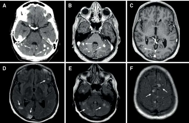
Figure 1.
Axial brain CT scan without contrast shows calcification in the cerebellar hemispheres (A). Axial T1-weighted brain
MRI shows corresponding areas of nodular and irregular enhancement by the gadolinium (B and C). Axial FLAIR brain MRI
demonstrates several areas of hyperintense signal in the cerebral cortex and cerebellum (D, E and F).
A
B
C
898
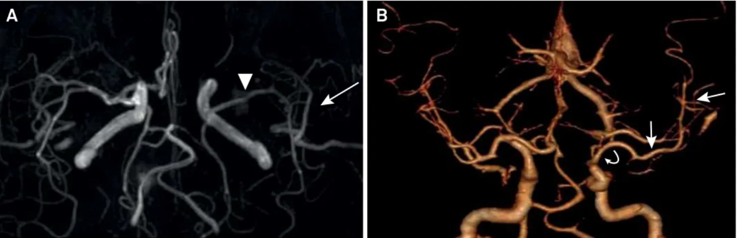
Arq Neuropsiquiatr 2017;75(12):897-898Figure 2.
Brain MRI angiography shows segmental narrowing of the left middle cerebral artery (arrowhead) with reduction of flow
in the opercular segments (arrow) (A). Follow up with brain CT scan angiography shows focal narrowing of the supraclinoid portion
of the left internal carotid artery (curved arrow) and reduction of the flow in the left middle cerebral artery (straight arrow) (B).
A
B
References
1. Hajj-Ali RA, Calabrese LH. Diagnosis and classification of central nervous system vasculitis. J Autoimmun. 2014;48-49:149-52. https://doi.org/10.1016/j.jaut.2014.01.007