ANALYSIS OF SURFACES FOR CHARACTERIZATION
OF FUNGAL BURDEN – DOES IT MATTER?
CARLA VIEGAS1,2, TIAGO FARIA1, MÁRCIA MENESES1, ELISABETE CAROLINO1, SUSANA VIEGAS1,2,
ANITA QUINTAL GOMES1,3, and RAQUEL SABINO1,4 1 Polytechnic Institute of Lisbon, Lisbon, Portugal
Environment and Health Research Group, Lisbon School of Health Technology
2 Universidade Nova de Lisboa, Lisbon, Portugal
Centro de Investigação em Saúde Pública, Escola Nacional de Saúde Pública
3 University of Lisbon, Lisbon, Portugal
Institute of Molecular Medicine, Faculty of Medicine
4 National Health Institute Doutor Ricardo Jorge, Lisbon, Portugal
Mycology Laboratory
Abstract
Objectives: Mycological contamination of occupational environments can be a result of fungal spores’ dispersion in the air and on surfaces. Therefore, it is very important to assess it in both types of the samples. In the present study we assessed fungal contamination in the air and in the surface samples to show relevance of surfaces sampling in complementing the results ob-tained in the air samples. Material and Methods: In total, 42 settings were assessed by the analysis of air and surfaces samples. The settings were divided into settings with a high fungal load (7 poultry farms and 7 pig farms, 3 cork industries, 3 waste management plants, 2 wastewater treatment plants and 1 horse stable) and a low fungal load (10 hospital canteens, 8 college canteens and 1 maternity hospital). In addition to culture-based methods, molecular tools were also applied to detect fungal burden in the settings with a higher fungal load. Results: From the 218 sampling sites, 140 (64.2%) presented different species in the examined surfaces when compared with the species identified in the air. A positive association in the high fungal load settings was found between the presence of different species in the air and surfaces. Wastewater treatment plants constituted the setting with the highest number of different species between the air and surface. Conclusions: We observed that surfaces sampling and application of molecular tools showed the same efficacy of species detection in high fungal load settings, cor-roborating the fact that surface sampling is crucial for a correct and complete analysis of occupational scenarios.
Key words:
Surface samples, Fungal burden assessment, High fungal load settings, Low fungal load settings, Air samples, Occupational environments
This work was financially supported by the Lisbon School of Health Technology – Polytechnic Institute of Lisbon and coordinated by Carla Viegas. Received: January 7, 2015. Accepted: August 27, 2015.
Corresponding author: C. Viegas, Polytechnic Institute of Lisbon, Lisbon School of Health Technology, Environment and Health Research Group, Nations Park, 1990-090 Lisbon, Portugal (e-mail: carla.viegas@estesl.ipl.pt).
INTRODUCTION
Recently, concerns about human exposure to microor-ganisms in indoor environments have been also focused on fungi. Interest in bioaerosol exposure has significantly increased because it is now recognized that exposure to fungal agents is associated with a wide range of adverse
health effects with a major impact on public health [1]. However, despite the division of fungi in different bio-safety levels [2], as well as the Directive 2000/54/EC [3], referring to the importance of the safety of workers ex-posed to biological agents, these classifications and docu-ments do not include toxic species that can be present in
In the present study we assessed fungal contamination in the air and in the samples of surfaces, and showed the rel-evance of the latter in complementing the results obtained only via analyses of the air samples.
MATERIAL AND METHODS Assessed settings
The sampling of several settings was performed between February 2010 and March 2014. The analyzed settings included 7 poultry farms and 7 pig farms, 3 cork indus-tries, 3 waste management plants, 2 wastewater treat-ment plants and 1 horse stable. Those settings were cho-sen because they have high probability of a high fungal load (HFL) [20]. The sampling sites selected for each of the settings mentioned above were chosen based on the amount of time spent by the workers in those places during their occupational activity. In some of those set-tings, in addition to conventional methods, also molecu-lar methods were applied to detect fungi (Table 1). This approach was performed to overcome some limitations of the culture-based methods and whenever specific species/ strains needed to be detected.
Settings with a low fungal load (LFL) were also analyzed. Those included 10 hospital canteens, 8 college canteens and 1 maternity hospital. All these settings were assessed using conventional methods.
Sample collection Conventional methodologies
Two hundred fifty-nine air samples were collected by the use of conventional methods. The amount of the col-lected air samples ranged from 20 l (from pig farms) to 500 l (from hospital wards). The air samples were collected by means of the impaction method with a flow rate of 140 l/min onto malt extract agar (MEA) supple-mented with chloramphenicol (0.05%), using the Mil-lipore Air Tester (MerckMilMil-lipore, USA). The samplers were placed at a height of 0.6–1.5 m above the floor, several occupational settings [4]. Additionally, there is no
classification that would consider, on the one hand, fungal ability to disseminate and, on the other, the principal in-take routes from their spores and metabolites taking into account each occupational setting.
Based on their small size and a large number, fungal spores are classified as bioaerosols. They are always pres-ent in the atmosphere and their concpres-entration changes depending on environmental conditions. Production and spore release varies drastically from species to species, in-fluencing its dissemination in the air/surfaces [5].
Aspergillus and Penicillium spores may remain indoors for
long periods of time, while Stachybotrys spores diminish their concentration/viability soon after being produced, conditioning interpretation of the results of air samples’ cultures [6]. The type of sporulation and spore charac-teristics (size, density, colony structure and roughness) must also be considered when analyzing fungal con-tamination of air. Importantly, additional environmental characteristics related to the surfaces, including surface vibrations, smoothness and a role as a substrate [7] may also influence fungal contamination. Cultures in the air samples are usually the only parameters used to assess indoor fungal contamination [8]. According to several authors [9–11], surfaces analysis complements microbio-logical characterization of the air and is used in order to identify contamination sources. It may also be used to evaluate efficacy of surface cleaning and disinfection procedures.
The number of studies where fungal contamination in workplaces has been characterized both in the air and in the surface samples is very limited [12–19]. In fact, in most of the studies on environmental fungal assessment, the results have been achieved only by air sampling. Be-cause mycological contamination can be a result of fungal spores’ dispersion in the air and on the surfaces, it is very important to assess it and evaluate a specific environment based on the results of both types of the samples.
phosphate-buffered saline with 0.05% Triton X-100, and the collection liquid was subsequently used for DNA ex-traction using the ZR Fungal/Bacterial DNA MiniPrep Kit (Zymo Research, USA), according to the manufac-turer’s instructions.
Sample preparation and analysis Conventional methodologies
All the collected samples were incubated at 27°C for 5–7 days. After laboratory processing and incubation of the collected samples, quantitative (colony-forming units – CFU/m3 and CFU/m2) and qualitative results were
obtained, with identification of the isolated fungal spe-cies. For species identification, microscopic mounts were performed using tease mount or Scotch tape mount and lactophenol cotton blue mount procedures. Morphologi-cal Identification was achieved through macro and micro-scopic characteristics as noted by de Hoog et al. [22]. approximately at the breathing zone level, and as close
as possible to the worker during a normal working day. An outdoor sample was also collected to be used as a ref-erence. Samples of the surfaces (231 samples) were col-lected by swabbing the surfaces of the same indoor sites, using a 10×10 cm2 stencil disinfected with 70% alcohol
solution between samples according to the International Standard ISO 18593 (2004) [21]. The obtained swabs were then streaked onto MEA.
Molecular methodologies
Molecular tools were applied to detect fungal presence in 44 samples from the settings with higher fungal loads. The samples collected using both conventional and mo-lecular methodologies are indicated in Table 1. The air samples of 250 l were collected using the impinger Coriolis μ air sampler (Bertin Technologies, USA), at 300 l/min air-flow rate. The samples were collected into 10 ml of sterile Table 1. Collected samples and detected fungal species
Setting air samples Conventional methods Molecular biology Fungal species (molecular biology)
[n] surface samples [n] air samples [n]
Poultry farms [34] 28 20 18 A. flavus complex (toxigenic strains)
A. fumigatus complex Stachybotrys chartarum
Pig farms [17] 56 48 – –
Wastewater treatment
plant (WWTP) [37] 12 12 11 A. flavus complex (toxigenic strains)A. fumigatus complex
Stachybotrys chartarum
Waste treatment plant (WTP) [18] 22 22 21 A. flavus complex (toxigenic strains)
A. fumigatus complex Stachybotrys chartarum
Cork industries 13 13 12 A. fumigatus complex
Penicillium glabrum Horse stable 6 3 – – Hospital canteen 50 41 – – College canteens 29 29 – – Maternity hospital 43 43 – – Total 259 231 44
RESULTS
The samples were collected from 133 (61%) sampling sites with a HFL and from 85 (39%) locations with a LFL, from a total of 218 sampling sites (Table 3). Of the 218 ob-served sites, 140 (64.2%) presented different species in surfaces when compared with the ones identified in the air (Table 3). One hundred and seven out of the 140 sampling sites (76.4%) that presented different species were from the group of HFL.
Figure 1 shows distribution of the sampling sites with HFL and LFL, namely the pig farms (36.1%), waste treat-ment plant (WTP) (23.3%), wastewater treattreat-ment plant (WWTP) (13.5%), poultry farms (15%), cork indus-tries (9.8%), horse stable (2.3%), maternity hospital (38.8%), college canteens (31.8%) and hospital canteens (29.4%). In the HFL settings, a positive association was detected between the presence of different species in the air and surfaces (Chi2 test, Chi2
1 = 32.197, p = 0.000 and
associa-tion coefficient F = – 0.384, p = 0.000). Molecular methodologies
Five milliliters of the collection liquid were centrifuged at 2500×g for 10 min, supernatant was removed and DNA was then extracted using the ZR Fungal/Bacterial DNA MiniPrep Kit (Zymo Research, USA), according to the manufacturer’s recommendations.
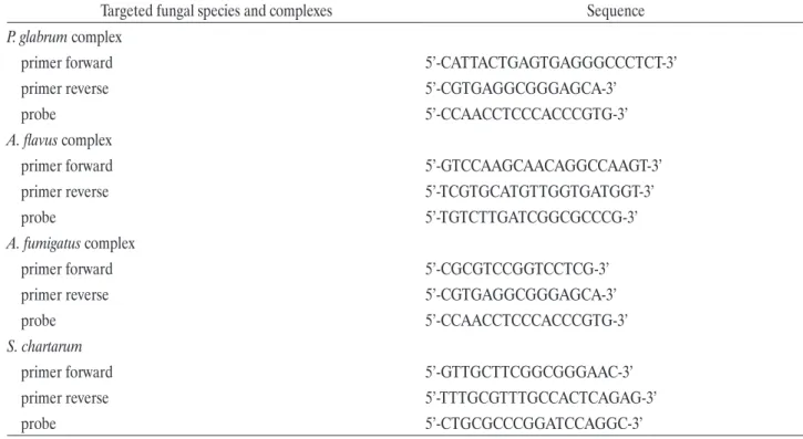
Molecular identification of different species/strains (Ta-ble 1) was achieved by the real time polymerase chain re-action (RT-PCR) using the Rotor-Gene 6000 qPCR Detec-tion System (Corbett, Germany). ReacDetec-tions included 1×iQ Supermix (Bio-Rad), 0.5 μM of each primer (Table 2), and 0.375 μM of TaqMan probe in a total volume of 20 μl. Amplification followed a 3-step PCR: 40 cycles with dena-turation at 95°C for 30 s, annealing at 52°C for 30 s, and extension at 72°C for 30 s. A non-template control was used in every PCR reaction. As positive controls for the spe-cies, DNA samples were obtained from reference strains from the Mycology Laboratory from the National Health Institute of Health Doutor Ricardo Jorge (INSA).
Table 2. Sequence of primers and TaqMan probes used for the real time polymerase chain reaction (PCR)
Targeted fungal species and complexes Sequence
P. glabrum complex
primer forward 5’-CATTACTGAGTGAGGGCCCTCT-3’
primer reverse 5’-CGTGAGGCGGGAGCA-3’
probe 5’-CCAACCTCCCACCCGTG-3’
A. flavus complex
primer forward 5’-GTCCAAGCAACAGGCCAAGT-3’
primer reverse 5’-TCGTGCATGTTGGTGATGGT-3’
probe 5’-TGTCTTGATCGGCGCCCG-3’
A. fumigatus complex
primer forward 5’-CGCGTCCGGTCCTCG-3’
primer reverse 5’-CGTGAGGCGGGAGCA-3’
probe 5’-CCAACCTCCCACCCGTG-3’
S. chartarum
primer forward 5’-GTTGCTTCGGCGGGAAC-3’
primer reverse 5’-TTTGCGTTTGCCACTCAGAG-3’
Statistically significant differences were found in the num-ber of different species between the air and surface sam-ples among the sample sites with HFL (Kruskal-Wallis test, Chi2
kw(5) = 11.403, p = 0.044). The multiple
com-parisons of the Kruskal-Wallis test detected significant differences between pig farms (p = 0.027), poultry farms (p = 0.033) and WWTP (p = 0.012) with cork industries sampling sites, with the first ones showing higher values. The setting with the major difference in the number of species found between the air and surface was the WWTP. In the HFL settings 55 species/genera were found only in the samples of surfaces. In the assessed sampling sites, Table 3. Comparison between fungal assessment performed using conventional and molecular methods
Setting (molecular biology)Fungal species
Conventional methods (species level) Molecular biology
air samples
[n] surface samples [n] air samples [n]
Poultry farms A. flavus complex
(toxigenic strains) 7 4 in 2 samples wasn’t found by 4
conventional methods
A. fumigatus complex 1 1
different sample from air in 7 samples wasn’t found by 8
conventional methods
S. chartarum 0 0 0
Wastewater treatment
plant (WWTP) A. flavus complex (toxigenic strains) 0 0 0
A. fumigatus complex 1 3
different samples from air in 6 samples wasn’t found by 7
conventional methods
S. chartarum 0 0 0
Waste management A. flavus complex
(toxigenic strains) 0 0 0
A. fumigatus complex 10 12
2 different samples from air in 1 sample wasn’t found by 15
conventional methods
S. chartarum 0 0 0
Cork industries A. fumigatus complex 1 3
2 different samples from air 0
P. glabrum complex 2 2
1 different sample from air in 6 samples wasn’t found by 10
conventional methods
poultry farms
pig farms horse stable cork industries wastewater treatment plant waste treatment plant college canteens hospital canteens maternity hospital Sampling sit es [n] 240 48 96 144 192 0 Setting high fungal load low fungal load
Analysis of the surface samples, in addition to the air sam-ples, was an important way to optimize sensitivity of sur-veillance in the areas that are presumed to have low con-tamination [29] such as hospital environment [14,29,30]. Nevertheless, this technique is not broadly applied, mainly in the HFL settings. This fact limits or even prevents data comparison between the settings. It’s important to point out that our main objective of collecting samples, if there was a suspicion of fungal contamination, was to detect, quantify and identify any fungi that might be present [31]. However, in most of the studies where environmental sampling is achieved only through air sampling, data re-garding fungal burden are probably underestimated. The percentage of sampling sites presenting different species in the surfaces than the ones identified in the air (64.2%), corroborates importance of surface analysis to complement mycological air characterization [9,12–15,32]. Fungal bur-den differences both in the air and surfaces could be ex-plained by differences in fungal spores dispersion, which varies according to the fungal characteristics and environ-mental variables [7,9,33,34]. In addition, some species with toxic potential (belonging to A. fumigatus complex, A.
versi-color complex and A. flavus complex) were isolated only in
the surfaces from the HFL settings, reinforcing the need to assess fungal species present on the surfaces. In the present study, we only compared differences between the air and surfaces in terms of the species level; we did not perform a complete molecular assessment since we applied the tools mentioned above only for specific species/strains targeting. Therefore, we believe that with a broader molecular as-sessment concerning 2 different sources (air and surfaces), the differences would be higher.
Although in the HFL settings the surface sampling was not widely applied, a positive association in these occupa-tional settings was found between different fungal species present in the air and on the surfaces. Several constraints may explain such results, and some of them are in line with conventional methods limitations, namely growth the most frequently found genera/species were:
Clad-osporium sp. (10%), Penicillium sp. (8.2%), A. fumigatus
complex (9.1%), A. versicolor complex (7.7%) and A.
fla-vus complex (5.9%).
Regarding the surfaces samples from the LFL scenar-ios, 22 different species/genera were identified only in the samples of surfaces, with Penicillium sp. (13.5%),
Chrysonilia sp. and Chrysosporium sp. (11.5%) being
the most frequently found species.
In the samples that were subjected to both conventional and molecular biology analyses, it was possible to amplify the spe-cies that were not detected in the cultures by quantitative PCR (qPCR) (molecular analysis) sequences. A reverse situ-ation was observed in the cork industry, where A. fumigatus complex was not detected using molecular methods and it was detected by means of cultural methods (Table 3). DISCUSSION
Significant fungal exposure occurs in agricultural and in-dustrial industries and may cause occupational respiratory diseases [23]. Fungi can affect human health in a variety of ways resulting in such health outcomes as: infections, al-lergic reactions (sensitization and immune overreaction), irritations and toxic reactions [24,25].
The lowest observed effect level of 100 000 spores/m3 for
non-pathogenic and non-mycotoxin producing fungal spe-cies has been proposed in a document with fungal criteria contamination levels based on inflammatory respiratory effects [26]. Several organizations have already proposed guidelines for fungi in indoor environments. However, the applied criteria have been suggested based on problems of indoor fungi assessment and they do not take health effects into consideration [27,28]. Additionally, none of the proposed guidelines mentioned surface fungal assess-ment. As we observed in some of the analyzed settings in this study, surfaces showed higher diversity in terms of the number of fungal species detected, as well as a higher fungal load, when compared with the air samples.
since both methodologies were not able to identify/detect species/strains in one setting (A. flavus complex identi-fication in surfaces samples from some poultry farms in comparison with air samples and A. fumigatus complex detection in cork industry in comparison with data from conventional methods). It is also important to point out that in the LFL it was possible to identify different species in the surface samples from the ones found in the air sam-ples (in 23.6% of the sampling sites), corroborating impor-tance of application of this resource due to the patients susceptibility (maternity hospital and hospital canteens as-sessed) and also due to final products safety criteria (food products from hospitals and college canteens assessed). CONCLUSIONS
In this study, fungal contamination from several settings was assessed based on the air and surface sampling, prov-ing the relevance of analysis of the latter samples in com-plementing the results obtained by air sampling. Addition-ally, we observed that samples of surfaces and molecular tools showed the same efficacy in the HFL settings, cor-roborating the fact that application of surfaces sampling is crucial not only in hospital wards but also in other oc-cupational scenarios with similar fungal loads.
ACKNOWLEDGMENTS
The authors are grateful to the Environment and Health Re-search Group from Lisbon School of Health Technology.
REFERENCES
1. Méheust D, le Cann P, Reboux G, Millon L, Gangneux J. In-door fungal contamination: Health risks and measurements methods in hospitals, homes and workplaces. Crit Rev Mi-crobiol. 2014;40:248–60, http://dx.doi.org/10.3109/1040841X. 2013.777687.
2. De Hoog G. Risk assessment of fungi reported from hu-mans and animals. Mycoses. 1996;39:407–17, http://dx.doi. org/10.1111/j.1439-0507.1996.tb00089.x.
inhibition in the air samples caused by other species be-cause of different growth rates [35,36]. We also observed that pig farms, poultry farms and WWTP presented signifi-cant differences in terms of the number of species isolated in the air and in the surface samples, in comparison with cork industries. Such a situation may have occurred due to the overgrowth of Crysonilia sitophilia [37] in both the air and surface samples of this latter setting, simultaneously hindering other species that we were not able to identify. High fungal load settings are occupational contexts with high dust contamination [18,19,38,39] and this environ-mental parameter can act as a fungi carrier and disseminat-ing agent [40]. Previous research work has presented data that relates high dust contamination to high fungi aeros-solization [41,42], corroborating this association. Regard-ing the WWTP settRegard-ing, differences regardRegard-ing the number of species isolated in the air and in the surface samples were the most notorious, probably due to the high levels of moisture that may reduce dust aerossolization and, conse-quently, also decrease the number of fungal species found in the air [43,44].
Molecular techniques have been often used to measure single species in the air using qPCR techniques [26]. Many studies have shown that concentrations of some species were 10–104 times higher when quantified using molecular
methods than when assessed by the use of culture meth-ods [45–47]. However, currently, there are no criteria for evaluation of occupational exposure measurements per-formed with those molecular methods [26]. This has been considered as an obstacle to achieve fungal assessment by the use of these tools. Due to this constraint, simple de-tection of a specific species/strain using molecular tools is essential to evaluate potential health effects and the risk to the workers posed by the discussed exposure, and it is crucial to overcome conventional methods limitations al-ready addressed in several publications [36,48].
We observed that the surface samples and molecular tools showed the same efficacy of detection in the HFL settings,
11. Faure O, Fricker-Hidalgo H, Lebeau B. Eight-year sur-veillance of environmental fungal contamination in hos-pital operating rooms and hematological units. J Hosp Infect. 2002;50(2):155–60, http://dx.doi.org/10.1053/jhin. 2001.1148.
12. Ramos C, Viegas C, Cabo Verde S, Wolterbeek HT, Al-meida SM. Characterizing the fungal and bacterial mi-croflora and concentrations in fitness centers. Indoor Built Environ. 2015 May 31:1–11, http://dx.doi.org/10. 1177/1420326X15587954.
13. Lu Z, Lu WZ, Zhang JL. Microorganisms and particles in AHU systems: Measurement and analysis. Build Envi-ron. 2009;44:694–8, http://dx.doi.org/10.1016/j.buildenv. 2008.05.014.
14. Brenier-Pinchart MP, Lebeau B, Mallaret MR. Mobile air-decontamination unit and filamentous fungal load in the hematology ward: How efficient at the low-activity mode? Am J Infect Control. 2009;37:680–2, http://dx.doi. org/10.1016/j.ajic.2008.12.006.
15. Cabo Verde S, Almeida SM, Matos J, Guerreiro D, Men-eses M, Faria T, et al. Microbiological assessment of in-door air quality at different hospital sites. Res Micro-biol. 2015;166(7):557–63, http://dx.doi.org/10.1016/j.resmic. 2015.03.004.
16. Sabino R, Faísca VM, Carolino E, Veríssimo C, Vie-gas C. Occupational exposure to Aspergillus by swine and poultry farm workers in Portugal. J Toxicol Environ Health A. 2012;75:1381–91, http://dx.doi.org/10.1080/15287 394.2012.721170.
17. Pinheiro C, Viegas C, Viegas S, Veríssimo C, Brandão J, Macedo M. Indoor air quality in Portuguese archives: A snapshot on exposure levels. J Toxicol Environ Health A. 2012;75:1359–70, http://dx.doi.org/10.1080/15287 394.2012.721168.
18. Viegas C, Carolino E, Sabino R, Viegas S, Veríssimo C. Fun-gal contamination in swine: A potential occupational health threat. J Toxicol Environ Health A. 2013;76(4–5):272–80, http://dx.doi.org/10.1080/15287394.2013.757205.
3. Directive 2000/54/EC of the European Parliament and of the Council of 18 September 2000 on the protection of workers from risks related to exposure to biological agents at work (7th individual Directive within the meaning of Ar-ticle 16(1) of Directive 89/391/EEC). Off J Eur Union L 262, p. 21–45 (Sep 18, 2000).
4. Gutarowska B, Skóra J, Stepien L, Twarużek M, Błajet-Kosicka A, Otlewska A, et al. Estimation of fungal con-tamination and mycotoxin production at workplaces in composting plants, tanneries, archives and libraries. World Mycotoxin J. 2014;7(3):345–55, http://dx.doi.org/10.3920/ WMJ2013.1640.
5. Buttner MP, Willeke K, Grinshpun SA. Sampling and analysis of airborne microorganisms. In: Hurst CJ, Knud-sen GR, McInerney MJ, Stetzenbach LD, Walter MV, editors. Manual of environmental microbiology. Washing-ton: ASM Press; 1997. p. 629–40.
6. Flannigan B, Miller JD. Health implications of fungi in indoor environments: An overview. In: Samson RA, Flan-nigan B, FlanFlan-nigan ME, editors. Health implications of fungi in indoor environments. Amsterdam: Elsevier Sci-ence Ltd.; 1994. p. 1–28.
7. Roussel S, Reboux G, Bellanger AP, Sornin S, Grenouillet F, Dalphin JC, et al. Characteristics of dwellings contaminated by mould. J Environ Monit. 2008;10(6):724–9, http://dx.doi. org/10.1039/b718909e.
8. Srikanth P, Sudharsanam S, Steinberg R. Bio-aerosols in in-door environment: Composition, health effects and analy-sis. Indian J Med Microbiol. 2008;26:302–12, http://dx.doi. org/10.4103/0255-0857.43555.
9. Stetzenbach L, Buttner M, Cruz P. Detection and enu-meration of airborne biocontaminants. Curr Opin Bio-technol. 2004;15:170–4, http://dx.doi.org/10.1016/j.copbio. 2004.04.009.
10. Klánová K, Hollerová J. Hospital indoor environment: Screening for micro-organisms and particulate matter. In-door Built Environ. 2003;12:61–7, http://dx.doi.org/10.1177/ 1420326X03012001010.
hospitals]. J Mycol Med. 2006;16:204–11, http://dx.doi. org/10.1016/j.mycmed.2006.10.002. French.
30. Sabino R, Veríssimo C, Parada H, Brandão J, Viegas C, Carolino E, et al. Molecular screening of 246 Portuguese Aspergillus isolates among different clinical and environ-mental sources. Med Mycol. 2014;52:517–27, http://dx.doi. org/10.1093/mmy/myu006.
31. Portnoy JM, Barnes CS, Kennedy K. Sampling for indoor fungi. J Allergy Clin Immunol. 2004;113:189–98, http:// dx.doi.org/10.1016/j.jaci.2003.11.021.
32. Viegas C, Alves C, Carolino E, Pinheiro C, Rosado L, Silva-Santos C. Assessment of fungal contamination in a group of Lisbon’s gymnasiums with a swimming pool. Ital J Occup Environ Hyg. 2011;2(1):15–20.
33. Kemp PC, Neumeister-Kemp HG, Esposito B, Lysek G, Murray F. Changes in airborne fungi from the outdoors to indoor air; large HVAC systems in nonproblem buildings in 2 different climates. Am Ind Hyg Assoc J. 2003;64: 269–75, http://dx.doi.org/10.1080/15428110308984817. 34. Górny RL. Filamentous microorganisms and their
frag-ments in indoor air – A review. Ann Agric Environ Med. 2004;11:185–97.
35. Zorman T, Jersek B. Assessment of bioaerosol con-centrations in different indoor environments. In-door Built Environ. 2008;17(2):155–63, http://dx.doi. org/10.1177/1420326X08089251.
36. Viegas C, Malta-Vacas J, Sabino R, Viegas S, Veríssimo C. Accessing indoor fungal contamination using conventional and molecular methods in Portuguese poultries. Environ Monit Assess. 2014;186(3):1951–9, http://dx.doi.org/10.1007/ s10661-013-3509-4.
37. Francuz B, Year H, Geraut L, Bensefa-Colas L, Nghiem ZH, Choudat D. Occupational asthma induced by Chrysonilia sit-ophila in a worker exposed to coffee grounds. Clin Vaccine Immunol. 2010;17(10):1645–6, http://dx.doi.org/10.1128/ CVI.00134-10.
38. Viegas C, Carolino E, Malta-Vacas J, Sabino R, Viegas S, Veríssimo C. Fungal contamination of poultry litter: A public 19. Viegas C, Quintal Gomes A, Abegão J, Sabino R, Graça T,
Viegas S. Assessment of fungal contamination in waste sorting and incineration – Case study in Portugal. J Toxicol Environ Health A. 2014:77(1–3):57–68, http://dx.doi.org/10. 1080/15287394.2014.865583.
20. European Agency for Safety and Health at Work. EU-OSHA Annual report 2007: Bringing safety and health closer to European workers. Luxembourg: Office for Official Publications of the European Communities; 2007.
21. ISO 18593:2004(E). Microbiology of food and animal feed-ing stuffs – Horizontal methods for samplfeed-ing techniques from surfaces using contact plates and swabs. Geneva: Inter-national Organization for Standardization; 2004.
22. De Hoog C, Guarro J, Gené G, Figueras M. Atlas of clinical fungi. 2nd ed. Utrecht: Centraalbureau voor Schimmelcul-tures; 2000.
23. Reboux G, Roussel S, Grenouillet F. [Fungi in agricultural environment]. J Mycol Med. 2006;16:248–62, http://dx.doi. org/10.1016/j.mycmed.2006.09.003. French.
24. Fischer G, Dott W. Relevance of airborne fungi and their secondary metabolites for environmental, occupational and indoor hygiene. Arch Microbiol. 2003;179:75–82.
25. McGinnis MR. Pathogenesis of indoor fungal diseases. Med Mycol. 2004;42:107–17, http://dx.doi.org/10.1080/136937804 10001661473.
26. Eduard W. The Nordic Expert Group for Criteria Documen-tation of Health Risks from Chemicals. 139. Fungal spores. Stockholm: National Institute for Working Life; 2006 [cit-ed 2014 Dec 15]. Available at: http://gupea.ub.gu.se/dspace/ bitstream/2077/4359/1/ah2006_21.pdf.
27. Rao CY, Burge HA, Chang JC. Review of quantitative stan-dards and guidelines for fungi in indoor air. J Air Waste Manage Assoc. 1996;46:899–908, http://dx.doi.org/10.1080/ 10473289.1996.10467526.
28. World Health Organization guidelines for indoor air qual-ity: Dampness and mould. Geneva: The Organization; 2009. 29. Gangneux JP, Bousseau A, Cornillet A, Kauffmann-La-croix C. [Control of fungal environmental risk in French
distribution on aerosol light extinction in the urban area of Guangzhou. Atmos Chem Phys. 2013;13:1115–28, http:// dx.doi.org/10.5194/acp-13-1115-2013.
45. Meklin T, Haugland RA, Reponen T. Quantitative PCR anal-ysis of house dust can reveal abnormal mold conditions. J Environ Monit. 2004;6:615–20, http://dx.doi.org/10.1039/ b400250d.
46. Yamamoto N, Kimura M, Matsuki H, Yanagisawa Y. Optimi-zation of a real-time PCR assay to quantitate airborne fungi collected on a gelatin filter. J Biosci Bioeng. 2010;109:83–8, http://dx.doi.org/10.1016/j.jbiosc.2009.06.015.
47. Zeng QY, Westermark SO, Rasmuson-Lestander A, Wang XR. Detection and quantification of Wallemia sebi in aerosols by real-time PCR, conventional PCR, and cul-tivation. Appl Environ Microbiol. 2004;70:7295–302, http:// dx.doi.org/10.1128/AEM.70.12.7295-7302.2004.
48. Viegas C, Malta-Vacas J, Sabino R. Molecular biology versus conventional methods – Complementary methodologies to understand occupational exposure to fungi. Proceeding of the 8th International Symposium on Occupational Safety and Hygiene; 2012 Feb 9–10; Guimarães, Portugal. Gui-marães: Minho University; 2012. p. 478–9.
health problem. J Toxicol Environ Health A. 2012;75(22– 23):1341–50, http://dx.doi.org/10.1080/15287394.2012.721165. 39. Viegas C, Almeida-Silva M, Gomes AQ, Wolterbeek HT, Al-meida SM. Fungal contamination assessment in Portuguese elderly care centers. J Toxicol Environ Health A. 2014;77 (1–3):14–23, http://dx.doi.org/10.1080/15287394.2014.861336. 40. Raulf M, Buters J, Chapman M, Cecchi L, de Blay F,
Doekes G, et al. Monitoring of occupational and envi-ronmental aeroallergens – EAACI position paper. Aller-gy. 2014;69(10):1280–99, http://dx.doi.org/10.1111/all.12456. 41. Crook B, Easterbrook A, Stagg S. Exposure to dust and bio-aerosols in poultry farming. Summary of observations and data. London: Health and Safety Executive; 2008.
42. Millner PD. Bioaerosols associated with animal production operations. Bioresource Technol. 2009;100:5379–85, http:// dx.doi.org/10.1016/j.biortech.2009.03.026.
43. Zhu K, Tan R, Ng WK, Shen S, Zhou Q, Heng P. Analysis of the influence of relative humidity on the moisture sorp-tion of particles and the aerosolizasorp-tion process in a dry powder inhaler. J Aerosol Sci. 2008;39:510–24, http://dx.doi. org/10.1016/j.jaerosci.2008.02.003.
44. Lin ZJ, Tao J, Chai FH, Fan SJ, Yue JH, Zhu LH, et al. Impact of relative humidity and particles number size
This work is available in Open Access model and licensed under a Creative Commons Attribution-NonCommercial 3.0 Poland License – http://creativecommons.org/ licenses/by-nc/3.0/pl/deed.en.