UNIVERSIDADE TÉCNICA DE LISBOA
Faculdade de Medicina Veterinária
ARRYTHMOGENIC RIGHT VENTRICULAR CARDIOMYOPATHY IN BOXER DOGS – RETROSPECTIVE STUDY (6 CASES)
DOROTEIA ISABEL VIEGAS FILIPE BOTA
DISSERTAÇÃO DE MESTRADO EM MEDICINA VETERINÁRIA
CONSTITUIÇÃO DO JÚRI ORIENTADOR
Dr José Paulo Pacheco Sales Luis Dr Babis Koffas
Drª Maria de São José Sousa Deyrieux Centeno CO – ORIENTADOR Profª Drª Ana Cristina Gaspar Nunes Lobo Vilela Profª Drª Ana Cristina
Gaspar Nunes Lobo Vilela
2009 LISBOA
Doroteia Bota Dissertação de Mestrado em Medicina Veterinária, FMV-UTL
i
Para ti, Didinha, que partiste tão cedo… Foste sem dúvida uma das “culpadas” pelas minhas conquistas e estar-te-ei sempre grata por isso. Fizeste-me sonhar, querer conhecer o mundo, gostar de livros e de História! Trago-te sempre comigo e sinto tanto, tanto a tua falta… À minha tia Ilda Viegas, uma das minhas quatro mães…
Doroteia Bota Dissertação de Mestrado em Medicina Veterinária, FMV-UTL
ii
“Onde há vontade, não falta caminho” (J.R.R. Tolkien)
À Professora Cristina Vilela, minha cúmplice desde o 3º ano de Faculdade. Sempre presente nas minhas aventuras fora de Portugal e meu pólo Norte em Lisboa. Muito obrigada pelos conselhos e ajuda que me tem dado ao longo de 6 anos.
Ao Dr Babis Koffas pela ajuda imensa na realização deste estudo, pelo companheirismo e aprendizagem enquanto sua interna em Inglaterra.
À minha mãe, Isabel, que mesmo longe está sempre presente na minha tomada de decisões, que me ensinou o valor de educar e a partilha de afectos. Que desdobra os meus problemas em simples equações e tanto me tem ajudado a ser quem sou.
Às minhas avós Valentina e Glória minhas segundas mães, os meus tesouros mais antigos e fonte de motivação. Minhas amigas de sempre para sempre.
Ao Carlos Gaspar, minha outra metade há quatro anos, o meu porto de abrigo, o meu melhor amigo, a minha fonte de tranquilidade e apoio permanente. Sem ele este trabalho não teria sido possível.
Doroteia Bota Dissertação de Mestrado em Medicina Veterinária, FMV-UTL
iii ABSTRACT
Arrythmogenic Right Ventricular Cardiomyopathy in Boxer Dogs – Retrospective Study (6 Cases)
Arrhythmogenic right ventricle cardiomyopathy (ARVC) is recognized in Boxers, cats and humans, is characterized clinically by ventricular tachyarrhythmias and histopathologically by fibrofatty replacement, mostly of the right ventricle.
Affected dogs may have syncope, exercise intolerance or heart failure associated signs. ARVC is an inherited adult onset disorder that can lead to sudden death which can also be the first and single clinical sign.
Ventricular premature complexes (VPC’s) with left bundle branch block morphology on the electrocardiogram (ECG) are typical for the disease. These VPCs can occur singly, in pairs, triplets or runs of ventricular tachycardia (VT). Routine ECG can be insensitive for the detection of ventricular arrhythmias when compared with a 24 hour Holter study.
The antiarrhythmic drugs most commonly used are sotalol or mexiletine-atenolol association. A 24 hour Holter study should be repeated in order to evaluate the efficacy of treatment or detect a proarrhythmic effect.
In this study, 4 out of 6 animals (66,6%) died suddenly, however owners refer an improvement in the animal’s quality of life once the treatment is initiated.
The prognosis of this disease is guarded due to sudden death risk or heart failure development.
Doroteia Bota Dissertação de Mestrado em Medicina Veterinária, FMV-UTL
iv RESUMO
Cardiomiopatia Arritmogénica do Ventrículo Direito em Boxers – Estudo Retrospectivo (6 Casos)
A cardiomiopatia arritmogénica do ventrículo direito (CAVD) é uma patologia reconhecida em cães de raça Boxer, gatos e humanos, caracterizada clinicamente por taquiarritmias ventriculares e histopatologicamente por substituição fibro-adiposa, sobretudo do ventrículo direito.
Os cães afectados podem apresentar síncope, intolerância ao exercício ou sinais associados a insuficiência cardíaca. CAVD é uma patologia hereditária do adulto com risco de morte súbita, que pode ser a primeira e única manifestação clínica da doença.
Complexos ventriculares prematuros (CVP) com morfologia de bloqueio de ramo esquerdo são achados típicos da doença no electrocardiograma (ECG). Estes CVP podem ocorrer singularmente, em pares, em grupos de três ou em runs de taquicardia ventricular. Um ECG de rotina pode ser insensível na detecção das arritmias que normalmente são mais evidentes num estudo Holter de 24 horas.
Os antiarritmicos mais frequentemente utilizados são o sotalol ou a associação mexiletina-atenolol. Para se avaliar a eficácia do tratamento e detectar um eventual efeito pro-arrítmico, um segundo Holter deve ser realizado.
Neste estudo, 4 dos 6 animais (66,6%) morreram subitamente, porém, segundo os proprietários a qualidade de vida do animal melhorou com o tratamento.
O prognóstico desta doença é reservado devido ao risco de morte súbita ou desenvolvimento de insuficiência cardíaca.
Doroteia Bota Dissertação de Mestrado em Medicina Veterinária, FMV-UTL vi ÍNDEXS GENERAL INDEX Page Dedicatória i Agradecimentos ii Abstract iii Resumo iv Indexs vi
Breve Descrição das Actividades Desenvolvidas no Estágio Curricular x
Introduction ... 1
PART ONE – Bibliographic Review: Arrythmogenic Right Ventricular Cardiomyopathy in Boxer Dogs Etiology ... 3 Clinical Presentation ... 3 Diagnosis ... 4 1 - Physical Examination ... 5 2 - Electrocardiography ... 5 3 - Holter Monitoring ... 7 4 - Echocardiography ... 10 5 - Biochemical Markers... 10
6 - Magnetic Ressonance Imaging ... 11
Pathology ... 12
Genetics ... 14
Treatment ... 15
Assymptomatic Boxers ... 17
ARVC vs Neurocardiogenic Bradicardia ... 18
Prognosis ... 19
Ventricular Dilatation and Systolic Dysfunction ... 19
PART TWO – Clinical Cases Case 1: Alice Joyner ... 21
Case 2: Oscar Eels ... 23
Case 3: Ralphie Nichols ... 25
Case 4: Sonny Thomas ... 27
Case 5: George Phillips ... 28
Case 6: Oscar Adsett ... 29
Conclusion ... 32
Doroteia Bota Dissertação de Mestrado em Medicina Veterinária, FMV-UTL
vii FIGURE INDEX
Figure 1 - Ventricular premature complex ... 3
Figure 2 - Left electrodes ... 8
Figure 3 - Connection to the device ... 8
Figure 4 - Right electrode... 8
Figure 5 - Holter ready ... 8
Figure 6 - ECG of Alice Joyner... 21
Figure 7 - Echocardiography of Alice Joyner ... 22
Figure 8 - ECG of Oscar Eels ... 24
Figure 9 - Echocardiography of Oscar Eels ... 24
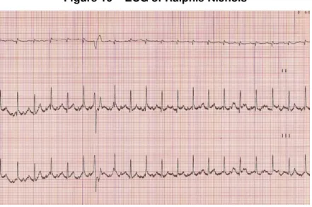
Figure 10 - ECG of Ralphie Nichols ... 25
Figure 11 - ECG of Ralphie Nichols ... 26
Figure 12 - Echocardiography of Ralphie Nichols ... 26
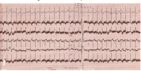
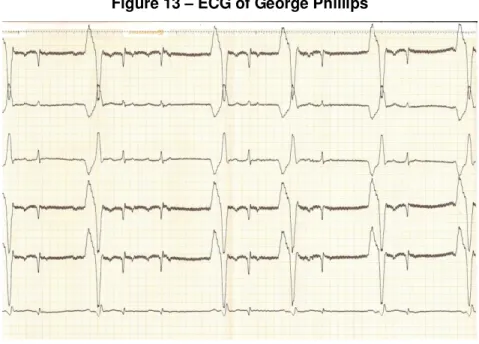
Figure 13 - ECG of George Philips ... 28
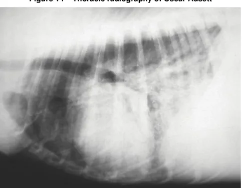
Figure 14 - Thoracic radiography of Oscar Adsett ... 29
Figure 15 - Echocardiography of Oscar Adsett ... 30
Figure 16 - Ascitic fluid of Oscar Adsett ... 30
TABLE INDEX Table 1 - Gross and Histopathological Characteristics of Boxer Dogs with ARVC ... 13
ANEX INDEX Anex I - CBC and Serum Biochemistry for Alice Joyner ... 37
Anex II - Holter of Alice Joyner ... 38
Anex III - CBC and Serum Biochemistry for Oscar Eels ... 40
Anex IV - CBC and Serum Biochemistry for Ralphie Nichols ... 41
Anex V - Holter of Ralphie Nichols ... 42
Anex VI - Holter of Sonny Thomas ... 46
Anex VII - CBC and Serum Biochemistry for George Philips ... 49
Doroteia Bota Dissertação de Mestrado em Medicina Veterinária, FMV-UTL
viii ABREVIATION INDEX
ALA: Alphalinoleic Acid ALT: Alaline Transaminase
ARVC: Arrythmogenic Right Ventricular Cardiomyopathy BB: Beta Blocker
BID: Bis In Die (twice daily) BNP: Brain Natriuretic Peptide BPM: Beats Per Minute CBC: Complete Blood Count CHF: Congestive Heart Failure CM: Cardiomyopathy
CTnI: Cardiac Troponin I DCM: Dilated Cardiomyopathy DHA: Docosahexaenoic Acid ECG: Electrocardiography EPA: Eicosapentaenoic acid ET: Ejection Time
FS: Fractional Shortening HR: Heart Rate
HRV: Heart Rate Variability
ICD: Implantable Cardioverter Defibrillator IMP: Index of Myocardial Performance LA: Left Atrium
LBBB: Left Bundle Branch Block LV: Left Ventricle
MRI: Magnetic Ressonance Imaging PO: Per Os
RA: Right Atrium
RBBB: Right Bundle Branch Block RV: Right Ventricle
RyR2: Cardiac Ryanodine Receptor
SAECG: Signal Average Electrocardiography SVT: Supra Ventricular Tachycardia
TID: Ter In Die (three times daily) VT: Ventricular Tachycardia
VPCs: Ventricular Premature Complexes VRun: Ventricular Run
Doroteia Bota Dissertação de Mestrado em Medicina Veterinária, FMV-UTL
x
Breve Descrição das Actividades Desenvolvidas no Estágio Curricular
O meu percurso desde 2007…
Quando me licenciei em Medicina Veterinária em 2007 cedo percebi que a minha formação tinha de continuar. Assim, decidi candidatar-me a um internato em França, no Centre Hospitalier Vétérinaire Frégis. Posso afirmar com certeza que cresci bastante a nível pessoal e profissional; fui responsável pelo serviço de urgências das 19h às 8h do dia seguinte durante 1 semana cada 8 semanas, fiz parte de um programa rotacional entre Medicina Interna, Cirurgia, Oftalmologia, Consultas e Exóticos. Aprendi que ser Interno é trabalhar muito e reclamar pouco e assim foi durante um ano.
Éramos 8 internos no total, 5 franceses, 1 espanhola, 1 luxemburguesa e eu. Todos me ajudaram imenso com o francês que eu não dominava de todo e como dizia o meu amigo José Luís “ a única coisa que sabes dizer é abat-jour! ” Assim passaram-se 12 meses de trabalho proporcional à aprendizagem.
Findo o internato, em Setembro de 2008, ingressei num segundo internato desta vez em Inglaterra. Muitas foram as razões para fazer um segundo internato mas a que mais pesou foi acreditar que a porta para a especialização poderia abrir-se mais facilmente neste país. O meu sonho é tornar-me numa especialista europeia em Medicina Interna e é para isso que luto desde 2007.
Aqui em Inglaterra a maneira de abordar a Medicina Veterinária é bastante diferente da Francesa e sobretudo da Portuguesa. Trabalho em North Kent Referrals (NKR), uma clínica de referência o que significa que só recebe casos referenciados dos “general practice”. Também aqui faço parte de um serviço rotacional entre Medicina Interna, Cirurgia, Oftalmologia, Imagiologia e Emergências. Contudo, o dia começa só as 9h, um pouco diferente de França que começava às 7h30 depois de já ter feito 1h de transportes.
Nós, internos, somos responsáveis pelos animais internados no nosso serviço, fazemos os exames complementares de diagnóstico, preparamos e ajudamos em cirurgia, recebemos as emergências fora de horas e somos responsáveis pela estabilização do animal durante a noite. Também aqui somos uma equipa muito internacional, com representantes da Austrália, Espanha, Grécia, França, África do Sul e claro, do Reino Unido.
Aqui as companhias de seguros têm uma presença muito forte e os preços praticados são bastante diferentes dos que eu estava habituada. O chá e as bolachas estão sempre disponíveis na sala de espera e esta nunca é muita porque “assim tem de ser num serviço de referência”. Uma primeira consulta pode demorar 1 hora e o contacto com o cliente faz-se pelo menos 2 vezes ao dia. A equipa de enfermagem está divida pelos faz-serviços e cada animal tem uma enfermeira.
Doroteia Bota Dissertação de Mestrado em Medicina Veterinária, FMV-UTL
xi
Com os especialistas fazemos um journal club e book club todas as semanas o que nos obriga a estar a par das mais recentes publicações. Uma apresentação mensal sobre 1 caso clínico também faz parte das nossas obrigações.
Este estudo em Boxers foi feito na clínica onde actualmente trabalho sob a supervisão do Dr Babis Koffas diplomado pelo colégio Europeu de Cardiologia.
A casuística que acompanhei ao longo destes 2 anos nas duas clínicas tem sido imensa. Estive presente em centenas de casos nas diferentes áreas. O trabalho tem sido muito e o tempo pouco e talvez seja essa a razão da minha demora na entrega deste trabalho.
Todas estas experiências têm sido muito intensas e gratificantes. Ao longo de 2 anos percebi o quão vasto é o mundo da Medicina Veterinária e acredito que tal como na Medicina Humana, deveríamos apostar na especialização. As exigências por parte dos donos são cada vez maiores e a qualidade dos serviços prestados têm de acompanhar esta evolução.
Tenho hoje, mais do que nunca, a certeza que escolhi a profissão correcta e que mais prazer me dá exercer. Considero fascinante tudo que já se faz pelos animais e assim espero que possa acontecer cada vez mais e melhor também no meu país.
Por isso mesmo, eu aposto na minha formação, que por enquanto tem de ser feita no exterior, para um dia mais tarde poder regressar a Portugal e aplicar o que tenho aprendido. Para já, não posso dizer que não estou a gostar de viver nos diferentes países em que já vivi e aprender a Medicina Veterinária nas diferentes línguas mas tão só com um objectivo: proporcionar os melhores cuidados de saúde aos nossos (verdadeiros) melhores amigos.
Doroteia Bota Dissertação de Mestrado em Medicina Veterinária, FMV-UTL
1 INTRODUCTION
In the early 1980s Dr Neil Harpster was the first to describe myocardial disease in the Boxer dog as the result of examination of 64 Boxers over a 15-year period with varying presentations of the condition (Harpster, 1983). He described it as being quite different from other large and giant breed cardiomyopathies as had been characterized in the Doberman and Great Dane in that the heart of the affected Boxers showed an absence of similar degrees of ventricular dilatation and the dogs rarely suffered from atrial fibrillation. Because the dogs that Harpster studied were closely related, he proposed an inherited origin for the condition. In the original group of 64 dogs studied a slight male predisposition (57.8%) was identified and the average age at the time of diagnosis was 8.2 years. In a 1991 report, which added another 48 dogs to the original study, the average age at diagnosis dropped to 6.9 years (Harpster, 1991).
Dr. Harpster divided the 64 dogs into 3 categories based on the clinical features of their disease. The first category included dogs that were asymptomatic. The second group had occasional episodes of fainting or weakness usually after a stressful event but were otherwise completely normal. The third group included dogs with signs of heart failure. The most common finding on the physical exams of all these dogs was the presence of a cardiac arrhythmia.
Electrocardiographic findings consistently showed ventricular premature complexes (VPCs) occurring singly, in pairs and in runs or ventricular tachycardia (VT).
Of the 64 dogs in the original study, only 18 were presented for necropsy. Affected hearts had extensive and diffusely distributed changes in the myocardium. The changes included replacement of muscle tissue by fibrous tissue and infiltration of fat.
The previously described Boxer cardiomyopathy (CM) has similarities to a human myocardial disease called Arrythmogenic Right Ventricular Cardiomyopathy (ARVC).
The term ARVC was adopted since clinical presentation is featured by VT and arrhythmic sudden death (“arrhythmogenic”). Also the morbid identity is a primary heart muscle disease of still unknown origin (“cardiomyopathy”) with a peculiar involvement of the “right ventricular” myocardium (Thiene & Basso, 2001).
Similarities with human ARVC in clinical presentation, pathologic findings and presumed etiologic basis supported reclassification of the disease as Boxer ARVC (Meurs, 2004). While Dr. Harpster saw equal numbers of each of the three categories of Boxer CM, we now seem to see more Boxers in the first 2 categories and very few with signs of congestive heart failure (CHF). Dr. Kathryn Meurs at The Ohio State University, currently the most active researcher in regard to ARVC, has placed the number of ARVC dogs with CHF at probably less than 10% (Meurs, 2004).
PART ONE
Bibliographic Review
Doroteia Bota Dissertação de Mestrado em Medicina Veterinária, FMV-UTL
3
ARRYTHMOGENIC RIGHT VENTRICULAR CARDIOMYOPATHY IN BOXER DOGS
ETIOLOGY
ARVC is a myocardial disease of unresolved pathogenesis characterized by progressive replacement of the right ventricle (RV) and, less commonly the left ventricle (LV), by adipose and fibrous tissue. The disease has been recognised mostly in dogs but can also affect cats (Fox, Maron, Basso, Liu & Thiene, 2000; Harvey, Battersby, Faena, Fews, Darke & Ferasin et al, 2005). In humans is a well-recognise cause of sudden death, especially in athletes during physical activity (Fauci et al., 2008)
In humans, ARVC is commonly inherited as an autosomal dominant manner and multiple mutations of several genes are responsible for causing the disease. It has been suggested that this mutations occur in genes that encode proteins that constitute desmosomes, which will cause detachment of myocytes followed by fibrofatty replacement (Fauci et al., 2008). Meurs, Spier, Willer, Lehmkuhj and Towbin (1999) and Basso et al. (2004) found a familial occurrence of the disease in Boxer dogs. The affected offspring may be quite variable which suggest that incomplete penetrance of the disease may be involved.
ARVC is an adult onset disease both in humans (with its peak between 16-35 years) and dogs.
Based on echocardiography most affected dogs present no changes in myocardial systolic function or chamber dimensions. These findings support a presence of an electrical disorder rather than a contractile one (Meurs, 2004).
CLINICAL PRESENTATION
Boxer ARVC is an adult-onset myocardial disease and, as originally proposed by Harpster, there seems to be 3 forms of the disease now referred as concealed, overt and myocardial dysfunction (Meurs, 2004).
The first form of the disease is characterized by an asymptomatic dog with variable number of VPCs (Figure 1).
Doroteia Bota Dissertação de Mestrado em Medicina Veterinária, FMV-UTL
4
The second form is characterized by the presence of tachyarrhythmia, syncope and/or exercise intolerance. Syncope is defined as a brief loss of consciousness due to inadequate cerebral blood flow and this is usually followed by spontaneous recovery. According to Thomason, Kraus, Surdyk, Fallaw and Calvert (2008), syncope in Boxer dogs is often due to VT associated with ARVC or due to neurocardiogenic bradycardia (vasovagal syndrome). VT in Boxers with ARVC can degenerate to ventricular fibrillation (V-fib) that is not compatible with life. This V-fib is the cause of sudden death in most Boxers with ARVC. In one study with 23 Boxer dogs, 39% of the dogs died suddenly and unexpectedly, during vigorous exercise (n=3), leisurely walking (n=4), or while sleeping (n=2). Syncope occurred in 6 of the 9 dogs. Of these 9 dogs that died suddenly, 1 had sustained VT, 2 had nonsustained VT with VPCs and 3 had VPCs alone (Basso et al., 2004).
The third form of ARVC in dogs is characterized by the development of myocardial systolic dysfunction, sometimes with evidence of CHF. CHF results from elevated cardiac filling pressures that cause pulmonary or systemic venous hypertension and extravasation of fluid into the interstitial space or body cavity.
Although it is likely that these 3 forms of ARVC represent a continuum of the disease this has not been well documented. Most Boxers affected with ARVC will ultimately die of their arrhythmia, not of CHF.
Some affected dogs have an abnormal degree of ectopy (VPCs) but never develop clinical signs, whereas some affected dogs with the same degree of ectopy gradually progress and develop more severe arrhythmias as they mature. Some Boxers have thousands of VPCs over 24 hours and a high grade of complexity and remain asymptomatic.
The factors that determine which dogs eventually progress to the clinical form of the disease are not known.
It has been estimated that ARVC accounts for as many as 5% of sudden cardiac deaths among young adult humans in the United States and has a prevalence of 1 in 5000 persons (Meurs, Ederer & Stern, 2007).
DIAGNOSIS
The diagnosis of canine ARVC can be challenging. Unfortunately, a single diagnostic test for ARVC does not exist. The diagnosis is based on the presence of a combination of factors: familial history, syncope or exercise intolerance, arrhythmia on auscultation and presence of upright VPCs on the ECG trace (lead I, II, III and aVF) and finally post-mortem finding of fibrofatty infiltration into the myocardium.
Doroteia Bota Dissertação de Mestrado em Medicina Veterinária, FMV-UTL
5 1 - PHYSICAL EXAMINATION
Many affected dogs have a normal physical examination. Heart murmurs are infrequently heard; however, many adult Boxers have a left basilar systolic heart murmur that may be physiologic or, in some cases, may be associated with subvalvular or valvular aortic stenosis. The myocardial dysfunction form of the disease may be responsible for the presence of a left apical systolic murmur, due to mitral insufficiency.
The erratic beats or VPCs may or may not be detected on thoracic auscultation. Whether or not they are detected depends on the frequency of the abnormal rhythm. These VPCs are heard as an extra beat or a skipped beat that do not have a corresponding pulse. In the normal functioning heart, there is a pulse for every beat that is heard.
2 - ELECTROCARDIOGRAPHY
Cardiac arrhythmias are variations of the cardiac rhythm from normal sinus rhythm. Some are benign and clinically insignificant whereas other may cause severe clinical signs such as syncope or degenerate into malignant arrhythmias leading to cardiac arrest and sudden death.
The presence of an upright VPC on a lead I, II, III and aVF (left bundle branch morphology) of the ECG is suggestive of ARVC.
In one study (Baumwart, Orvalho & Meurs, 2005) some dogs with ARVC had VPCs with morphology consistent with right bundle branch block (RBBB) while others had VPCs with morphology consistent with both RBBB and left bundle branch block (LBBB). In another study with 23 Boxer dogs, VPCs with LBBB morphology were documented by 24-hour Holter ECG in 19 animals. VPCs of RBBB morphology were also detected in 4 of these 19 dogs. It is possible that dogs with VPCs with RBBB morphology had a ventricular septal site of origin for the ventricular ectopy. It has been suggested that portions of the ventricular septum may produce VPCs similar in morphology to those originating in the LV (Kraus, Moïse, Rishniw, Dykes & Erb, 2002).
Supraventricular tachyarrhythmias also have been reported in Boxers with myocardial disease. In the original description of Boxer CM made by Harpster (1983) approximately 13% of the affected dogs had atrial fibrillation or supraventricular tachycardia (SVT). In a more recent study, dogs from category III ARVC had a high prevalence of arrhythmias of supraventricular and ventricular origin as well as a high prevalence of CHF (Baumwart et al., 2005).
Some affected dogs have a different morphology to their VPCs or may not have any VPCs detected on an ECG. However, “a normal electrocardiogram does not exclude a diagnosis of ARVC because of the intermittent nature of the arrhythmia” (Meurs, 2004).
Doroteia Bota Dissertação de Mestrado em Medicina Veterinária, FMV-UTL
6
The electrocardiographic findings in affected dogs and humans with ARVC are variable. Studies in humans affected by ARVC showed that individuals with more than 3000 VPCs in 24 hours have a 29% chance of having a normal random ECG, those with 1000-3000 VPCs in 24 hours have a 50% chance of having a normal ECG and those with less than 300 VPCs have almost 100% chance of having a normal ECG (Waller, 2006).
The mechanism of the arrhythmias in Boxers is unknown. There are 3 mechanisms postulated for the genesis of ventricular tachyarrhythmias, including enhanced automaticity, triggered activity and re-entry. In humans, this last type is believed to be the predominant mechanism of ventricular tachyarrhythmias resulting in sudden death (Fauci et al., 2008). Reentrant circuits are created when adjacent myocardial tissues have various electrophysiologic properties, allowing for the development of abnormal impulse conduction. This may be caused by structural abnormalities or functional changes. Anatomic abnormalities, including hypertrophy, fibrosis and ischemia create areas of different excitability and electrophysiologic properties resulting from abnormal tissue. Functional reentry occurs in the absence of any identifiable anatomic substrate, presumably as the result of inherent electrophysiologic differences in membrane and ionic properties of excitable tissue.
The QT interval, measured from the surface ECG, is an electrocardiographic variable of ventricular repolarization. The duration of the QT interval is not identical in all leads and this interlead difference between the maximum and minimum QT duration is known as QT dispersion. It is hypothesized that the degree of variation in QT dispersion reflects regional differences of ventricular repolarization: increased QT dispersion reflects increased inhomogeneity of ventricular repolarization (Fauci et al., 2008). In humans, QT dispersion has been used with some success for identifying patients at risk for developing ventricular tachyarrhythmias associated with a number of cardiovascular diseases, including CHF, hypertrophic CM, myocardial ischemia, aortic stenosis and mitral valve prolapse.
Spier, Meurs, Muir, Lehmkuhl and Hamlin, (2001) hypothesized that QT dispersion may be an indicator of reentry and serve as a noninvasive mean to identify electrical dispersion in Boxers with ARVC. This could provide evidence for an arrhythmogenic substrate and could be useful in identifying dogs at risk for arrhythmia development and sudden cardiac death. However, this study failed to identify a relation between QT dispersion with total number of VPCs, arrhythmia grade or fractional shortening (FS = difference between diastolic and systolic LV dimensions). Most of dogs had QT dispersion equal to 0 showing that substantial dispersion of repolarization was not observed.
Signal-average ECG (SAECG) has showed to be a useful tool for evaluation of Boxers with ARVC and at risk for cardiac-related death. SAECG is a non invasive electrocardiographic technique that enables detection of abnormal conduction by identifying late potentials in the QRS complex, which are otherwise hidden in baseline noise during standard ECG. These
Doroteia Bota Dissertação de Mestrado em Medicina Veterinária, FMV-UTL
7
late potentials can be identified as high-frequency, low voltage potentials that occur in the terminal portions of the QRS complex. Late potentials are believed to represent areas of abnormal conduction that may contribute to re-entry. So, late potentials may serve as a pathophysiologic marker of a re-entrant substrate and identify individuals at risk for fatal arrhythmias. Spier and Meurs (2004) studied 94 Boxers with ARVC and 49 clinically normal non Boxers (as controls) using SAECG. More severely affected dogs showed more abnormal SAECG findings associated with high sensitivity, specificity and negative predictive value for cardiac-related sudden death or death secondary to CHF.
However, veterinary cardiologists believe that the best way to diagnose ARVC is by means of 24-hour Holter recording.
In fact, Meurs, Spier, Wright and Hamlin (2001) studied 188 Boxer dogs trying to determine the ability of in-hospital ECG (between 2 and 3 minutes of recorded electrical activity) to detect VPCs compared with 24-hour Holter monitoring. By their nature, ventricular arrhythmias are intermittent. It has been estimated that a typical 3-minute rhythm strip records approximately 0.2% of total depolarizations that occur during the day; therefore, an arrhythmia would need to occur frequently for detection. The results of this study suggest that performing brief in-hospital ECG is an insensitive technique for detection of intermittent ventricular arrhythmias.
As conclusion, the presence of VPC on the ECG should be a strong indication of additional evaluation of the dog and the Holter monitoring has greater sensitivity for detection of intermittent arrhythmias.
3 - HOLTER MONITORING
An important part of the diagnosis and management of ARVC is the Holter monitoring. A Holter study can provide the best assessment of overall frequency and complexity of the arrhythmia and serve as an important guide for treatment monitoring.
When assessing a 24-hour Holter recording the following parameters should be examinated: number or abnormal beats relative to overall number of beats, the complexity of arrhythmia (eg, single, couplets, triplets, VT), the average heat rate (HR), the amount of time when the HR is greater than 120 or lesser 50 beats per minute (bpm) as well as the presence and length of pauses (Meurs, 2004).
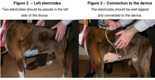
It is very easy to attach the Holter device to the dog (Figure 2, 3, 4 and 5). The dog should keep the device during all the study and a diary kept by the client with the sleep/awake activity or events such as syncope will help to correlate to ECG changes on the Holter.
Doroteia Bota Dissertação de Mestrado em Medicina Veterinária, FMV-UTL
8
Figure 2 – Left electrodes Figure 3 – Connection to the device
Two electrodes should be placed in the left The electrodes should be well tapped side of the thorax. and connected to the device.
Figure 4 – Right electrode Figure 5 – Holter ready
One electrode should be placed and tapped in The device is protected in a sock ventrally to the right side of the thorax. the dog’s neck and a final net is placed.
As many as 50% of clinically normal humans have ectopic ventricular activity with several thousand VPCs per 24h. Decades ago, it was suggested that the occurrence of arrhythmias in clinically normal dogs is rare. Results of a more recent assessment of the electrical activity in clinically normal dogs suggest that clinically normal dogs do not have the same frequency of ectopic ventricular activity that is seen in the clinically normal humans (Spier & Meurs, 2004).
Although it has been suggested that a small number of VPCs may be normal, especially in old dogs, it is uncommon for a healthy dog to have VPCs. In one study, healthy adult large breed dogs had a median of 2 VPCs in 24h (Meurs, Spier, Hamlin, Wright & 2001). In other study in 300 asymptomatic adult Boxers, 75% of the population was found with less than 75 VPCs in 24h (Meurs, 2004). Twenty-four hour Holter monitoring of 228 Beagles by Ulloa,
Doroteia Bota Dissertação de Mestrado em Medicina Veterinária, FMV-UTL
9
Houston and Altrogge et al (1995) documented VPCs in approximately 20% of the dogs. However, in all but 4 dogs the total number of VPCs per day was less or equal to 9.
Some breed variation may exist and certain breeds (including Boxers) normally may have a higher number of VPCs per 24h.
The identification of frequent ventricular ectopy (>100 VPCs over 24h) in an adult Boxer is strongly suggestive of a diagnosis of ARVC, particularly if there is significant complexity to the arrhythmia – couplets, triplets, bigeminy or VT (Meurs, 2004).
When performing a Holter monitoring the number of VPCs is tabulated and the complexity of the ventricular arrhythmia graded as: grade 0 (no VPCs are detected), grade 1 (only single VPC detected), grade 2 (single VPCs are detected and are present either in a bigeminal or trigeminal pattern, or multiform complexes are detected) grade 3 (couplets or triplets are detected) or grade 4 (runs of VT > or equal to 4 beats) or the RonT phenomenon is detected (superimposition of an ectopic beat on the T wave of a preceding beat, or simply R waves interrupting T waves).
Most asymptomatic animals have single VPCs interspaced with their normal beats throughout the 24h period. If a Holter shows many runs of VPCs, this means that this animal may be at higher risk for syncope or sudden death (Meurs et al., 1999).
Sometimes, a strong suspicion of ARVC exists but the Holter monitor reading is not clearly abnormal. This may be due to the significant day-to-day variability in VPC number and affected untreated Boxers. Spier and Meurs (2004) performed a 24 hour Holter monitoring for 7 days in 11 Boxer dogs. This study showed that variability in the frequency of ventricular arrhythmias in dogs with > 500 VPCs/24 h during the study period did not exceed 80%. Therefore, changes in VPC frequency of < or equal to 80% may be within the limits of spontaneous variability. This degree of variability should be considered in evaluations of ambulatory ECG recording, particularly in the assessment of the efficacy of anthiarrhythmic drugs.
The assessment of heart rate variability (HRV) in Boxers using a 24h ambulatory ECG was studied by Spier and Meurs (2004). They used 24 Boxer dogs divided in affected and unaffected (<2 VPCs/24h) dogs, 10 clinically normal non-Boxers to assess the ability of HRV analysis to identify differences in Boxers on the basis of severity of their arrhythmia and evaluate the use of HRV to determine whether persistently high sympathetic tone is present in these dogs. Sympathetic and parasympathetic tones not only impact HRV but also affect heart rhythm; increase sympathetic tone exacerbates the ventricular arrhythmias and increased parasympathetic tone has a protective effect. This study showed less HRV in Boxers with CHF and did not reveal any differences between affected and unaffected Boxers. This might be due to the fact that these dogs didn’t have persistently high sympathetic innervations associated with their arrhythmia; it is likely that sympathetic modulation is important in the development of ventricular arrhythmias but this may occur
Doroteia Bota Dissertação de Mestrado em Medicina Veterinária, FMV-UTL
10
intermittently. Also in humans with CHF, high sympathetic tone has been associated with low HRV (like in Boxers with CHF) and the reduction of HRV in humans with CHF is predictive for death (Spier & Meurs, 2004).
4 - ECHOCARDIOGRAPHY
Although ARVC is a myocardial disease, most of the myocardial changes are abnormalities noted at histologic examination as opposed to abnormalities obvious on gross examination of the ventricle. Most affected dogs have normal echocardiograms, particularly with regard to evaluation of the size and function of the LV (Meurs, 2004). In some cases, careful echocardiographic evaluation of the RV may detect RV enlargement and, possibly, RV dysfunction. A small percentage of adult Boxers with tachyarrhythmias are observed to have LV dilation with systolic dysfunction. In one study 4 dogs out of 5 with type III ARVC had LV dilation and decreased LV systolic function (Baumwart et al, 2005).
Baumwart and Meurs (2008) studied 12 Boxers with ARVC and 10 control Boxers trying to use an index of myocardial performance (IMP) to assess RV function in Boxers with ARVC. Inflow velocity through the tricuspid valve and RV outflow velocity were recorded separately by use of a pulsed-wave Doppler. The IMP value was calculated by use of the equation (isovolumetric contraction + isovolumetric relaxation)/Ejection Time (ET). This study failed to identify a significant difference between the 2 groups. IMP values were within normal limits for both groups.
5 - BIOCHEMICAL MARKERS
Brain natriuretic peptide, atrial natriuretic peptide, endothelin and C-reactive protein are used to detect the presence of cardiac disease in dogs.
Brain natriuretic peptide (BNP) is secreted in response to stretching of the myocytes in the ventricles of the heart; in humans with ARVC, plasma concentrations of BNP are increased, compared with clinically normal individuals (Baumwart & Meurs, 2005). Brain natriuretic peptide has natriuretic, diuretic, hypotensive and smooth muscle relaxant effects (Baumwart & Meurs, 2005). Baumwart and Meurs (2005) studied 13 Boxers with ARVC, 9 clinically normal Boxer dogs and 5 hound dogs with systolic dysfunction to determine whether Boxers with ARVC had increased plasma concentration of BNP. There was no significant difference in BNP concentration between the 2 groups of Boxers. A significant correlation between plasma BNP concentration and number of VPCs per 24h in the ARVC-affected Boxers was not identified. This may indicate that evaluation of plasma BNP concentration is not an accurate indicator of ARVC in Boxers.
Doroteia Bota Dissertação de Mestrado em Medicina Veterinária, FMV-UTL
11
Cardiac troponins are increasingly used as a biomarker by clinical cardiologists. Cardiac troponin consists of 3 subunits: troponin C, troponins I and troponin T. Measurements of troponin I and troponin T are used to specifically identify cardiac muscle injury in humans. An increase in serum cardiac troponin concentration is an important marker for the diagnosis of acute myocardial infarction in humans and in dogs with experimentally induced disease. In dogs, as in humans, the normal range of plasma cTnI is similar: 0.0-0.04ng/mL (Sleeper, Clifford & Laster, 2001).
In dogs, cardiac troponin concentrations increase with mitral valve disease, dilated cardiomyopathy, sub aortic stenosis, pericardial effusion, babesiosis, suspected cardiac contusions and gastric dilatation and volvulus (Baumwart et al., 2007).
Boxer ARVC, because it is a disease characterized by myocardial atrophy with fibrofatty replacement, should have increased serum cardiac troponin I concentrations, compared with clinically normal dogs. This hypothesis was tested by Baumwart, Orvalho and Meurs (2007) when 10 Boxers with clinical diagnosis of ARVC, 10 control Boxers and 10 control non-Boxers were analysed for serum cTnI concentration. In this study, non-Boxers with ARVC had significantly greater serum concentrations of cTnI. However, serum cTnI concentrations in control Boxers were also higher than that of control non-Boxers. This mean concentration of serum cTnI for clinically diagnosed ARVC dogs is similar to that previously described for a large group of dogs (Doberman Pinschers, Great Danes, Boxers, Rottweilers, Labrador Retrievers) with CM (median concentration of 0.14 ng/mL). A significant correlation was found between the serum cTnI concentration and the number of VPCs/24h as well as between serum cTnI concentration and grade of ventricular arrhythmia for the Boxer groups. A recent study done by La Vecchio, Marin, Baumwart, Iazbik, Westendorf and Couto et al., (2009) for serum cTnI concentration in retired racing Greyhounds showed that this breed has a higher cTnI concentration (mean concentration 0.10ng/mL) than other breeds but not significantly different from normal Boxers (mean concentration 0.08ng/mL) or Boxers with ARVC (mean concentration 0.12ng/mL).
All these studies suggest that cTnI may be an indicator of stage or severity of disease.
6 - MAGNETIC RESSONANCE IMAGING
Magnetic resonance imaging (MRI) can both quantify functional abnormalities and characterize morphological changes such as fatty infiltration of the RV, without the limitations of echocardiography or the risks of myocardial biopsy.
Human hearts affected by ARVC may display high transmural signal intensity in the anterolateral and/or infundibular regions of the RV when fatty replacement is present (Basso et al., 2004). However, in a recent study with 5 Boxers with clinical diagnosis of ARVC and 5 healthy control dogs, MRI identified no definitive fatty changes of the RV myocardium in the
Doroteia Bota Dissertação de Mestrado em Medicina Veterinária, FMV-UTL
12
affected dogs (Baumwart, Meurs & Raman, 2009). Probably, findings of fatty infiltrations may be present only in more chronic, advanced cases then those used in this study. In the above study RV ejection fraction was significantly lower in dogs with ARVC, consistent with myocardial disease, usually difficult to assess by imaging.
PATHOLOGY
Post mortem findings can be helpful in the evaluation of sudden cardiac death in the Boxer. Many affected dogs have a grossly normal appearance to their heart at the time of death; however, some cases may show evidence of RV enlargement and, in some cases, LV enlargement. Careful histologic evaluation should identify fatty, and sometimes, fibrofatty, segmental or diffuse replacement of the RV free wall from the epicardium toward the endocardium. Occasionally, the atrial walls, the interventricular septum and LV free wall are also involved. Both the fatty and fibrofatty forms are characterized by residual surviving myocytes embedded within regions of fat and fatty and fibrous tissue, respectively.
The pathological hallmark in human ARVC is the loss of RV myocytes with replacement by fat or fibrofatty tissue. In one study (Basso et al., 2004), allthe 23 dogs examined, presented histopathological lesions that closely resembled those characteristic of human patients with ARVC.
As in the young ARVC patients who died suddenly, the presence of diffuse RV myocytes loss, fatty replacement and residual myocytes embedded within fat suggests a substrate predisposed to sudden death.
A likely consequence of this fibrofatty replacement is the development of VPCs however, a correlation between VPCs and the degree of fibrofatty infiltrate into the myocardium has not been determined, and such a correlation is unlikely to be a linear relationship (Baumwart & Meurs, 2008).
It is extremely difficult to noninvasively assess fibrofatty infiltration into the myocardium, and methods to quantify infiltration are lacking. Evaluation of endomyocardial biopsy specimens may also be used to identify fibrofatty infiltrate in the myocardium of the RV but cannot be used to assess the extent of infiltrate in the ventricle.
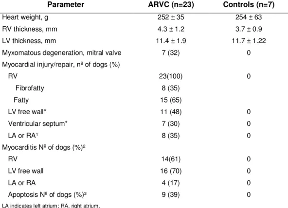
In one recent study, Basso et al. (2004) identified gross and histopathological characteristics of Boxer dogs (n=23) with ARCV in comparison with clinical normal control dogs (n=7) (Table 1).
Doroteia Bota Dissertação de Mestrado em Medicina Veterinária, FMV-UTL
13
Table 1. Gross and Histopathological Characteristics of Boxer Dogs with ARVC (adapted from Basso et al., 2004)
Parameter ARVC (n=23) Controls (n=7)
Heart weight, g 252 ± 35 254 ± 63 RV thickness, mm 4.3 ± 1.2 3.7 ± 0.9 LV thickness, mm 11.4 ± 1.9 11.7 ± 1.22 Myxomatous degeneration, mitral valve
Myocardial injury/repair, nº of dogs (%) RV Fibrofatty 7 (32) 23(100) 8 (35) 0 0 Fatty LV free wall* Ventricular septum* LA or RA¹ Myocarditis Nº of dogs (%)² RV LV free wall LA or RA Apoptosis Nº of dogs (%)³ 15 (65) 11 (48) 7 (30) 8 (35) 14(61) 16 (70) 4 (17) 9 (39) 0 0 0 0 0 0 0
LA indicates left atrium; RA, right atrium.
*Focal, fibrous tissue replacement with mild adipose replacement. ¹Fatty (n=4) or fibrofatty (n=4) replacement
²Significant inflammatory infiltrates by immunohistochemistry quantitative criteria ³Mean apoptotic index in positive dogs was 4.0 ± 3 in RV and 2.0 ± 0.7 in LV.
A substantial replacement of RV cardiac myocytes by adipose or fibrous tissue: fatty form was found in 15 dogs and fibrofatty form in 8 dogs. Fatty tissue replacement was more extensive in the RV of ARVC Boxer dogs than in controls and replacement of RV myocardium by fat was more likely to be diffuse than segmental. Myocarditis characterized by focal or multifocal lymphocytic infiltrates (identified in this study by immunohistochemistry) and associated with myocytes death was identified in the RV, LV and atrium of only ARVC dogs. Myocarditis and fibrofatty myocardial injury and repair were characteristic of Boxer dogs with ARVC that died suddenly, suggesting that myocardial inflammation may also play a role in arrhythmogenesis.
Some investigators consider fatty and fibrofatty patterns to be consecutive stages of disease, mediated by myocarditis, in which the fatty form is an early feature and fibrofatty repair results from myocarditis-induced injury. Consistent with this hypothesis, areas of myocarditis were small and uncommon when associated with purely fatty replacement and in animals with the fibrofatty form, RV inflammatory infiltrates were pronounced (Oxford et al., 2007).
Doroteia Bota Dissertação de Mestrado em Medicina Veterinária, FMV-UTL
14 GENETICS
Meurs et al. (1999) studied 2 families of Boxers with ventricular arrhythmias and provided evidence that familial ventricular arrhythmia is inherited in Boxers as an autosomal dominant trait. The pattern of inheritance was defined as autosomal dominant because affected individuals occurred in every generation, sex distribution was equal and affected parents were able to produce unaffected dogs.
The genetic evaluation in humans has identified 9 different loci and 6 different causative genes. Because 4 of the reported genes that contain causative mutations encode desmosomal proteins, a component of the intercalated disc, it has been proposed that ARVC is a disease of the desmosome (Meurs et al., 2007). Causative mutations have been identified in the 3 genes that encode desmoplakin, plakoglobulin and plakophilin-2; these desmosomal proteins are responsible for the mechanical coupling of the myocytes and provide a continuous cell-to-cell connection. A fourth gene containing causative mutations is desmoglein-2, one of the essential transmembrane components of desmosomes. Studies in Boxer dogs failed to identify mutations in the 4 desmosomal genes associated with the human form of ARVC (Meurs et al., 2007).
Oxford et al. (2007) studied hearts from 12 Boxers with confirmed ARVC and 2 control dogs for the molecular composition of the intercalated disc structure, essential for the coupling between cardiac cells. This study found a loss of gap junction plaques (the Connexion 43 protein) in ARVC hearts suggestive of a loss of cell-cell communication, which may be one of the substrates for arrhythmogenesis in ARVC.
Causative mutations have also been found in 2 genes that encode the nondesmosomal proteins TGF-β3 and the cardiac ryanodine receptor (RyR2). Mutations in the RyR2 have been identified in human families with a specific form of ARVC, ARVC2 that is characterized by effort-induced polymorphic ventricular tachycardia (Lenhart, Wehrens, Kushnir & Marks, 2004). The RyR2 is an intracellular calcium release channel and altered RyR2 function and abnormal calcium cycling have been linked to electrical instability, contractile dysfunction, heart failure and death. Because of the similarities between the human form of the disease and the canine form, Meurs, Laombe, Dryburgh, Fox, Reiser and Kittleson, (2006) studied the expression of the RyR2 in normal and ARVC canine hearts.
When compared with control dogs (Beagles), in ARVC dogs (Boxers) the message RNA and protein expression of the RyR2 was significantly decreased in all three cardiac regions (RV, LV, interventricular septum) with no significant difference between the three regions. This study showed no genetic linkage of canine ARVC to the RyR2 gene which may suggest that one of the genes that encodes for a protein assisting in RyR2 stabilization could be the primary problem and that the observed RyR2 changes found in this study are a secondary associated defect.
Doroteia Bota Dissertação de Mestrado em Medicina Veterinária, FMV-UTL
15
The activity of the RyR2 is modulated from several molecules and calstabin2, also known as FKBP12.6, is one of them (Marks, 2001; Lenhart et al., 2004).
Oyama et al. (2008) examined the presence and effect of calstabin2-deficiency in Boxers with ARVC. This study identified a molecular mechanism involving RyR2 dysfunction due to depressed calstabin2 expression and intracellular calcium leak in ARVC Boxers. These results indicate that Boxer dogs with calstabin2 deficiency may represent a potentially important abnormality in Boxer ARVC. However, in this study a causative mutation of calstabin2 was not identified.
It is possible that the canine form of ARVC is attributable to a mutation that is common to that in affected humans but is in a yet to be identified gene. As a matter of a fact, only about 50% of humans with ARVC have an identifiable genetic cause (Oyama et al., 2008). Thus, ARVC in Boxers may corresponds to the human condition and be caused by a nucleotide sequence alteration in a locus that has not been identified in humans.
TREATMENT
The criteria for initiating treatment for ventricular arrhythmias, particularly in a dog without clinical signs, are uncertain and ill-defined. There is little information linking the number of VPCs or their arrhythmia grade with development of clinical signs or risk of sudden death in dogs.
In dogs, it has been suggested that when there are > 30 VPCs/min, multiple complex QRS forms, VPC with short intervals or indications of decrease cardiac output (syncope), pharmacologic control should be started. Also, it is recommended always to obtain an echocardiogram before making specific antiarrhythmic drug recommendations due to the association of VT with ARVC and myocardial systolic dysfunction.
Treatment considerations for affected dogs are generally directed toward the use of ventricular antiarrhythmics, because most affected dogs do not have systolic dysfunction and do not seem to progress to heart failure.
In human medical literature the list of prescribed drugs most commonly chosen to treat ventricular arrhythmia are quinidine, procainamide, mexiletine, atenolol, propanolol, sotalol and amiodarone (Fauci et al., 2008)
Sotalol (Class III antiarrhythmic drug) is the treatment of choice for VT in Boxers, if the myocardial function is normal or mildly decreased, otherwise it may reduce contractility and lead to worsening of heart failure. In these cases, mexiletine with atenolol can be used (Tilley, Smith, Oyama & Sleeper, 2008). The atenolol dosage can be started at a lower dose, to limit the beta blocker effect on myocardial contractility. For refractory VT and recurrent syncope the combination of sotalol with mexiletine is useful. For those cases where VT persists or the dog does not tolerate sotalol or mexiletine/atenolol, amiodarone (Class III)
Doroteia Bota Dissertação de Mestrado em Medicina Veterinária, FMV-UTL
16
may be a solution. However, monthly measurement of liver enzymes is recommended to monitor the possibility of hepatopathy associated with amiodarone.
Meurs et al. (2002) compared the effects of 4 antiarrhythmic treatment protocols on number of VPCs, severity of arrhythmia, HR and number of syncopal episodes in Boxers with ventricular tachyarrhythmias. Forty nine Boxer dogs with > 500 VPCs/24h were treated with atenolol, procainamide, sotalol or mexiletine and atenolol for 21 to 28 days.
The mean doses used were: atenolol 0.3-0.6mg/kg PO BID, procainamide 20-26mg/kg PO TID, sotalol 1.5-3.5mg/kg PO BID, mexiletine 5-8mg/kg PO TID with atenolol 0.3-0.6mg/kg PO BID. Treatment efficacy was defined as > 85% reduction in number of VPCs. A proarrhythmic effect was attributed to treatment if the number of VPC increased > 85%. Sotalol and the combination of mexiletine and atenolol were well tolerated and significantly reduced the number of VPCs and the grade of the arrhythmia when compared with procainamide or atenolol alone. None of the individual treatments significantly reduced the incidence of syncope, although for all treatments combined, overall incidence of syncope was significantly reduced. In this study, only one dog showed proarrhythmic effects under sotalol.
In this study, atenolol alone did not decrease the number of VPCs, the grade of arrhythmia, the HR or syncope. However, in humans a reduction in mortality rate is observed even when reduction in VPCs is minimal (Meurs et al., 2002). Procainamide has been used in the past to treat Boxers with ARVC and is still frequently used. In this study, 5 of the 11 dogs treated with this drug had > 85% increase in VPCs but none had an increase in syncope. Procainamide was the only drug associated with noncardiac side effects (eg, gastrointestinal upset, depression) and this has already been documented in humans as well.
Because of the day-to-day variability in frequency of VPC, the reduction itself in the number of VPCs/24h is not an accurate method to assess drug efficacy. It has been suggested that to differentiate drug effect from day-to-day variability, a reduction of >85% should be observed (Meurs et al., 2002).
Ideally, therapy should be managed by a pre-treatment and post-treatment (3-4 weeks later) Holter monitoring study. Comparing the pre-treatment and post-treatment studies should help to evaluate the efficacy of treatment and detect a proarrhythmic effect.
Cardiomyopathies appear to have a heritable basis but only a few studies have investigated the manner and extent to which environmental factors might influence the expression or progression of cardiomyopathies or ventricular arrhythmias. In humans, it is clear that dietary factors play an important role in the management of cardiac disease.
Studies in humans showed a possible cardioprotective role of long chain omega-3 fatty acids. There are 2 major Omega-3 fatty acids: eicosapentaenoic acid (EPA) and docosahexaenoic acid (DHA), the predominant fatty acids in the fish. In humans, studies with healthy men demonstrated a significant, inverse relationship between whole blood omega-3
Doroteia Bota Dissertação de Mestrado em Medicina Veterinária, FMV-UTL
17
fatty acids concentrations and sudden death from cardiac causes. This relation is more likely to be due to their antiarrhythmic effect rather than to anti-atherosclerotic effects (Smith, Freeman, Rush, Cunningham & Biourge, 2007).
Smith et al. (2007) studied the effect of Omega-3 fatty acids (DHA, EPA and alphalinoleic acid – ALA) in asymptomatic Boxers with documented ventricular arrhythmia. During 6 weeks, 91 dogs were supplemented either with fish oil (EPA/DHA), flax oil (ALA) or sunflower oil (control group). The number of dogs that had a decrease in VPC number was not different between groups however, only the fish oil group had a significant reduction in VPC number. Significant correlations between plasma fatty acid concentrations after supplementation and percentage change in VPC frequency were present only in the fish oil group. Although this study did not evaluate mortality, changes in HR or blood pressure, it shows that administration of fish oil providing 780mg/dL EPA and 497mg/dL DHA per day is associated with decreased ventricular arrhythmia in Boxers with ARVC.
Nelson, Lahmers, Schnieder and Thompson (2006), used an implantable cardioverter defibrillator (ICD) in a Boxer dog to control clinical signs of ARVC. This study was done in a 2 year old Boxer with sustained VT at approximately 300bpm and with failure to response to oral treatment with different drugs.
ICD were designed to solve the delay in providing therapy to ambulatory individuals with life-threatening ventricular arrhythmias. However, after a couple of weeks the dog received 6 maximal energy defibrillations while was actively playing and he behaved fearfully to the defibrillations by fleeing and hiding. When investigating the reason why this happened, T-wave sensing resulting in doubling the HR, triggered tachycardia detections. When the dog was playing, the sympathetic is stimulated and as the HR increases the amplitude of T-waves also increases, creating a bigger chance of T-wave sensing. In this dog the ICD was finally removed due to inflammation of the implantation site.
ASYMPTOMATIC BOXERS
Screening of breeding dogs for ARVC is of particularly importance for Boxer breeders. The absence of a perfect diagnostic test for ARVC makes the screening of an asymptomatic dog a true challenge. Important factors are a family history of ARVC and an abnormal Holter study.
Therapy in an asymptomatic affected Boxer is not well understood. Some dogs die from their arrhythmia before developing clinical signs however, the absence of clinical signs does not mean that there is no risk of sudden death.
If an arrhythmia is detected on routine examination in an asymptomatic dog, the next step should be a Holter monitoring to evaluate for frequency and complexity of the arrhythmia.
Doroteia Bota Dissertação de Mestrado em Medicina Veterinária, FMV-UTL
18
Meurs (2004) established a possible system for screening asymptomatic dogs might include the following Holter monitoring criteria:
1. None to 20 single VPCs over 24 hours: within normal limits;
2. Twenty to 100 VPCs over 24 hours: indeterminate, repeating in 6-12 months is suggested;
3. One hundred to 300 single VPCs over 24 hours: suspicious, out of breeding program for 1 year and repeating Holter study;
4. One hundred to 300 VPCs over 24 hours with increased complexity (couplets, triplets, VT) or 300 to 1000 single VPCs over 24 hours: likely affected;
5. More than 1000 VPCs over 24 hours: affected, treatment should be considered. The factors that determine which dogs eventually progress to the clinical form of the disease are not known and are a reason of frustration for screening for this disease.
Holter monitoring is strongly recommended and significant emphasis on a single Holter monitor reading is not advised. As said before, ARVC is an adult-onset disease thus, a single Holter monitoring performed in a young adult may not reflect an affected dog in the future.
ARVC vs NEUROCARDIOGENIC BRADYCARDIA
Neurally mediated bradycardia is referred to by numerous synonyms including neurocardiogenic syndrome, cardiogenic syncope and vasovagal syncope and is distinct from that of advanced heart blocks.
As it was said before, neurocardiogenic bradycardia is a frequent cause of syncope in Boxer dogs. It is less common and is not associated with ARVC. When a Holter recording performed soon after a syncopal event in a Boxer contains no or few VPCs, syncope secondary to bradycardia should be strongly considered.
Vasovagal syncope is associated with both sympathetic withdrawal (vasodilatation) and increased parasympathetic activity (bradycardia). Neurocardiogenic syncope often occurs in the setting of increased peripheral sympathetic activity and venous pooling. Under these conditions, vigorous myocardial contraction of a relatively empty left ventricle is thought to activate myocardial mechanoreceptors and vagal afferent nerve fibers that inhibit sympathetic activity and increase parasympathetic activity. The resultant vasodilatation and bradycardia induce hypotension and syncope. Neurocardiogenic bradycardia can coexist with ARVC in Boxers because both conditions are common. Β-blocker or sotalol treatment unlike mexiletine, administered for VPCs may uncover or aggravate neurocardiogenic bradycardia. Boxers with VPCs that are treated with sotalol and then experience new or exacerbated syncopal episodes may be experiencing neurocardiogenic bradycardia. Usually, the episodes are infrequent and treatment of neurocardiogenic bradycardia is often not required. Avoiding what triggers the episode (exertion or excitement) is, most of the times,
Doroteia Bota Dissertação de Mestrado em Medicina Veterinária, FMV-UTL
19
what is advised. For those patients that the syncopal episodes are very frequent, the implantation of a pacemaker can be an option.
Neurocardiogenic bradycardia is not believed to be fatal for Boxers (Thomason et al., 2008).
PROGNOSIS
Dogs with ARVC are always at risk of sudden death. However, many affected dogs live for several years even without treatment as well as many other may be managed for years on antiarrhythmics.
A small percentage of dogs may develop ventricular dilation and systolic dysfunction (Meurs, 2004).
VENTRICULAR DILATION AND SYSTOLIC DYSFUNCTION
A small number of Boxers with tachyarrhythmias may have systolic dysfunction and, in a few cases, with left or biventricular heart failure.
When performing an echocardiogram ventricular dilation and systolic dysfunction is present, treatment for canine DCM is necessary.
Presenting complains in dogs with myocardial dysfunction may include cough, depression, dyspnoea, weight loss, panting, syncope and polydipsia. Clinical presentation commonly includes signs of left-sided or biventricular CHF, i.e. dyspnoea caused by pulmonary oedema and/or pleural effusion, abdominal distention caused by ascites and irregular heart rhythm with pulse deficits. Pulmonary oedema is the most common finding on thoracic radiographs. Atrial fibrillation is the most commonly diagnosed electrocardiographic abnormality (Tilley et al., 2008).
Death is either due to advanced CHF or inadequate response to medical treatment.
Echocardiographic evaluation of left ventricular systolic performance reveals increased end-systolic and end-diastolic dimensions, dilation of the left atrium (LA) and decreased FS %. Treatment involving diuretics, inodilators, angiotensin-converting enzyme (ACE) inhibitors and dietary salt restriction is described elsewhere (Tilley et al., 2008). Ventricular arrhythmias and atrial fibrillation requires use of specific antiarrhythmics, as already described.
PART TWO
Doroteia Bota Dissertação de Mestrado em Medicina Veterinária, FMV-UTL
21 CLINICAL CASES
I am going to present 6 cases of ARVC that I followed with Dr Babis Koffas while I was at North Kent Referrals.
We always strongly advised owners performing a 24-hour Holter monitoring before and after initiation of the treatment. However, sometimes the ECG was strongly suggestive of ARVC and this allowed us to start the treatment without performing the pre treatment Holter. Other times the cost of a 24-hour Holterwas the reason to decline performing this test.
Case 1 (Alice Joyner)
Alice was a 4-year-old female spayed Boxer. Alice was examined at NKR in November 2008for investigation of a history of collapse occurring mainly on exertion. Alice was previously diagnosed, at the age of 2, with a small atrial septal defect.
On physical examination she was bright and alert. Her mucous membranes were pink and the capillary refill time was < 2 seconds. The femoral pulses were strong and no deficits were detected. The HR was 120 bpm and the cardiac rhythm was regular. Auscultation of the heart and the lungs was unremarkable.
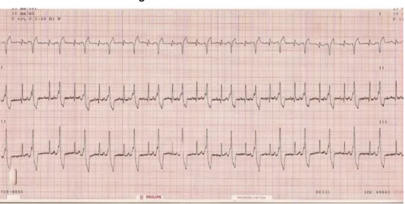
On electrocardiography frequent VPCs originating from the RV (positive in leads I-II-III-aVF) were present (Figure 6).
Figure 6 – ECG of Alice Joyner
On echocardiography a small atrial septal defect was present. Otherwise the contractility of the LV and the size of all cardiac chambers were within normal limits (Figure 7). Colour flow Doppler echocardiography showed a small mitral insufficiency (1+/4+).
Doroteia Bota Dissertação de Mestrado em Medicina Veterinária, FMV-UTL
22
During the echocardiographic examination several VPCs were recorded. The atrial septal defect was small, not haemodynamically significant and was not responsible for Alice's clinical signs.
Figure 7 – Echocardiography of Alice Joyner
Blood pressure: measurement with the Doppler technique revealed a systolic pressure of 160 mm Hg.
Complete Blood Count (CBC), serum biochemistry, measurement of Total T4 and TSH were done.
The thyroide profile was within normal limits. This measurement was done whenever we suspected a systolic disfunction. This can be due to hypothyroidism, which can lead to DCM, as well as when the animal shows lethargy or high levels of cholesterolemia. The other blood analysis showed a slight increase for urea of 7.6mmol/L (1.7-7.4), creatinine of 121µmol/L (0-106) and ALT of 120UL (0-25). (Anex I). These results could be compatible with pre-renal azotemia but a urianalysis was not performed to confirmed it. Mild increasing in ALT could be compatible with hypoxia due to decreased perfusion and/or damage to heart muscle.
We have suggested a 24-hour Holter recording prior to initiation of treatment and then 2 to 3 weeks post-initiation of treatment, in order to quantify the antiarrhythmic effect of the treatment. In this case, this was agreeded by the owners. In the meanwhile treatment with sotalol 1mg/kg orally (PO) twice daily (BID) was iniciated. The first Holter was not possible to localize till the end of this study so the results here described were taken from the referral report of Dr Babis Koffas to Alice’s referring vet.
Pre-treatment 24-hour Holter monitoring showed 1421 VPCs per 24h. Periods of paroxysmal VT at 300 bpm, single VPCs, couplets and triplets were also detected.
The first 24-hour Holter recording showed that Alice experienced frequent periods of fast VT that most likely were the cause of her collapsing episodes. The above findings were, then, compatible with ARVC. Alice’s owners were advised to not over-exercise her or over-excite
Doroteia Bota Dissertação de Mestrado em Medicina Veterinária, FMV-UTL
23
Alice in order to prevent further collapsing. The second 24-hour Holter recording was performed 2 weeks post initiation of treatment with sotalol (Anex II). This second study showed 1891 VPCs per 24h, some of them were noted as bigeminal, trigeminal, coupled and triplet. Occasional to frequent episodes of primarily monomorphic Vrun / VT to maximum of 77 beats duration were present. Maximum HR was 326 bpm. Some of them noted as non-Torsades de Pointes type polymorphic VT.
The results from this second Holter showed that treatment with sotalol at 1mg/kg PO BID was not able to control effectively the arrhythmia in Alice (a reduction > 85% in the frequency of VPCs during 2 subsequent Holter recordings is essential in order to acquire a significant antiarrhythmic effect, as said before). Based on the above results the increase of sotalol dose to 1,5mg/kg PO BID was advised. Despite the suboptimal results of the recent Holter recording it was encouraging that Alice stopped collasping since the initiation of treatment with sotalol.
A third 24-hour Holter recording was not necessary because Alice did not collapse again. I contacted Mrs Joyner when I finished this study and I was told that Alice unfortunately died 7 months after we first saw her. For Mrs Joyner there was a clear improvement of Alice condition under treatment and she died after a walk in the garden. Mrs Joyner also contacted Alice’s breeder who told her that 2 dogs from the same litter of Alice died at 6 months of age. However, the cause of death was unknown.
Case 2 (Oscar Eels)
Oscar was a 7-year-old male neutered Boxer dog. Oscar was examined at NKR in January 2009 for further investigation of a recent history of worsening collapsing episodes. He first started collapsing when he was 6 months old but these episodes became more frequent only recently which prompted to a cardiology referral to NKR.
On physical examination he was bright and alert. The mucous membranes were pink and the capillary refill time was < 2 seconds. The femoral pulses were strong but deficits were detected. The HR was 120 bpm and the cardiac rhythm was irregular. No cardiac murmur was detected.
On electrocardiography normal sinus rhythm predominated, and ventricular bigeminy was also present(Figure 8).