Article
J. Braz. Chem. Soc., Vol. 29, No. 9, 1796-1802, 2018 Printed in Brazil - ©2018 Sociedade Brasileira de Química
*e-mail: [email protected]
Determination of Colchicine in Pharmaceutical Formulations and Urine by
Multiple-Pulse Amperometric Detection in an FIA System Using Boron-Doped
Diamond Electrode
Débora A. R. Moreira,a,b Fernando M. de Oliveira,b,c Dilton M. Pimentel,b
Tiago J. Guedes,b Rita C. S. Luz,d Flávio S. Damos,d Arnaldo C. Pereira,a
Rodrigo A. B. da Silvae and Wallans T. P. dos Santos*,f
aDepartamento de Ciências Naturais, Universidade Federal de São João Del-Rei,
36301-160 São João Del-Rei-MG, Brazil
bDepartamento de Química, Universidade Federal dos Vales do Jequitinhonha e Mucuri (UFVJM),
Rodovia MGT 367, km 583, No. 5000, Alto da Jacuba, 39100-000 Diamantina-MG, Brazil
cInstituto Federal de Minas Gerais (IFMG), Rua Itamarati, 140,
Bairro São Caetano, 32677-564 Betim-MG, Brazil
dDepartamento de Química, Universidade Federal do Maranhão, 65080-805 São Luís-MA, Brazil
eInstituto de Química, Universidade Federal de Uberlândia, 38400-902 Monte Carmelo-MG, Brazil
fDepartamento de Farmácia, Universidade Federal dos Vales do Jequitinhonha e Mucuri (UFVJM),
Rodovia MGT 367, km 583, No. 5000, Alto da Jacuba, 39100-000 Diamantina-MG, Brazil
This work presents a simple and fast method for colchicine (CO) determination in pharmaceutical formulations and urine by multiple pulse amperometry (MPA) with flow injection analysis (FIA) system using a boron-doped diamond electrode. In optimized conditions, it was possible to quantify CO in urine samples without interference of uric acid and ascorbic acid. The working linear range for CO quantification was achieved from 1.0 × 10⁻⁷ to 0.5 × 10⁻³ mol L−1 with a low limit
of detection of 2.14 × 10⁻⁸ mol L−1. Furthermore, the proposed method showed high repeatability
for 10 consecutive injections of 1.0 × 10⁻⁴ mol L−1 CO (relative standard deviation = 1.28%) and
good analytical frequency (30 determinations per hour). The addition and recovery studies in all
pharmaceutical samples were approximately 100% and the results for CO determination were compared by high performance liquid chromatography (HPLC) with UV detection.
Keywords: colchicine,boron-doped diamond,multiple pulse amperometry, flow-injection analysis
Introduction
Colchicine (CO) is an anti-inflammatory used in treatment of acute gouty arthritis. This drug has low therapeutic index, so the therapeutic effective concentration range is very narrow. Inadequate dose of CO presents some side effects, such as severe gastrointestinal distress, hepatocellular insufficiency, central nervous system
dysfunction, among other problems.1-5 Thereby, CO
quantification in pharmaceutical and biological samples is very important to monitor the treatment of patients and
perform pharmacological studies of this drug, beyond performing an efficient quality control of its formulations.5-7
The structural formula of CO is presented in Figure 1. Identification tests of CO in pharmaceutical formulations described in pharmacopoeias are based in infrared
absorption spectrophotometry, absorption in UV range or change of color in reaction solution containing ferric chloride.8-10 The International Pharmacopoeia10 suggests
UV absorption for calculating tablet contents of CO. Quantification of this drug in urine samples and other matrices in general is performed by high performance liquid
chromatography (HPLC) with UV detection.11-13 Some
methods for CO determination by HPLC coupled with mass spectrometry14,15 and spectrophotometry,16 as well as
immunoassay17 were also found in the literature. Despite
HPLC technique having advantages as high selectivity and reliability, it is not so suitable for routine analysis in pharmaceutical industries due to high costs and low sample throughput. In this context, the electroanalytical methods have been widely applied to such samples, providing a
simple, fast and low-cost analysis.18-20 These advantages
can justify the number of works that have already been reported for electrochemical behavior studies of CO and its determination using different working electrodes.3,21-30
Among these, the boron-doped diamond (BDD) electrode was used for CO determination in pharmaceuticals and
human serum samples.30 The BDD electrode is widespread
in electroanalytical methods with some advantages in comparison with other electrodes, such as high stability
and low background current.31-37
Although electroanalytical methods have presented the above-mentioned advantages for CO determination, these methods did not present a highly selective determination of CO in biological samples with presence of large excess of interferants, such as ascorbic acid (AA) and uric acid (UA). Furthermore, the quality control of drugs requires faster and simpler methods for routine analysis.
According to Felix and Angnes,38 the flow injection
analysis (FIA) system with amperometric detection has been used as an interesting alternative to improve speed and simplicity of drug electroanalysis. Additionally, multiple-pulse amperometry (MPA) can provide advantages over classic amperometry (constant potential) such as improved stability (constant cleaning of working electrode surface during the measurements) and selectivity (detection of analyte in a potential without interferants). FIA-MPA detection was used for the first time by dos Santos et al.39 for
simultaneous determination of AA and paracetamol. This method has been used in other simultaneous determinations, such as for two synthetic colorants in food samples,40 two41
or three42 synthetic antioxidants in food samples, and two
or three drugs in pharmaceutical samples.43-45 Moreover,
MPA detection has been used to improve the selectivity in the quantification of electroactive compounds,46,47 and
improve precision by the addition of an internal standard in flow analysis48 or by the reduction of the contamination
of the working electrode.49,50 The MPA can also be used for
oxidation (or reduction) products detection of an analyte.51
Thereby, this paper presents a simple and fast strategy for selective determination of CO in pharmaceutical formulations and urine samples (recovery studies) using FIA-MPA system and BDD as working electrode.
Experimental
Reagents and solutions
CO was obtained from Sigma-Aldrich (São Paulo, Brazil); AA, UA, sulfuric acid and sodium dihydrogen phosphate/sodium hydrogen phosphate from Vetec (Duque de Caxias, Brazil); and acetic acid/sodium acetate and boric acid/borate sodium from Merck (Rio de Janeiro, Brazil). All reagents were of analytical grade and used without any further purification. Stock solutions of CO, AA and UA were freshly prepared with deionized water (Milli-Q system, Millipore, Bedford, MA, USA) with resistivity of 18.2 MΩ cm at 298 K. Sulfuric acid solutions
(0.1 and 0.3 mol L−1) and Britton-Robinson buffer with
different values of pH (2.0 to 12.0) were used as supporting electrolytes in electrochemical measurements. The
Britton-Robinson buffer was composed of 0.1 mol L−1 boric
acid, 0.1 mol L−1 acetic acid and 0.1 mol L−1 phosphoric
acid. The buffer pH values were adjusted using sodium hydroxide and hydrochloric acid.
Human urine samples were collected from healthy volunteers and diluted (100 times) in supporting electrolyte without any sample pre-treatment for the FIA-MPA detection. The pharmaceutical samples of CO were purchased at local pharmacies in Diamantina-MG (Brazil). The tablets (n = 20) of CO were powdered in a mortar, and a weight corresponding to one tablet was dissolved in supporting electrolyte using an ultrasonic bath for 10 min prior the FIA-MPA detection.
Instrumentation and apparatus
The BDD film (8000 ppm of doping level) was acquired from Neo Coat SA (La Chaux-de-Fonds, Switzerland). A homemade electrochemical wall-jet cell was used for the electrochemical experiments, with a Pt wire as auxiliary
electrode, and an Ag/AgCl (3.0 mol L−1 KCl) reference
electrode. The area of the BDD working electrode was 0.13 cm2 (delimited by an O-ring with diameter of 0.4 cm).
Before the measurements, different pretreatments were performed in the working electrode placed in FIA cell containing 0.5 mol L−1 H
2SO4 solution. For anodic activation
applied −30.0 mA during 360 s. Such pretreatment was carried out once a day.
In the FIA analysis, a single-line system was employed using polyethylene tubing of 1.0 mm (i.d.). The injection system consisted of a manual acrylic injector with polyethylene tubes of 0.5 mm (i.d.). The flow rate was controlled by a peristaltic pump (Gilson Minipuls 3, Villiers-le-Bel, France). The flow rates were evaluated from
0.5 to 5.0 mL min−1, and the injection volumes from 50 to
400 µL. All electrochemical measurements were carried out at room temperature in absence of oxygen (previously removed with nitrogen gas bubbling).
Results and Discussion
Electrochemical study of CO
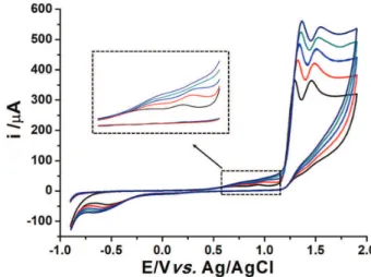
Cyclic voltammetry (CV) was used to evaluate the electrochemical behavior of CO on the surface of BDD working electrode in Britton-Robinson buffer solutions at different pH (2 to 12) and H2SO4 solution (pHapparent 1). As
shown in Figure 2, 0.1 mol L−1 H
2SO4 supporting electrolyte
presented a better sensitivity to CO electrochemical processes with four oxidation peaks at about +0.70, +0.93, +1.20 and +1.40 V and one reduction peak at –0.60 V. The first two oxidation processes for CO are more clearly presented in Figure 3. In contrast, Stankovićet al.30 noticed
only two processes on BDD electrode (cathodically pretreated) in pH 2 to 10 and some reduction process.
It can also be verified in Figure 2 that the electrochemical behavior of CO on BDD after the cathodic or anodic pretreatments presented similar oxidation currents and
potential peaks. However, the cathodic treatment was chosen due to its better cleaning of BDD electrode surface after some measurements of CO by the proposed method.
The dependence on CO electrochemical processes was evaluated. When the scan was performed in potential range from −1.0 to +1.0 V no reduction processes were noted (not shown), suggesting CO is not directly reduced in this potential range on BDD electrode. However, when the scan was performed only until oxidation process of CO at +1.2 V, the reduction peak (–0.6 V, as presented in Figure 2) was observed in the reverse scan of this study, which indicates the reduction process is due to the generated product of the third CO oxidation.
In addition, the effect of the scan rate (v) over currents for all oxidation peaks of CO was studied by changing the
scan from 50 to 150 mV s–1 (Figure 3) and the regression
equations revealed a linear behavior between the square root of the scan rate (v1/2) and the peak current (Ip) for all
processes, suggesting the CO mass transport process is controlled by diffusion on the BDD electrode.
The electrochemical behavior of CO has already been reported in other carbon-based electrodes by authors such
as Zhang,21 who performed only a process of irreversible
oxidation of CO by CV at approximately +1.12 V on glassy
carbon electrode in 0.1 mol L−1 perchloric acid medium,
and no reaction mechanism was proposed. Bodoki et al.22
showed that it is possible to obtain a quasi-reversible system
using a graphite-based electrode in solution of perchloric and phosphoric acids (pH 2.05). These authors observed two well-defined oxidation peaks at 1.06 and 1.22 V and one peak of reduction at –1.04 V.
A systematic mechanistic study for the oxidation and reduction processes of CO was carried out by Bodoki et al.3,24 For the mechanism study of CO oxidation,24
Figure 2. Cyclic voltammograms in 0.1 mol L−1 H
2SO4 supporting electrolyte (black line) at BDD electrode and in the presence of 1.0 mmol L−1 CO after cathodic (blue dashed line) and anodic (red dotted line) treatment. Scan rate: 50 mV s−1.
Figure 3. Cyclic voltammograms of 5.0 mmol L−1 CO at BDD electrode in 0.1 mol L−1 H
electrochemistry coupled to mass spectrometry with two different types of electrolytic cells (aqueous or non-aqueous medium) and different working electrodes (glassy carbon,
gold, platinum and BDD) were used.24 The main product
observed for CO oxidation at around +1.0 V (vs. Pd/H2)
in a large pH range was the 7-hydroxy derivative of CO. The authors also reported several other generated
oxidation products for CO at +1.0 V (vs. Pd/H2), which
could explain the two first oxidation peaks noticed in this work (Figure 3). When potentials above +1.4 V (vs. Pd/H2)
were applied, Bodoki et al.24 reported the second oxidation
process of CO as being due to epoxidation (and/or multiple hydroxylation). For the mechanism study of CO reduction, Bodoki et al.3 showed the possibility of CO direct reduction
at a diamond working electrode. On the other hand, this work presents a reduction process dependent on oxidation
process at +1.0 V on BDD electrode (vs. Ag/AgCl),
suggesting a different reduction process than that reported by these authors. Therefore, the reduction process mechanism for the product generated by CO oxidation (as presented in this work) requires a deeper investigation.
Optimization parameters of FIA-MPA detection
For MPA detection, two potential pulses were applied in sequence on BDD electrode (chosen with basis on the electrochemical behavior of CO, Figure 2): +1.7 V for 500 ms for CO oxidation and –1.1 V for 30 ms for reduction of CO oxidation products (generated in third oxidation process). The pulse times, as well as the injection volume and flow rate (FIA parameters), were optimized considering the best compromise among sensitivity, selectivity and sampling rate. AA and UA are electroactive interferants commonly found in urine sample, but both do not exhibit reduction peaks on BDD electrode. Thus, a study was conducted to find out what concentration of AA and UA would not affect the analytical signal of CO at –1.1 V during the analysis. However, it was observed that AA and UA reacted with CO oxidized at the electrode surface, lowering the peak reduction signal at –1.1 V. This behavior has been reported by dos Santos et al.39 for determination
of dopamine in the presence of AA. The authors evaluated that the chemical reaction between AA and dopamine was inhibited in more acidic media, and an electrolyte of 0.2 mol L−1 H
2SO4 was used to minimize interference of
AA. Similarly, an evaluation regarding decrease of the CO reduction peak signal was performed as a function of acid concentration of the electrolyte in the presence of AA and UA. The best results were obtained with 0.3 mol L−1 sulfuric
acid, as can be seen in Figure 4, where the CO signal is not significantly attenuated (3%) in the presence of AA and
UA. Figure 4 shows the FIA-MPA responses after duplicate injections of solutions containing only CO, only AA, only UA and a solution containing a mixture of CO, AA and UA at the same concentration. However, a biological sample, such as urine, has high concentrations of AA and UA, and thus the current signal of CO was evaluated in the presence of high concentrations of these interferants (Table 1). As can be seen in Table 1, the CO signal remained constant even in the presence of AA or UA in concentration higher than 100-fold.
Repeatability studies
A repeatability study of the proposed method was evaluated, in which 10 consecutive injections of 1 × 10⁻⁴ mol L–1 CO solution were analyzed in the FIA-MPA
under the optimized conditions. As presented in Figure 5, the relative standard deviation (RSD) value (n = 10) found for reduction peaks (acquired at –1.1 V) was only 1.28%, demonstrating an outstanding precision of the proposed method. Using the optimized conditions, the FIA-MPA system provided an analytical frequency of 30 injections
per hour, suitable for application in routine analysis.
Table 1. Current signal relation for CO detection by FIA-MPA obtained after triplicate injections of solutions containing only 2.0 µmol L–1 CO and with increasing concentrations of AA and UA
[Interferant] / [CO] CO current signal for [AA] / [CO] relation / %
CO current signal for [UA] / [CO] relation / %
1 100.5 100.1
50 101.6 98.6
100 104.2 105.0
CO: colchicine; AA: ascorbic acid; UA: uric acid.
Figure 4. Amperometric responses obtained by FIA-MPA method after duplicate injections of solutions containing only CO, AA, UA and a mixture of CO + AA + AU (10 µmol L−1 for all analytes). Electrolyte: 0.3 mol L−1 H
The low RSD value obtained by the proposed method can be justified by association of three factors: (i) BDD,
a working electrode with a highly stable surface; (ii) FIA,
a highly reproducible hydrodynamic system that allows a continuous cleaning of the working electrode during
the analyses; and (iii) MPA detection, that improves the
stability of the electrochemical signal due to the permanent application of cleaning pulses.
Analytical parameters
Analytical parameters of the FIA-MPA method were evaluated for CO determination in pharmaceutical formulations as well as in human urine. After optimization of all parameters of the proposed method, the calibration curve was constructed by the injection of CO (triplicate) in the concentration range of 1.0 × 10⁻⁷ to 0.5 × 10⁻³ mol L−1
(Figure 6). Two linear ranges were observed from this
study: 0.1-2.0 and 20-500 µmol L−1. The respective linear
regressions were: i (A) = (8 ± 5) × 10−7 + (11.3 ± 0.5) × [CO]
(mol L−1) with R = 0.997 and i (A) = (6.4 ± 0.7) × 10−5 +
(0.315 ± 0.003) × [CO] (mol L−1) with R = 0.999. The limits
of detection (LOD) and quantification were calculated from the range with smaller concentrations and the values were 0.021 and 0.071 µmol L−1, respectively.
The addition-recovery studies in pharmaceutical
samples were performed to verify the accuracy of proposed method. The obtained results for the recovery of CO in pharmaceutical formulations (n = 3) and human urine (n = 3) were 100.2 ± 1.0% and 95.0 ± 5.0%, respectively. The obtained values were close to 100%, indicating the absence of matrix effect in these samples. The obtained results for the determination of CO in pharmaceutical
samples using the proposed method versus the official
method (HPLC-UV) are presented in Table 2. Statistical tests (F and Student’s t) were carried out comparing the
results obtained by both methods with a confidence level of 95%. The calculated values of statistical tests were smaller (F = 12.96 and t = 2.17) than the tabulated critical values
(F = 19.00 and t = 2.78), so the results can be considered
similar for both methods.
Table 3 shows some analytical parameters obtained by the proposed method for determination of CO in comparison with others reported in literature. As shown in Table 3, the proposed method presented a wider linear range with LOD and RSD close to or lower than the modified electrodes used for CO determination. The lowest LOD and RSD for CO determination were achieved at mercury electrode using adsorptive stripping voltammetry (ASV),27,28 but the use of this working electrode presents
environmental drawbacks. Moreover, two linear ranges were also obtained in another work employing the BDD Figure 5. Amperogram obtained at –1.1 V for 30 ms by FIA-MPA of 10
consecutive injections of 10 µmol L−1 CO. Electrolyte: 0.3 mol L−1 H2SO4; flow rate: 3.0 mL min−1; injected volume: 330 µL. Potential pulse at 1.7 V for 500 ms was applied for oxidation of CO (not shown).
Table 2. Determination of CO in pharmaceutical formulation by FIA-MPA and official method (HPLC-UV). The studies were performed in triplicate
Sample Ingredient Labeled mass / mg Mass (FIA-MPA) / mg Mass (HPLC-UV) / mg
Capsules CO 0.500 0.541 ± 0.001 0.543 ± 0.004
FIA-MPA: flow injection analysis coupled to multiple-pulse amperometry; HPLC-UV: high performance liquid chromatography with UV detection; CO: colchicine.
Figure 6. Amperogram obtained by FIA-MPA at –1.1 V for 30 ms after
triplicate injections of standard solution containing CO [(a)-(j): 0.1 to 500 µmol L–1]. Respective calibration curves are shown in the inset. Electrolyte: 0.1 mol L−1 H
electrode,30 but the proposed method (FIA-MPA) presented
lower LOD and RSD than the ones reported. This can be justified by the higher sensitivity obtained in the FIA system, since the flow decreases the Nernst diffusion layer in working electrode surface. Moreover, in an FIA system the solution (electrolyte) continuously passes over the working electrode surface, allowing a better cleansing of this electrode and improving its stability.
Conclusions
The proposed FIA-MPA method using the bare BDD working electrode showed some advantages compared to other reported methods for CO quantification in pharmaceutical formulation and urine samples (recovery studies), such as simple, fast and accurate analysis and low waste generation. Furthermore, the pulsed amperometric detection provided a selective determination of CO in urine sample even in the presence of high concentrations of AA and UA, without sample treatment.
Acknowledgments
The authors are grateful to the Brazilian agencies Fundação de Amparo à Pesquisa do Estado de Minas Gerais (FAPEMIG, APQ-03637-16) and Conselho Nacional de Desenvolvimento Científico e Tecnológico (CNPq, 309208/2015-7 and 475276/2013-2) for financial support.
References
1. Leung, Y. Y.; Hui, L. L. Y.; Kraus, V. B.; Semin. Arthritis Rheum.
2015, 45, 341.
2. Gilman, A. G.; Hardman, J. G.; Limbird, L. E.; Goodman & Gilman: as Bases Farmacológicas da Terapêutica, 10a ed.;
McGraw-Hill: Rio de Janeiro, 2003.
3. Bodoki, E.; Vlase, L.; Sãndulescu, R.; Electrochem. Commun.
2015, 56, 51.
4. Deftereos, S.; Ginnopoulos, G.; Papoutsidakis, N.; Panagopoulou, V.; Kossyvakis, C.; Raisakis, K.; Cleman, M. W.; Stefanadis, C.; J. Am. Coll. Cardiol. 2013, 62, 1817.
5. Wallace, S. L.; Semin. Arthritis Rheum. 1974, 3, 369. 6. Slobodnick, A.; Shah, B.; Pillinger, M. H.; Krasnokutsky, S.;
Am. J. Med. 2015, 128, 461.
7. Petterman, C.; Bem-Chetrit, E.; Caraco, Y.; Levy, M.; Semin. Arthritis Rheum. 1991, 21, 143.
8. European Pharmacopoeia Commission; E u ro p e a n Pharmacopoeia, 5th ed.; Council of Europe: Strasbourg, 2005.
9. British Pharmacopoeia Commission; British Pharmacopoeia; Stationery Office: London, 2008.
10. World Health Organization; The International Pharmacopoeia: Tests and General Requirements for Dosage Forms; Quality Specifications for Pharmaceutical Substances and Tablets, vol. 5, 3rd ed.; World Health Organization: Geneva, 2003.
11. Fernandez, P.; Bermejo, A. M.; Tabernero, M. J.; Lopez-Rivadulla, M.; Cruz, A.; Forensic Sci. Int. 1993, 59, 15. 12. Ko, R. J.; Li, W. Y.; Koda, R. T.; J. Chromatogr. 1990, 525,
411.
Table 3. Comparison of the proposed method with electrochemical methods reported for CO determination
Technique Electrode Linear range / (µmol L−1)
LOD / (µmol L−1)
RSD (n) /
% Sample Reference
LSV PRDE and GRDE 2000-10000 – – – 29
DPV GCE/PoPD/SWNTs 0.1-40 0.04 5.3 (10) biological 23
DPV GCEs/AB-DHP 0.1-10 0.035 5.3 (5) pharmaceutical 21
DPV SPEs 0.21-3.0 0.103 – pharmaceutical 22
ASV HDME 0.05-0.4 0.000026 1.8 (3) pharmaceutical and biological 27
DPV MWCNTs/CPE 0.01-25 0.008 6.85 (10) pharmaceutical 26
ASV SMDE 0.01-0.1 0.00013 1.1 (12) biological 28
SWASV GO/Nafion/GCE 0.05-20 0.015 2.8 (10) pharmaceutical 25
DPV BDD 1.0-10 and 10-100 0.26 1.7 (10) pharmaceutical and biological 30
FIA-MPA BDD 0.1-2 and 20-500 0.021 1.28 (10) pharmaceutical and biological this work
13. Bartusik, D.; Tomanek, B.; Lattová, E.; Perreault, H.; Tuszynski, J.; Fallone, G.; Bioorg. Chem. 2009, 37, 193.
14. Tracqui, A.; Kintz, P.; Ludes, B.; Rouge, C.; Douibi, H.; Mangin, P.; J. Chromatogr. B: Biomed. Sci. Appl. 1996, 675, 235.
15. Hamscher, G.; Priess, B.; Nau, H.; Panariti, E.; Anal. Chem.
2005, 77, 2421.
16. Singh, D. K.; Srivastava, B.; Sahu, A.; J. Indian Chem. Soc.
2004, 81, 171.
17. Poulev, A.; Deus-Neumann, B.; Bombardelli, E.; Zenk, M. H.; Planta Med. 1994, 60, 77.
18. Uslu, B.; Ozkan, S. A.; Anal. Lett. 2011, 44, 2644.
19. Ozkan, S. A.; Uslu, B.; J. Pharm. Biomed. Anal. 2016, 130,
126.
20. Couto, R. A. S.; Lima, J.; Quinaz, M. B.; Talanta 2016, 146,
801.
21. Zhang, H.; Bioelectrochemistry 2006, 68, 197.
22. Bodoki, E.; Laschi, S.; Palchetti, I.; Sândulescu, R.; Mascini, M.; Talanta 2008, 76, 288.
23. Zhang, X. H.; Wang, S. M.; Jia, L.; Xu, Z. X.; Zeng, Y.; Sens. Actuators, B 2008, 134, 477.
24. Bodoki, E.; Chira, R.; Zaharia, V.; Sandulescu, R.; Electrochim. Acta 2015, 178, 624.
25. Wang, F.; Zhou, J.; Liu, Y.; Wu, S. J.; Song, G.; Ye, B.; Analyst
2011, 136, 3943.
26. Zhang, K.; Zhou, J.; Liu, J.; Li, K.; Li, Y.; Yang, L.; Ye, B.; Anal. Methods 2013, 5, 1830.
27. Kasim, E. A.; Anal. Lett. 2002, 35, 1987. 28. Wang, J.; Ozsoz, M.; Talanta 1990, 37, 783.
29. Bishop, E.; Hussein, W.; Analyst 1984, 109, 623.
30. Stanković, D. M.; Švorc, L.; Mariano, J. F. M. L.; Ortner, A.; Kalcher, K.; Electroanalysis 2017, 29, 2276.
31. Luong, J. H. T.; Male, K. B.; Glennon, J. D.; Analyst 2009,
134, 1965.
32. Peckova, K.; Musilova, J.; Barek, J.; Crit. Rev. Anal. Chem.
2009, 39, 148.
33. Zhou, Y.; Zhi, J.; Talanta 2009, 79, 1189.
34. Chailapakul, O.; Siangproh, W.; Tryk, D. A.; Sens. Lett. 2006, 4, 99.
35. Pleskov, Y. V.; J. Anal. Chem. 2000, 55, 1045.
36. Einaga, Y.; Foord, J. S.; Swain, G. M.; Mater. Res. Bull. 2014,
39, 525.
37. Macpherson, J. V.; Phys. Chem. Chem. Phys. 2015, 17, 2935.
38. Felix, F. S.; Angnes, L.; J. Pharm. Sci. 2010, 99, 4784. 39. dos Santos, W. T. P.; Almeida, E. G. N.; Ferreira, H. E. A.;
Gimenes, D. T.; Electroanalysis 2008, 20, 1878.
40. Medeiros, R. A.; Lourenção, B. C.; Rocha-Filho, R. C.; Fatibello-Filho, O.; Talanta 2012, 99, 883.
41. Medeiros, R. A.; Lourenção, B. C.; Rocha-Filho, R. C.; Fatibello-Filho, O.; Anal. Chem. 2010, 82, 8658.
42. Bavol, D.; Economou, A.; Zima, J.; Barek, J.; Dejmkova, H.; Talanta 2018, 178, 231.
43. Chaves, S. C.; Aguiar, P. N. C.; Torres, L. M. F. C.; Gil, E. S.; Luz, R. C. S.; Damos, F. S.; Munoz, R. A. A.; Richter, E. M.; dos Santos, W. T. P.; Electroanalysis 2015, 27, 2785.
44. Freitas, J. M.; Oliveira, T. C.; Gimenes, D. T.; Munoz, R. A. A.; Richter, E. M.; Talanta 2016, 146, 670.
45. Gimenes, D. T.; Cunha, R. R.; Ribeiro, M. M. A. C.; Pereira, P. F.; Munoz, R. A. A.; Richter, E. M.; Talanta 2013, 116, 1026.
46. Alecrim, M. F.; Oliveira, F. M.; Guedes, T. J.; Neves, C. D. C.; Mendonça, V. A.; Gil, E. S.; Verly, R. M.; dos Santos, W. T. P.; Electrochim. Acta 2016, 222, 331.
47. Gimenes, D. T.; dos Santos, W. T. P.; Tormin, T. F.; Munoz, R. A. A.; Richter, E. M.; Electroanalysis 2010, 22, 74.
48. Gimenes, D. T.; dos Santos, W. T. P.; Munoz, R. A. A.; Richter, E. M.; Electrochem. Commun. 2010, 12, 216.
49. Lopes Jr., A. C. V.; Luz, R. C. S.; Damos, F. S.; Santos, A. S.; Franco, D. L.; dos Santos, W. T. P.; J. Braz. Chem. Soc. 2012, 23, 1800.
50. Guedes, T. J.; Andrade, G. A. R.; Lima, A. B.; da Silva, R. A. B.; dos Santos, W. T. P.; Electroanalysis 2017, 29, 1.
51. Guedes, T. J.; Alecrim, M. F.; Oliveira, F. M.; Lima, A. B.; Barbosa, S. L.; dos Santos, W. T. P.; J. Solid State Electrochem.
2016, 20, 2445.
Submitted: January 10, 2018
Published online: March 20, 2018