Evaluation of the impact of an oil spill on the coast of Peniche
using the bioindicator Paracentrotus lividus (Lamarck, 1816).
Monique Santos Sarly da Silva
Evaluation of the impact of an oil spill on the coast of Peniche
using the bioindicator Paracentrotus lividus (Lamarck, 1816).
Monique Santos Sarly da Silva
Dissertation for the master’s degree in Biotechnology of Marine Resources.
Dissertation of Master performed under the orientation of Doctor Sílvia Gonçalves and co-orientation of Doctor Ana Pombo.
ii This page was intentionally left blank.
iii Title: Evaluation of the impact of an oil spill on the coast of Peniche using the bioindicator
Paracentrotus lividus (Lamarck, 1816).
Título: Avaliação do impacto de um derrame de fuelóleo na costa de Peniche utilizando o bioindicador Paracentrotus lividus (Lamarck, 1816).
Copyright © Monique Santos Sarly da Silva
The School of Tourism and Maritime Technology and the Polytechnic Institute of Leiria have the right, forever and without geographical limits, to file and publish this project work through printed copies reproduced on paper or in digital form, or by any other means known or to be invented, and to disclose it through scientific repositories and to admit its copying and distribution for non-commercial educational or research purposes, provided the author and publisher are given credits.
iv This page was intentionally left blank.
v
Agradecimentos
“Aprendi que se depende sempre, de tanta, muita, diferente gente… Toda pessoa sempre é as marcas das lições diárias de outras tantas pessoas. E é tão bonito quando a gente entende que a gente é tanta gente onde quer que a gente vá. E é tão bonito quando a gente sente que nunca está sozinho por mais que pense estar (…)”
Caminhos do coração – Gonzaguinha Ao melhor amigo, Deus, agradeço pela oportunidade que me deu de cursar o mestrado e por tudo ter sido exatamente do jeito que foi.
À Escola Superior de Turismo e Tecnologia do Mar/IPL e ao seu corpo docente agradeço pela oportunidade de hoje poder descortinar um horizonte maior construído a partir do conhecimento que encontrei em vocês. Aos colegas do CETEMARES, muito obrigada por todo suporte e disposição em ajudar.
Especialmente à minha orientadora, professora Sílvia Gonçalves, agradeço por ter acreditado na minha capacidade de desenvolver esse trabalho diante das muitas (lê-se “muuuitas” com tonicidade; rs) adversidades, pelo seu empenho intransponível, suas correções e comentários, por ter ouvido pacientemente as minhas questões sempre que solicitei e ter humildemente compartilhado sua experiência comigo. Muito obrigada também à minha coorientadora e professora Ana Pombo, por toda dedicação, por todas sugestões, pela competência profissional e pelo apoio em todos os momentos que precisei.
À Carmen Pedro, muito obrigada por tudo que me ensinou, pela confiança, pelos desafios propostos e pela disposição em compartilhar o que sabe. Eu não sei o que seria desse último ano sem a sua ajuda.... provavelmente “não seria”. rs! Obrigada à Catarina Bruno, pela parceria, amizade verdadeira e momentos partilhados nesta jornada (desde as horas subordinadas à AAS até os dias à procura de Hediste em Óbidos). À Andreia Raposo, agradeço pela disponibilidade, dicas no Excel, programas estatísticos, toda paciência e orientação nas análises bioquímicas e histológicas. Obrigada mesmo, pessoal!
Obrigada aos meus pais e ao meu irmão pelo amparo e por me presentearem diariamente com a certeza de que posso contar com o amor de vocês. À minha vó e ao meu vô pelas
vi orações frequentes, por toda educação, preocupação, orientação e cuidado. Aos meus tios (as) e primos (as), muito obrigada pela torcida e pelo carinho.
À Thiago Viana, pelo companheirismo, apoio, amor e presença perseverante, mesmo à quilômetros de distância.
A todos que torceram, contribuíram, se sensibilizaram e apoiaram das diversas maneiras existentes: muito obrigada!
Divido a alegria dessa experiência com vocês, confiante de que o futuro nos reserva muito mais.
vii
Resumo
A contaminação de águas costeiras por poluentes pode ter impactos em diferentes aspetos do sucesso reprodutivo e no desempenho de diversos organismos marinhos podendo, consequentemente, afetar a manutenção dos ecossistemas. Neste sentido, muitos trabalhos de monitorização ambiental têm sido desenvolvidos com o intuito de estudar os efeitos da presença de contaminantes nas áreas costeiras utilizando organismos bioindicadores.
Na costa de Peniche, em julho de 2017, ocorreu acidentalmente uma descarga de fuel óleo na Praia do Abalo, proveniente do sistema de alimentação de uma caldeira de uma empresa. Perante este cenário, o principal objetivo do presente estudo foi contribuir para a avaliação do impacto ambiental desse derrame, usando o ouriço-do-mar Paracentrotus lividus (Lamarck, 1816) como indicador ambiental da presença de contaminantes. Visando este objetivo, 30 ouriços-do-mar foram recolhidos na Costa Rochosa do Impacto e numa Costa Rochosa de Referência nos meses de julho, agosto e setembro de 2017. Foram realizadas análises para quantificação dos elementos Cd, Pb, Ni, Fe, Mn, Zn e Cu nas gónadas de ouriços-do-mar por espectrometria de absorção atómica, análises biométricas, histológicas e bioquímicas para investigar se o derrame afetou as respostas fisiológicas e bioquímicas dos animais em estudo. Foram também realizadas análises para detetar a presença de anomalias, lesões ou outras alterações histopatológicas na aparência das gónadas que possam ser indicativas de um cenário de poluição ambiental.
As variáveis biométricas estudadas variaram significativamente entre as estações de amostragem e entre os meses de estudo, sendo maioritariamente mais elevadas, na Costa Rochosa Impactada, exceto o Índice Gonadossomatico, que foi significativamente maior na Costa Rochosa de Referência. Os resultados demonstraram que houve presença de contaminantes nas duas estações de amostragem ao longo dos meses estudados, com exceção do Cd em setembro, o qual não foi detetado em nenhuma das praias. O estudo identificou também um efeito significativo da interação entre os locais e os meses para as concentrações de Cd obtidas. Além disso, os resultados demonstram que as concentrações médias dos metais Zn e Cd foram significativamente menores nas gónadas masculinas do que nas gónadas femininas dos ouriços do mar. A maioria dos índices histopatológicos registados não variou significativamente entre as estações de amostragem, apresentando valores muito
viii semelhantes, exceto para os índices IHPA dilatação da membrana e a IHPA atrofia, que em julho na Costa Rochosa Impactada foram significativamente maiores que na Costa Rochosa de Referência. Cd e Cu apresentaram correlação positiva com as IHPAs reabsorção, dilatação e atrofia. Observou-se ainda um atraso no desenvolvimento do ciclo gametogénico dos ouriços do mar, sobretudo na Costa Rochosa Impactada. Por fim, observaram-se correlações negativas entre os metais Cd, Cu, Ni e o conteúdo em ácidos gordos das gónadas.
Diante dos resultados observados neste estudo, não foram observadas evidências fortes e diretas de contaminação em P. lividus pelo derrame de nafta ocorrido em julho de 2017, embora tenham sido detetados efeitos ligeiros na função reprodutiva. Supõe-se, portanto, que o evento não tenha produzido impactos negativos fortes no ecossistema em questão, uma vez que os possíveis efeitos prejudiciais desse acidente foram atenuados com sucesso com as medidas de controle imediato tomadas pela empresa envolvida e pelos órgãos ambientais responsáveis. Ainda assim, o presente estudo contribui com um conjunto de dados e resultados que podem servir, no futuro, como referência para estudos ecotoxicológicos utilizando Paracentrotus lividus como indicador ambiental na costa de Portugal.
Palavras-chave: Paracentrotus lividus, bioindicador, nafta, avaliação ambiental, metais traços, respostas fisiológicas, respostas bioquímicas, biologia reprodutiva.
ix
Abstract
Pollutant-contaminated coastal waters can impact different aspects of the reproductive success and performance of various marine organisms and, consequently, may affect ecosystem maintenance. In this sense, many environmental monitoring works have been developed in order to study the effects of the presence of contaminants in coastal areas using bioindicator organisms.
On the coast of Peniche, in July 2017, there was an accidental fuel oil spill at Abalo Beach, which originated in a company's boiler feed system. Given this scenario, the main objective of this study was to contribute to the assessment of the environmental impact of this accidental discharge, using Paracentrotus lividus (Lamarck, 1816) as an environmental indicator of the presence of contaminants. For this purpose, 30 sea urchins were collected at the Impacted Shore (Abalo) and at a Reference Shore in July, August and September of 2017. The presence of the trace metals Cd, Pb, Ni, Fe, Mn, Zn and Cu in sea urchin gonads was quantified by atomic absorption spectrometry, and several biometric, histological and biochemical analyses were performed in order to investigate whether the spill affected the physiological and biochemical responses of the animals at those two locations. In addition, analyses to detect the presence of anomalies, lesions or other histopathological changes in the appearance of the gonads, that may be indicative of an environmental pollution scenario, were also performed.
The biometric variables studied varied significantly between sampling stations and between the months of study, being mostly higher at the Impacted Shore, except for Gonadosomatic Index, which was significantly higher at the Reference Shore. The results showed that contaminants were present in both sampling stations over the months studied, except for September regarding the metal Cd, which was not detected in either sampling site. Also, the concentrations of Cd were significantly influenced by both the Shore and the Months of study. Moreover, significantly different concentrations of Zn and Cd were observed between male gonads and female gonads of sea urchins. Most of the histopathological indexes recorded did not vary significantly between the rocky shores, presenting very similar values, except for the IHPAs dilation and atrophy, which in July at the Impacted Shore were significantly higher than at the Reference Shore. Cd and Cu
x exhibited a positive correlation with the IHPAs reabsorption, dilation and atrophy. The results also suggest a delay in the development of the gametogenic cycle of the sea urchins at IS. Finally, significant correlations between the metals Cd, Cu and Ni and the fatty acid content of the gonads were observed.
As a whole, according to the results observed in this study, strong and direct evidences of contamination in P. lividus by the oil spill that occurred in July 2017 were not observed, although slight effects were detected in the reproductive function. Therefore, it can be assumed that this accident has not produced strong negative impacts on the ecosystem in question, since the possible harmful effects of this accident were successfully mitigated with the immediate control measures taken by the company involved and by the responsible environmental agencies. Nevertheless, this study brings together a set of data and results that may serve, in the future, as a reference for ecotoxicological studies using Paracentrotus
lividus as an environmental indicator on the coast of Portugal.
Keywords: Paracentrotus lividus, bioindicator, naphtha, environmental assessment, trace metals, physiological responses, biochemical responses, reproductive biology.
xi
Index
Resumo………....…vii Abstract……….………ix Index………...…xi List of Figures………xiii List of Tables………...…xvList of Abbreviations & Acronyms……….xix
1. Introduction ... 1
1.1 Marine environments and pollution ... 1
1.2 Marine pollution by oil products ... 2
1.3 Bioindicators as a tool for the monitoring and evaluation of marine pollution……….…….5
1.4 The sea urchin Paracentrotus lividus as a bioindicator ... 6
1.5 Objectives of the study ... 8
2. Material and Methods ... 10
2.1 Study Area ... 10
2.2 Collection of Sea Urchins and Determination of Biometric Data ... 11
2.3 Analysis of Concentration of Metals ... 13
2.3.1 Acid digestion of biological samples by microwave radiation…………..13
2.3.2 Analysis of metals by Atomic Absorption Spectrophotometry ... 13
2.4 Histological Analysis ………..16 2.5 Biochemical Analysis…...……….... 19 2.5.1 Total lipids………...19 2.5.2 Proteins……….……….…..20 2.5.3 Fatty acids………....20 2.6 Data analysis………...…...21
xii 3. Results………23 3.1 Biometric data ……….23 3.2 Presence of metals ... 26 3.3 Histology ... .33 3.4 Biochemical analysis ………...36
3.5 The influence of the presence of metals on the biological data ………..37
4. Discussion ... 53
5. Conclusion………...71
6. Future Perspectives ... 74
xiii
List of Figures
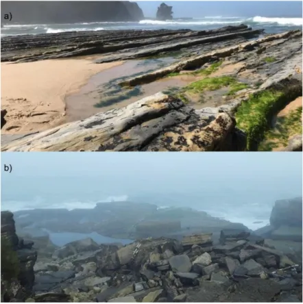
Figure 1 – The sea urchin Paracentrotus lividus………..………..7 Figure 2 – Map of Peniche with the indication of the study sites ( Impacted Shore, 39°22'12.69"N;009°23'7.07"W; Reference Shore, 39°22'02.4"N;9°24'08.07"W) where the specimens of Paracentrotus lividus were sampled……….10 Figure 3 - Sea urchins collection stations. a) Abalo’s Beach, the main point of the oil spill; b) Reference Shore, located 1.5 km away from the rocky shore hit by the oil spill………...………11 Figure 4 - Material used to determine biometric data and to remove the gonads from the sea urchins.. ... 12 Figure 5 - High-performance microwave digestion system (Milestone connect, MA182-001 Ethos up, Italy). ... .13 Figure 6 - Atomic Absorption Spectrometer (Thermo Scientific™ iCE™ 3500 Atomic Absorption spectrometer, Thermo Unicam, Portugal). ... 15 Figure 7 - Total wet weight (WW) and diameter of the carapace (DC) average values of
Paracentrotus lividus at the Impacted Shore (IS) and at the Reference Shore (RS) (Peniche,
Portugal) collected in July, August and September of 2017, after an oil spill event. a) Total wet weight in grams (g); b) Carapace Diameter in centimetres (cm). All values were expressed as mean ± standard deviation. Different letters (a and b) indicate significant differences between sites and different symbols (*, #, +) indicate significant differences within months (Tukey HSD test; p < 0.05)...27 Figure 9 - Relative frequency of Paracentrotus lividus males and females at the Impacted Shore (IS) and at the Reference Shore (RS) (Peniche, Portugal) collected in July, August and September of 2017, following an oil spill event.………..………….…26 Figure 10 - Metal concentrations (in milligram per kilogram, dry weight) on the gonads of
Paracentrotus lividus at the Impacted Shore (IS) and at the Reference Shore (IS) (Peniche,
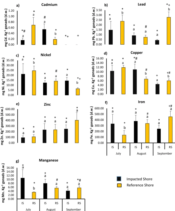
Portugal) collected in July, August and September of 2017, after an oil spill event. a) Cadmium b) Lead, c) Nickel, d) Copper, e) Zinc, f) Iron and g) Manganese. All values were expressed as mean ± standard error. Different letters (a and b) indicate significant differences between sites and different symbols (*, #, +) indicate significant differences within months (Tukey HSD test; p < 0.05)………... 27
xiv Figure 11 - Variation on the gametogenic stages of Paracentrotus lividus at the Impacted Shore (IS) and at the Reference Shore (RS) (Peniche, Portugal) collected in July, August and September of 2017, after an oil spill event. I - Initial; II - Growth; III - Premature; IV - Matures; V - Posture; VI - Post-posture…….………....……...…………....34 Figure 12 - Percentage of lipids and proteins in Paracentrotus lividus gonads at the Impacted Shore (IS) and at the Reference Shore (RS) (Peniche, Portugal) collected in July, August and September of 2017, after an oil spill event. Different symbols (*, #, +) indicate significant differences within months (Tukey HSD test; p < 0.05)……….37 Figure 13 - Percentage of lipids and proteins in female and male gonads of Paracentrotus
lividus gonads at the Impacted Shore (IS) and at the Reference Shore (RS) (Peniche,
Portugal) collected in July, August and September of 2017, after an oil spill event. Different letters (a and b) indicate significant differences between males and females at each sampling station (Tukey HSD test; p < 0.05)...……….………38
xv
List of Tables
Table I: Concentrations of Cd, Pb, Cu, Zn, Ni, Fe and Mn in the gonads of males and females of Paracentrotus lividus collected in the Impacted Shore and in the Reference Shore at Peniche (Portugal) in July, August and September of 2017, following an oil spill event. All values were expressed as mean ± standard error.………..…...32 Table II: Index of histopathological alterations (IHPA) in the gonads of Paracentrotus
lividus collected in the different sampling areas: Impacted Shore (IS) and Reference Shore
(RS) (Peniche, Portugal) in July, August and September of 2017, after an oil spill event. Different letters (a and b) indicate significant differences between sites, different roman numbers (i and ii) indicate significant differences between sexes and different symbols
(#, *, +) indicate significant differences within months (Tukey HSD test; p < 0.05)......36
Table III: Gonad fatty acid composition (%) of Paracentrotus lividus males and females collected at the Impacted Shore and the Reference Shore (Peniche, Portugal) in July, August and September of 2017, after an oil spill event. Values are mean ± standard deviation………..…..……….………...42 Table IV - PERMANOVA tests results for the fatty acids content in gonads of
Paracentrotus lividus collected in the sampling sites at Peniche (Portugal) during the study
period. Only the variables that presented significant results were represented (ρ-value < 0.05). The effects of different sampling stations (Impacted Shore and Reference
Shore) and the different sexes (Male and Female) were considered as factors. The following symbols stand for: df - degrees of freedom; MS - Mean Square..………..………45 Table V: PERMANOVA and Pairwise Method Comparisons tests results for the fatty acids content in gonads of Paracentrotus lividus collected in the sampling sites at Peniche (Portugal) during the study period. Only the variables that presented significant results were represented (ρ-value < 0.05). The effects of different sampling stations (Impacted Shore and Reference Shore) and different months (July, August and September) were considered as factors. The following symbols stand for: df - degrees of freedom; MS –
Mean Square.………...……..….45
Table VI: Spearman correlation matrix (ρ spearman) for metal concentrations [Cd], [Pb], [Ni], [Zn], [Cu], [Fe] and [Mn], and biometric variables in gonads of Paracentrotus lividus,
xvi namely the diameter of the carapace (DC), total wet weight (WW), gonadal weight (GW) and gonadosomatic index (GSI). Significant correlations with p < 0.05 are highlighted in bold (N = 96)…………..………..……….. 47 Table VII: Spearman correlation matrix (ρ spearman) for metal concentrations [Cd], [Pb], [Ni], [Zn], [Cu], [Fe] and [Mn], and the index of histopathological lesions in gonads of
Paracentrotus lividus during the study period. Significant correlations with p < 0.05 are
highlighted in bold (N = 96). IHPA dilat = Index of membrane dilation; IHPA reabs = Index of sex cell resorption; IHPA llp= Index of lipofuscin-like pigments
accumulation; IHPA hyper = Index of hypertrophy in nutritive phagocytes; IHPA atro = Index of atrophy in nutritive phagocytes...………...….49
Table VIII:Spearman correlation matrix (ρ spearman) for metal concentrations [Cd], [Pb], [Ni], [Zn], [Cu], [Fe] and [Mn], and the percentages of lipids and proteins in the gonads of
Paracentrotus lividus during the study period. Significant correlations with p < 0.05 are
highlighted in bold (N = 72).………..……….………50 Table IX:Spearman correlation matrix (ρ spearman) for metal concentrations [Cd], [Pb], [Ni], [Zn], [Cu], [Fe] and [Mn], and the percentages of Saturated Fatty Acids in the gonads of Paracentrotus lividus during the study period. Significant correlations with p < 0.05 are highlighted in bold (N = 72)………50 Table X: Spearman correlation matrix (ρ spearman) for metal concentrations [Cd], [Pb], [Ni], [Zn], [Cu], [Fe] and [Mn], and the percentages of Monounsaturated Fatty Acid in the gonads of Paracentrotus lividus during the study period. Significant correlations with p < 0.05 are highlighted in bold (N = 72).……….……….51 Table XI: Spearman correlation matrix (ρ spearman) for metal concentrations [Cd], [Pb], [Ni], [Zn], [Cu], [Fe] and [Mn], and the percentages of Polyunsaturated Fatty Acid in the gonads of Paracentrotus lividus during the study period. Significant correlations with p < 0.05 are highlighted in bold (N = 72)..………..………...……...52
xvii
List of Abbreviations & Acronyms
Cd – Chemical element Cadmium Pb - Chemical element Lead Ni - Chemical element Nickel Cu - Chemical element Copper Fe - Chemical element Iron Zn - Chemical element Zinc
Mn – Chemical element Manganese PAHs - Polycyclic aromatic hydrocarbons EC50 - Half maximal effective concentration GSTs - Glutathione transferases
AChE – Acetylcholinesterase EEZ - Exclusive Economic Zone IS – Impacted Shore
RS – Reference Shore DC – Diameter of carapace GW - Gonads weight GSI - Gonadosomatic index WW - Total wet weight mg - Milligrams
ml – Milliliter g - Grams
mg/l – Milligrams per liter g/l – Grams per liter mol/l – Mol per litre µl – Microliter
µg/l –Microgram per liter μm –Micrometer
mg/kg -Milligram per kilo rpm -Rotations per minute
xviii GFAAS - Graphite Furnace Atomic Absorption Spectrometry
HCL - Hollow cathode lamps NP - Nutritive phagocytes OV - Vitellogenin oocyte OR - Remnant mature oocytes
IPHA - Index of pathological alterations LLP - lipofuscin-like pigments
IHPA Dilat - Index of membrane dilation IHPA Reabs - Index of sex cell resorption
IHPA LLP – Index of lipofuscin-like pigments accumulation IHPA Hyp - Index of hypertrophy in nutritive phagocytes IHPA Atro - Index of atrophy in nutritive phagocytes FA - Fatty acids
SFA - Saturated fatty acids
MUFA - Monounsaturated fatty acids PUFA - Polyunsaturated fatty acids C14 – Myristic acid
C15 - Pentadecanoic acid, C16 – Palmitic acid C16:1 - Palmitoleic acid C18 – Stearic acids C18:1 trans - Elaidic acid C18:1 cis - Oleic acid C18:2n6 - Linoleic acid C18:3n6 - Gamma-linolenic acid C18:4n3 - Stearidonic acid C20:4n6 – Arachidonic acid C20:1n9 - Gondoic acid C20:1n11 - Gadoleic acid C20:2n6 – Eicosadienoic acid C20:3n6 - Homo-γ-Linolenic acid C21 - Henicosanoic acid C22:1n9 - Erucic acid
xix EPA - Eicosapentaenoic acid
ALA - Alpha linolenic acid NO - Nitric oxide
CoA – Coenzyme A
PCA - Principal Component Analysis AChE – Acetylcholinesterase
1. INTRODUCTION
______________________________________________
1 1. Introduction
1.1 Marine Environments and Pollution
Marine ecosystems are the result of millions of years of several evolutionary processes and represent the largest reservoirs of water on Earth, covering over 70% of the planet's surface. Because they are the largest and oldest of the Earth’s ecosystems, they have almost the double of the number of organisms, serving as shelter for a high variety of species (Boeuf, 2011; Goulletquer et al., 2014). The marine domain is extremely important for maintaining life on the planet, since their ecosystems carry out about 50% of global primary production, have important ecological functions (such as control over climate) and they have resources which are essential for the social and economic development of the world, mainly for coastal communities (Cicin-Sain et al., 2015; Aslan et al., 2018).
In the marine domain, ecosystems are composed basically of habitats ranging from the seas to the coastal zones. In the coastal zone there are several habitats with unique characteristics, and ecological and commercial importance such as coral reefs, mangroves and intertidal systems (rocky, sandy, and muddy shores) (Baker et al., 2009). These ecosystems have a high biodiversity value, can play a key role in mitigating global climate change by its ability to store carbon and are responsible for much of the maintenance of productivity (especially fisheries) (Torres and Hanley, 2016). Coastal populations interact with these systems in different forms, mainly through artisanal fishing. Despite their importance, indiscriminate use of their resources and services by humanity is a fact and it has great direct and indirect impacts in those environments (Halpern et al., 2008).
In the last decades, with increasing population growth and the consequent increase in human activities, coastal marine waters have entered an intense process of pollution (Waldichuk, 1977; Dong, 2017). The World Health Organization (2000) defines coastal pollution as “the introduction by man, directly or indirectly, of substances or energy into the marine environment, including estuaries, which results or is likely to result in such deleterious effects such as harm to living resources and marine life, hazards to human health, hindrance to marine activities, including fishing and other legitimate uses of the sea, impairment of quality for use of sea water and reduction of amenities”. The reasons why
2 pollution occurs can be diverse, however the most frequent causes are associated to plastic debris, sewage and industrial effluents, oil spill and “no-point source”; this last refers to a source of pollution that issues from widely distributed or pervasive environmental elements. Aslan et al. (2018) affirms that since the sea became the final recipient of these elements, besides organic matter, many contaminants such as hydrocarbons, inorganic and chemical elements can be found accumulated in sediments or tissues of organisms (Martinho, 2016; Dong, 2017).Some of these substances are biodegradable while others are persistent. Their cumulative impact to the coastal marine environment over a long period could be quite harmful. This scenario can have devastating consequences for marine life and habitats on which marine organisms depend on (Elliott, 2003; Mearns et al., 2013).
The accumulation of contaminants in the marine environment can lead to a range of consequences in different levels (Gómez et al., 2004; Lohmann & Belkin, 2014). Some pollutants can be extremely toxic or fatal to some aquatic organisms during a short period of exposure, even at low concentrations; on the other hand, other pollutants may present a strenuous effect, that is, they do not kill organisms immediately, however they can cause chronic sub-lethal effects such as growth retardation, physiological stresses, and reproductive failures (Waldichuk, 1977; Dong, 2017; Hylland et al., 2017; Tornero and Hanke, 2017). Within this context, humans are also susceptible to toxicity through the consumption of contaminated seafood, having their health compromised with, for example, neurological development deficits, and disturbances of the hormone system (Waldichuk, 1977).
The issue of marine environments pollution is serious and has made it increasingly worrisome as it is rising at an alarming rhythm. Solving this problem of pollution in the oceans is a difficult and comprehensive task, since it is necessary to understand which are the main causes and the main effects (Cross et al., 1985).
1.2 Marine Pollution by Oil Products
Among the environmental problems that strongly affect the health of oceans, pollution by oil or its derivatives stands out (Moore & Dwyer, 1974; Martinho, 2016). Marine oil pollution comes mainly from oil spill, offshore oil production, marine transport and urban
3 pollution (ITOPF, 2011). According to the World Energy Balance (2018), millions of tons of oil are spilled into the sea annually; these numbers tend to become larger as the oil industry expands.
The impact of oil or its derivatives on marine fauna is relative, depending on the viscosity, toxicity, amount of oil and the sensitivity of different animals to contact with the substance (IPIECA, 1995). Crude oil is composed by a complex mixture of organic compounds, basically consisting of hydrocarbons (PAHs), some heterocyclic compounds and some heavy metals (Dupuis & Ucán-Marín, 2015); mainly due to its composition and properties, the consequences of marine contamination can be diverse and drastic.Because it is a hydrophobic substance, the oil does not mix with water and it forms a film on it; thus, there is a blockage of light incidence necessary for photosynthesis by algae and other photosynthetic organisms (Moore & Dwyer, 1974). This impact affects the entire marine ecosystem and causes damage to the environment, such as direct death of organisms, alterations in the food chain and interference with the reproduction rates of animals (Lee and Page, 1997).
Heavy metals and PAHs contained in oil spill represent a significant source of contamination, being highly toxic to organisms (Curl & O'Donnell, 1977; Vikas & Dwarakish, 2015; Lu et al., 2018). Metals can be classified into two different classes: essential (manganese, cooper, iron, zinc) and non-essential (nickel, cadmium and lead) (USFDA, 1993; Burger et al., 2002; Veiga, 2015). This classification is associated with the biological functions that these elements perform in organisms. Essential metals play important roles in the metabolic processes of organisms when minimally present; on the other hand, non-essential metals have high toxicity and generally do not play any important role in organisms (Jakimska et al., 2011).The absorption or direct contact of some metals with the tissues of organisms can lead to damage and disturbance of cell walls and cellular functions at the molecular level. If the concentration and duration of exposure to these toxic contaminants is high enough, the organisms can undergo potential physiological damage, with structural changes at the cellular level that can culminate in death (Curl & O'Donnell, 1977;Soualili et al., 2008; Vikas & Dwarakish, 2015; Lu et al., 2018).
4 There are several serious cases of oil spills around the world. A study carried out by Fernández et al. (2006) evaluated the toxicity of the soluble fraction of fuel oil spilled on the embryo development of the sea urchin Paracentrotus lividus (Lamarck, 1816) when an oil tanker sank in November 2002 on the coast of Galicia. Analyses of PAHs and four metals (copper, cadmium, lead and zinc) were conducted. The study observed that embryo development was strongly inhibited when associated with a fuel oil content in water and the effective concentration which provoked a delay in the successful embryogenesis of 50% of the population (EC50) was 2.3% of fuel oil. Some studies of the composition of samples collected in the oil spill area showed an increase of Zn, Cu and Pb concentration in seawater after the spill (CSIC, 2003). In this work, despite the observed metal concentrationswere below the median effective concentrations reported, the authors did not rule out the possibility of a synergistic effect.When combined with some organic substances, metals can form highly toxic metal-organic complexes (Veiga, 2015); this event has often been observed for different chemical mixtures and marine organisms, including the sea urchin
Paracentrotus lividus (Lamarck, 1816)
(Vanegas et al., 1997; Fernández & Beiras, 2001).
Another serious accident happened in December 2000 on the Portuguese coast, in which a ship ran aground and spilled a large quantity of fuel oil into the sea. Sometime after the oil spill, mussels collected at stations near the ship had higher and lower values of glutathione transferases (GSTs) and acetylcholinesterase (AChE) activity, respectively (Moreira et al., 2004). GSTs are enzymes that act on the biotransformation of xenobionts (Beiras, 2018). In turn, acetylcholinesterase is an enzyme responsible for the hydrolysis of the neurotransmitter acetylcholine and the completion of transmission of nerve impulses in cholinergic synapses; its inhibition has important implications in the health of organisms (Castellanos et al., 2018). In this sense, besides demonstrating the efficacy of these biomarkers as indicators of exposure to this kind of pollution, these results confirm the contamination by the oil. Similarly, in January 2017 an Indian coastal city named Chennai was impacted by a large oil spill caused by a collision that occurred between two cargo ships, spreading tons of heavy oil in the Bay of Bengala. Subsequent studies indicated that samples collected in the spill area showed relatively high levels of heavy metals and highly toxic polycyclic aromatic hydrocarbons (PAHs) (Han et al., 2018).
5 Portugal is a country with almost 1000 square kilometres of coast which, together with the archipelagos of Azores and Madeira, form the third largest Exclusive Economic Zone (EEZ) in the European Union. Because of all this extension, the exposure to the potential risks of accidents is large and there is an urgent need to develop techniques for environmental monitoring and forecasting of future damages (Borges, 2015). Disaster planning for oil spills requires learning from past events. However, it is always a challenge as the consequences depend on the geographic, ecological, social and temporal contexts in which the disaster occurs. Therefore,it is important to increasingly understand the mechanisms involved and the potential consequences to the ecosystem caused by the spill.
1.3 Bioindicators as a tool for the monitoring and evaluation of marine pollution
In the last years, many environmental quality evaluation works have been carried out, using bioindicator organisms as a tool (Branco et al., 2008; Soualili et al., 2008; Chiarelli &
Roccheri, 2014). According to McGeoch (1998), the term bioindicator refers to organisms
that are resistant to some levels of contamination, that is, they do not die when exposed to toxic agents and derivatives; instead, they provide assertive information about the environmental conditions of the places where they live (Gonçalves et al., 2013). This happens because the response of each organism is strongly influenced by the physical, chemical and biological conditions of the environment (e.g. temperature, humidity, winds and radiation) as well as by physiological, structural, nutritional and morphological conditions. In short, when the environment is contaminated, the bioindicator organism responds by changing some aspect(s) of its physiology; these changes, in turn, can be measured and may represent significant disturbances in the environment in which these organisms live (Market et al., 2003).
Marine invertebrates have been widely used as bioindicators in environmental monitoring under different levels of anthropic impact and in assessments of ecosystem health (Rainbow, 2002). Among the features that justify the frequent choice of invertebrates as biological indicators, the facts that these organisms have long life cycles, being sessile or with low mobility, are easy to sample with relatively low costs and are sensitive to different concentrations of environmental pollutants, providing a wide range of responses to different
6
levels of environmental contamination, stand out (Hodkinson and Jackson, 2005). Furthermore, invertebrates occupy a key position as intermediate consumers in the pelagic and benthic food chains of aquatic ecosystems; once affected, even if in the long term, the ecosystem would be impacted (Chiarelli & Roccheri, 2014).
1.4 The Sea urchin Paracentrotus lividus as a bioindicator
Several studies indicate that sea urchins living in or exposed to water contaminated by hydrocarbons or metals, can present alterations associated with their reproductive, physiological and morphological aspects, which vary according to the levels of pollution (Danis et al., 2005; Branco et al., 2008; Soualili et al., 2008; Schäfer and Kohler, 2009; Chiarelli & Roccheri, 2014). The sea urchin Paracentrotus lividus, in special, has been qualified as an excellent bioindicator species of contaminants at sea. This qualification is justified by its tolerance to physiological pressure, resistance to high levels of salinity, temperature, and desiccation or food shortage for long periods of time (Allain, 1975; Hereu, 2005). In addition, P. lividus is a widely distributed and abundant species, establishing very well in specific regions, thus allowing an analysis of micro regions. Finally, they are animals easy to collect, to handle and to maintain in the laboratory. Its value as a bioindicator for heavy metals and polycyclic aromatic hydrocarbons contaminations has been documented in several laboratory and field studies (Warnau et al., 1995a, 1996, 1998; Geffard et al., 2001; Soualili et al., 2008; Rocha et al., 2018; Ternengo et al., 2018).
Paracentrotus lividus (Figure 1) is a species of sea urchin in the Parechinidae family,
which lives in rocky substrates and in seagrass meadows, from shallow water to about 20 m depth (Tenuzzo et al., 2012). These organisms feed mainly on aquatic vascular plants and algae but may also feed on debris in the water column (Boudouresque & Verlaque, 2001). This species has a natural and wide distribution in the rocky bottoms of the shores of the eastern Atlantic, from Ireland to the south of Morocco and across the Mediterranean Sea. In countries such as France and Japan, P. lividus gonads are widely consumed (Fernandez & Boudouresque, 2000; Boudouresque & Verlaque, 2001), and aspects such as color, size, texture and granularity of the gonads greatly influence their trade and consumption (Blount & Worthington, 2002; Pearce et al., 2002). In the winter and spring months, the gonads reach a larger volume, a better texture and flavor, and thus these animals are harvested more
7 intensely (Ouréns et al., 2013). In recent years, its commercial exploitation has grown, and the wild populations have decreased drastically in some areas (Ceccherelli et al., 2011; Pais
et al., 2011; Bertocci et al., 2014; Sartori et al., 2016).
Figure 1: The sea urchin Paracentrotus lividus.
Like most echinoids, P. lividus are gonochoristic organisms; that is, they have separate sexes. These animals are devoid of sexual dimorphism and perform external fertilization, releasing simultaneously their gametes in the water. According to Boudouresque and Verlaque (2001), in a general way, reproduction occurs once or twice a year; however, it can vary among geographic areas depending on the environmental conditions.
The gonads of P. lividus are considered a source of proteins, lipids, fatty acids and minerals (Volpe et al., 2018). This whole nutritional apparatus is closely associated with the gender and the different gametogenic stages of the organism. At the beginning of the gametogenic process, there is a higher nutritional need and macrophagic cells - called nutritive phagocytes - synthesize proteins and other essential nutrients to provide energy and to promote the growth and maturation of the gametes (Carboni, 2013). As the gametes are released, the nutritional need decreases. Aspects of reproduction and gametogenic cycle can be altered by unfavorable conditions and limitation of food supply (Lozano et al., 1995).
Paracentrotus lividus is abundant on the Portuguese coast, colonizing different biotopes
(Girard et al., 2012; Jacinto et al., 2013; Domínguez et al., 2015). Their gonads make it a highly appreciated seafood in the country and, as previously presented, this echinoid is used as an excellent bioindicator of pollution in the marine environment. Parallel to these facts,
8 the Lusitanian coast has been increasingly exposed to anthropogenic impacts, mainly due to the occupation of areas close to coastal zones. In this sense, it is important to understand the importance of this species as a bioindicator of pollution in the habitats where they live. Thus, analyses performed on P. lividus can represent useful tools to evaluate the effects of pollutants in marine environments and when the results demonstrate a relationship with the concentration of these pollutants, the interpretation of toxicities and exposure conditions becomes clearer.
1.5 Objectives of the study
The increasing installation of human activities in coastal ecosystems has arguably a strong environmental impact, which is represented not only by the increasing degradation of coastal zones but also by the increased risk of serious environmental damage. In addressing this issue, the need for environmental assessment studies is recognized in order to ensure the permanence of the biota and to analyse the impacts that these problems may have on the long and short term for human life. Environmental assessment studies, therefore, are relevant tools that can aid in the management and prevention of environmental accidents, having also a fundamental role in planning and as a support in decision-making processes. In short, works such as these make it possible to understand the sensitivity of ecosystems to changes, the resulting impacts on biodiversity, and the risks to public health directly or indirectly related to environmental quality in those areas.
In July 2017, an accidental spill of 3 tons of naphtha occurred in Abalo’s Beach, on the coast of Peniche (Portugal). The fuel oil spill was originated in the system of feeding a boiler of a company. In this scenario, the main objective of the present study is to contribute to the environmental impact assessment of this oil spill by using Paracentrotus lividus as a bioindicator of the presence of contaminants. To achieve the main purpose of this work, the following specific objectives are proposed:
a) To perform analysis for the quantification of the elements Cd, Pb, Ni, Fe, Mn, Zn, Cu in the gonads of the sea urchins, in order to assess the degree of contamination derived from the oil spill.
9 b) To perform biometric, histological and biochemical analysis to investigate if the spill affected the following physiological and biochemical responses in P. lividus:
• The size and weight of the individuals (carapace diameter and individual wet weight); • The gonadal somatic index; the development of the gonads and the stages of gametogenesis;
• The characterization and quantification of total lipids, proteins and fatty acids; c) To detect the presence of anomalies, lesions or other histopathological changes in the appearance of the gonads that could be indicative of an environmental pollution scenario.
2.
MATERIALS AND METHODS
10 2. Materials and methods
2.1 Study Area
This study was performed on 2 rocky shores of the coastal zone of Peniche (Portugal). The specimens of P. lividus were collected at two distinct sampling areas: the first one, Abalo’s Beach (Impacted Shore - IS - 39°22'12.69"N;009°23'7.07"W), the central point where the fuel oil spill happened; the second area was not directly impacted by the spill,
functioning in the present study as a reference area for comparison (Reference Shore - RS -
39°22'02.4"N;9°24'08.07"W). The reference area is located 1.5 km to the west of the Impacted Shore (Figure 2).
Figure 2: Map of Peniche with the indication of the study sites ( Impacted Shore, 39°22'12.69"N;009°23'7.07"W; Reference Shore, 39°22'02.4"N;9°24'08.07"W) where the specimens of Paracentrotus lividus were sampled.
Abalo’s beach is characterized by a very irregular topography, presenting a very heterogeneous relief, due to the existence of large rocky fragments with a diverse morphology. In this beach, three distinct zones can be considered and easily discernible by their specific content: the supralittoral zone, the eulittoral zone and the sublittoral zone, as in most rocky shores of the world. The rocky shore of Abalo has a low hydrodynamics, is very fragmented and has also a small sandy area. Abalo’s beach is located next to an area where there are a series of industrial activities and industrial companies, with intense
11 movement. In addition, it is common to observe in the area shellfish harvesters distributed by the rocky shores at low tide; all these features make this site more susceptible to human impacts (Figure 3a).
The area used as a reference in this work, on the other hand, has exposed rocky shores, formed by smooth seawalls, which present a strong hydrodynamism due to the high impact of waves; therefore, this area possibly has less habitat diversity and a high rate of primary productivity due to an high nutrients flux (Satyam & Thiruchitrambalam, 2018). There is no sandy area in this location and the site’s accessibility is difficult compared to the Abalo’s Beach (Figure 3b).
Figure 3: Sea urchins collection stations. a) Abalo’s Beach, the main point of the oil spill; b) Reference Shore, located 1.5 km away from the rocky shore hit by the oil spill.
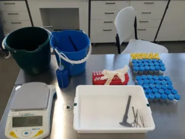
2.2 Collection of Sea Urchins and Determination of Biometric Data
On each rocky shore, 30 sea urchins were collected once a month during the months of July (immediately after the spill), August and September of 2017. The individuals were
12 collected manually and at random in the morning, during low tide of spring tides. As the tidal level was in the infralittoral zone, it was possible to perform the activities between the rocks and in the several intertidal pools left by the ebbing tide. After collection, the animals were transported to the aquaculture laboratory of MARE-IPLeiria - Marine and Environmental Sciences Centre, Campus of the Polytechnic of Leiria, inside buckets with marine water. In the laboratory, the horizontal diameter of the carapace (DC
;
± 0.1 mm accuracy) of each specimen was measured with the help of a caliper. Also, the total wet weight (WW; ± 0.01 g) and the gonads weight (GW; ± 0.01 g) of each individual were obtained using an analytical balance (AE ADAM PGL 3002, Milton Keynes, England) (Figure 4). With this information, it was possible to calculate the gonad somatic index (GSI) from the following formula (Marsh et al., 2013):Gonad somatic index (GSI) = ( Gonad weight
Total weight of individual) x100
Figure 4: Material used to determine biometric data and to remove the gonads from the sea urchins.
All 30 individuals (from each month) were histologically analysed; for that purpose, one of the five gonads in each individual was removed, fixed and preserved in 4% buffered formalin for further histological analysis (James et al., 2018). The other four gonads were stored in individual tubes at - 80°C and later lyophilized for biochemical analysis and for the determination of the presence of metals.
13 2.3 Analysis of the Concentration of Metals
2.3.1 Acid digestion of biological samples by microwave radiation
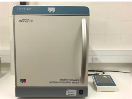
For the determination of the concentration of the metals cadmium (Cd), lead (Pb), nickel (Ni), iron (Fe), manganese (Mn), zinc (Zn) and copper (Cu), 16 sea urchins (8 males and 8 females), from each sampling moment and from each rocky shore, were randomly selected. The samples of the gonads of these sea urchins were first acid digested. Approximately 100 mg of P. lividus gonadal samples were accurately weighed into a Teflon microwave digestion tube. Then, the samples were digested using 10 ml of HNO3 (69.6%, AnalaR NORMAPUR, VWR pro lab chemicals, France) in a high-performance microwave digestion system (Milestone connect, MA182-001 Ethos up, Italy) (Figure 5).After being digested, the samples were transferred to a 50 ml plastic bottle and stored in a freezer until analysis by Atomic Absorption Spectrometry.
Figure 5: High-performance microwave digestion system (Milestone connect, MA182-001 Ethos up, Italy).
2.3.2 Analysis of metals by Atomic Absorption Spectrometry
The concentrations of non-essential (Cd, Ni and Pb) and essential (Fe, Mn, Zn and Cu) metals in the samples of the gonads were determined by Atomic Absorption Spectrometry (AAS) (Thermo Scientific™ iCE™ 3500, Thermo Unicam, Portugal) (Figure 6). The two
14 types of atomizers most used in AAS are the flame and the graphite furnace. The choice between one technique and another is directed according to the levels of metals expected in the samples. Atomic Absorption Spectrometry by flame is the technique most used for analysis at milligrams per liter levels, while the Atomic Absorption Spectrometry with electrothermal atomization in a graphite furnace (GFAAS) is used to quantify at low concentrations (micrograms per liter) (García & Báez, 2012). In this sense, Fe, Zn and Mn concentration in gonads were determined using flame atomic absorption spectrometry, using Acetylene and compressed air, while the quantifications of Cd, Cu, Ni, and Pb were carried out by AAS with graphite furnace (Thermo Scientific™ iCE™ 3500 Atomic Absorption spectrometer, Thermo Unicam, Portugal), using Argon.
Standard solutions of Cadmium (Cadmium standard solution (1000±0.0002 g/l AA Panreac Quimica SLU, European Union); Lead (AAS Standard Lead 1000 mg/l in nitric acid 2%, AVS TITRINORM, VWR international Haasrode, Belgium); Nickel (Nickel standard solution traceable to SRM from NIST Ni(NO3)2 in HNO3 0.5 mol/l, 1000 mg/l Ni contains nickel (II) nitrate Certipur®, Merck KGaA, Darmstadt Germany EMD Milipore Corporation); Iron (Iron standard solution traceable to SRM from NIST Fe(NO3)2 in HNO3 0.5 mol/l 1000 mg/l); Manganese (Manganese standard solution traceable to SRM from NIST Mn(NO3)2 in HNO3 0.5 mol/l 1000 mg/l Mn Certipur®, Merck KGaA, Darmstadt Germany EMD Milipore Corporation); Zinc (Zinc standard solution traceable to SRM from NIST Zn(NO3)2 in HNO3 5 mol/l 1000 mg/l Zn Certipur®, Merck KGaA, Darmstadt Germany EMD Milipore Corporation); and Copper (Copper standard solution traceable to SRM from NIST Cu(NO3)2 in HNO3 0.5 mol/l 1000 mg/l Cu Certipur®, Merck KGaA, Darmstadt Germany EMD Milipore Corporation) were used to construct the calibration curve for each metal and all standards and reagents were diluted using ultra-pure water.
For GFAAS, a volume of 20 µl of magnesium nitrate (Magnesium Matrix Modifier, Magnesium nitrate, Matrix Modifier Solution, 1% Mg (NO3) 2 in 2% HNO3 Specture, ThermoFisher (KAndel) GmbH, Karlsruhe, Germany) was added in each sample aliquot. The matrix modifier is added to samples to reduce analyte loss by decreasing its volatility or increasing matrix volatility during the decomposition reaction which occurs due to high temperatures (Voth, 1983). Magnesium Nitrate was added in all metals analysed in this work except Nickel.
15 As radiation source for the analysis of each metal, hollow cathode lamps (HCL) were used, where each cathode consists of a different element. The detection limits of the techniques used were 0.008 µg/l for Cd, 0.4 µg/l for Cu, 0.1 µg/l for Fe, 0.06 µg/l for Pb, 0.035 µg/l for Mn, 0.3 µg/l for Ni and 0.1 µg/l for Zn for this equipment.
Figure 6: Atomic Absorption Spectrometer (Thermo Scientific™ iCE™ 3500 Atomic Absorption spectrometer, Thermo Unicam, Portugal).
The determination of metal concentrations was performed from the method of standard additions which is a quantitative method generally used when the sample of interest has several components that result in matrix effects, so that additional components can reduce or enhance the analyte absorbance signal (Smith, 1983). Samples of sea urchin gonads were determined on a dry weight basis, as milligrams per kilogram, and the determination of concentrations was performed with 3 replicates. In all quantifications, a blank (1% HNO3) was subtracted and mean values and standard deviations were calculated (Pedro et al., 2013).
To verify the accuracy of the method, a known amount of analyte was added to the natural test sample matrix and its response was measured (recovered) in the assay compared to an identical elevation in the standard diluent. The objective of this procedure was to evaluate if the methodology used in this work was valid, thus allowing the repeatability of the analysis.
16 2.4 Histological Analysis
For histological analysis, the gonads were fixed in 4% buffered formalin for 24h and then inserted into a solution of 70% ethanol (also during 24h). Subsequently, three steps were followed: dehydration, clarification and impregnation. The gonads were processed in a tissue processor (Leica TP 1020, Nussloch, Germany), in which they passed through different ethanol solutions with increasing concentrations. After going through the dehydration step, the gonads were clarified in order to remove the dehydrating agent by replacing it with a liquid miscible with the impregnation medium (liquid paraffin). The intermediate agent used in the diaphanization was xylol. Lastly, gonads were impregnated in paraffin, which gave structure to their tissues. After being processed, the gonads were included in paraffin to obtain blocks that posteriorly allowed obtaining sections without destruction of the tissues. For this, the tissues were embedded in a medium which supports them: paraffin at 60°C. With the material included in paraffin, microtomy was performed on an Accu-Cut® SRM ™ 200 Rotary Microtome obtaining 5 µm thickness sections. The sections were dried and preserved in a kiln at 37°C (Binder, Tuttlingen, Germany) for 24 hours, until subjected to the staining technique. The staining was performed according to the Hematoxylin-Eosin staining technique.
For completion of the gonadal tissue preparations, the Coverquick 2000 Path® assembling medium (San Francisco, USA) was used and the slides were dried for 24 hours at room temperature. Finally, after obtaining the slides, the gonads of Paracentrotus lividus were observed under a composite optical microscope (Leica DM 2000 LED, Wetzlar, Germany), photographed using a microscope camera (Leica® MC170 5MP HD) and the combined LAS V4.4.0 software (Leica Application Suite) for monitor display (Leica Microsystems GmbH). From there, it was possible to determine the sex of individuals, which allowed the calculation of the sex ratio and to characterize the different stages of gametogenesis (Santos et al., 2019).
The characterization of the gametogenic cycle of sea urchins was performed according to Byrne (1990) and Spirlet et al. (1998). The growth pattern of both testicles and ovaries has been divided into six stages and were used to document the major events of the
17 individuals’ spermatogenic and oogenic cycle. The stages were identified based on the following characteristics:
• Stage I - Early Phase: In this phase, the gonad lumen is completely filled with nutritive phagocytes (NP) - the non-germinal accessory cells - which vary in size and colour. It is possible to observe the presence of primary oocytes along the ovarian/testicular ascinal wall, which in turn is covered by a thin basophilic layer. Eventually, the presence of remnant oocytes, not expelled in the last spawning, can be observed and are later reabsorbed by NP.
• Stage II - Growth: It is from this stage that cell growth gradually begins. The lumen of the gonad is occupied by nourishing phagocytes, and the pre-yolk oocytes, which are still along the ascending wall of the ovary/testis, begin to grow as they absorb nutrients supplied by nourishing phagocytes (NPs). The female gamete assumes the typical form of vitellogenin oocyte (OV). In males, sperm begin to protrude centrally and the basophilic layer increases.
• Stage III - Pre-maturation: In this phase there is a reduction in the presence of NPs and an increase in oocytes in number and size. It is still possible to observe pre-yolk oocytes attached to the gonadal tissue wall, surrounded by nutritive phagocytes (NPs). As vitellogenesis occurs, there is also a migration of mature oocytes to the centre of the acino. At this stage vitellogenesis is a continuous process and oocytes in all states are present. When primary oocytes reach their maximum size, they immediately mature and the eggs accumulate in the ovarian lumen. In males, premature testicles contain spermatocyte columns along the ascinal wall and sperm accumulates in the lumen.
• Stage IV - Maturation: A large number of mature oocytes (90 μm in diameter) occupy the lumen of the acini. Eventually, nutritive phagocytes can be observed forming a very thin layer near the ascinal wall, as well as some small vitellogenin oocytes (10 to 60 μm diameter) present in the ascinal wall, indicating that the vitellogenin process has not yet been completed. In males, the mature testes are full of sperm and the nutritive phagocytes are limited to the ascinal wall.
18 • Stage V - Posture: At this stage, the acini contracts and is emptied, however remnant mature oocytes (OR) can eventually be observed, which will later be reabsorbed by nutritive phagocytes (NPs). In females there are spaces between the unreleased oocytes. In males, this stage has a very similar aspect to stage IV, differing only by the presence of spaces in the lumen and the smaller amount of sperm. Also, the ascinal wall looks thin and sperm may be present in the gonoduct.
• Stage VI – Spent stage: At this stage, the ovaries have thin ascinal walls, lose their internal structure leading to disorganization, and it is possible to observe mature oocytes and pre-vitelline oocytes, which have detached from the ascinal wall. Any oocytes present in the ovary at this stage will probably be reabsorbed. One may also observe a meshwork of nutritive phagocytes around the periphery that may have begun to sequester reserves for the next oogenic cycle. In males, thin ascinal walls and a meshwork of nutritive phagocytes are observed on the periphery of the testis, as well as spaces created by the absence of sperm.
During the identification of the gametogenic stages of the sea urchins, careful observations to detect the presence of anomalies, lesions or other histopathological changes in their appearance that could be indicative of an environmental pollution scenario, were also performed. Thus, an index of histopathological alterations (IPHA) was determined by analysing the gonads lesions and classifying these lesions on a scale from 0 (no lesions) to 3 (severe) in the observed slides (Vaschenko et al., 2012). The changes observed were: oocyte resorption, accumulation of lipofuscin-like pigments (LLP), nutritive phagocyte hypertrophy or atrophy. High amounts of lipofuscin are easily observed in extracellular spaces and in the oocytes of some individuals, since the colour of large globular inclusions are from gold–yellow to brown–yellow. For the identification of hypertrophy of nutritive phagocytes (NPs), some accumulation of larger cells is observed due to the increased synthesis of their basic constituents and their volume. NPs atrophy, in turn, is found when smaller cells are identified as a result of the decreased nutrition, metabolism, and synthesis needed to renew their structures. Finally, resorption of oocytes (phagocytosing of atretic oocytes by NPs) is found when a higher volume density of atretic oocytes are observed. To
19 calculate the IPHA, the total score of the different pathologies observed in the gonads was divided by the number of individuals analysed (Vaschenko et al., 2012).
2.5. Biochemical Analysis
For the biochemical analysis, 12 individuals (6 males and 6 females) were randomly chosen for each month. All the samples were homogenized (Ystral ® D-79282, Ballrechten-dottingen, Germany) and all following analyses were performed in duplicates.
2.5.1. Total lipids
The determination of the total lipids present in the gonads was performed based on the protocol of Bligh and Dyer (1959). About 20 mg of each gonad sample were weighed into eppendorf’s in an analytical balance (Sartorious); then 500 µl of chloroform (CHCl3, Prolabo ® VWR, Lyndhurst, South Africa), methanol (CH3OH, HiPerSolv. CHROMANORN, Prolabo ® VWR, Fontanay-Sous-Bois, France) and ultra-pure water (18,02 MΩ, HiPerSolv. CHROMANORN, Prolabo ® VWR, Fontanay-Sous-Bois, France) were added. After addition of these reagents, with the objective of separating the layers, centrifugation was carried out at 2000g during 10 minutes at 4 °C (Eppendorf Centrifuge 5810 R, Billerica, EUA). Then, 100 µl of the organic phase were withdrawn into glass tubes. For the determination of the amount of lipids, a standard curve was prepared using a stock solution of known initial concentration of tripalmitin (C51H98O6, Alfa Aesar, Karlsruhe, Germany) in chloroform (3.2 mg/ml -1). Thereafter, 500 µl of sulfuric acid (95% H2SO4, Chem-Lab, Zedelgem, Belgium) was added in both standards and samples. Finally, all tubes were placed in a 200 °C kiln (Memmert oven, Schwabach, Germany) for 15 minutes. After removal from the kiln, and after cooling, 1.5 ml of ultrapure water was added to all tubes. Finally, each sample was divided into 4 x 300 µl into a microplate. A plate reader (Synergy H1 Hybrid Reader Biotek ® Winooski, USA) was used for reading the plates and absorbance was read at 375 nanometres.
20 2.5.2. Proteins
Protein determination in the gonad samples was performed according to the method of Lowry et al. (1951). To perform this extraction, approximately 0.02 to 0.03g of P. lividus gonad samples were weighed into glass tubes, using an analytical balance (Sartorius). 500 µl of saccharide solution in EDTA was added and then the tubes were placed on the shaker for 5 minutes (130 rpm). Thereafter, 4.5 ml of N/100 Sodium Hydroxide (NaOH Prolabo® VWR, Belgium) was added to make a total of 5 ml. The tubes were shaken by inversion (5x) and then they were placed in the refrigerator for 30 minutes. After this time, a filtration was performed by the pleat filter. 200 µl of the filtrate were transferred to glass tubes, and then 4.8 ml of N/100 Sodium Hydroxide were added to make a total of 5 ml. Once this was done, it was stirred again by inversion (5x).
For the determination of proteins, 300 µl were transferred into Eppendorf tubes. Parallel to this, two blanks were prepared with 300 µl of N/100 Sodium Hydroxide and standards. The standards were made from a solution of bovine albumin 1 mg/ml (CAS-No 90604-29-8, USA) and ultra-pure water. Thereafter, all samples, blanks and standards were treated with an alkaline solution of copper (1 ml in each tube), vortexed and incubated at 37 ° C for 3 minutes in the oven (Memmert oven, Schwabach, Germany). Then, 100 µl of the Folin-Colciteau reagent (diluted 1:1) was added and the tubes were vortexed again and incubated under the same conditions. Finally, 200 µl were transferred to wells in microplates and the reading was carried out at 750 nanometres in a plate reader (Synergy H1 Hybrid Reader Biotek ® Winooski, USA).
2.5.3. Fatty acids
For the quantification of fatty acids, the protocol established by Lepage and Roy (1986) was followed. Samples were weighed into glass tubes on an analytical balance (Sartorius, Göttingen, Germany). 2 ml of a solution of methanol (CH3OH) + sulfuric acid (2%) (H2SO4, Merk, Darmstadt, Germany) were added to each sample and then these were placed in 80 °C bath for 120 minutes. After the samples cooled, 1 ml of ultrapure water (18.02 MΩ, HiPerSolv CHROMANORN, Prolabo ® VWR, Fontanay-Sous-Bois, France) and 2 ml of heptane (C7H16, Fisher Scientific, Bishop Meadow Road, England) were added. Finally, the
21 samples were centrifuged (Eppendorf Centrifuge 5810 R, Billerica, USA) at 1500 g, for 5 minutes for subsequent collection of the organic phase (1 ml of supernatant) and transfer to vials. The samples in the vials were analyzed by Gas Chromatography (GC) (Finnigan TraceGC ultra Massachusetts, USA), according to Masood et al. (2005). Fatty acid characterization was then performed using two commercial standards (Polyunsaturated Fatty Acid Mix No. 3 analytical standard from menhaden fish oil [PUFA n-3], Supelco®, Bellefonte, USA; Supelco 37 component FAME mix, Sigma-Aldrich ®, St. Louis, USA).
2.6. Data Analysis
The statistical techniques used in this study were performed with the statistical software IBM® SPSS® Statistics 26, except for the PERMANOVA, which was performed in PRIMER7. In the statistical analysis of the data, the significance level α = 0.05 was assumed. Prior to any statistical analysis, all data were tested for normal distribution (using the non-parametric test Kolmogorov-Smirnov) and homogeneity of variance (Levene's test) and, when necessary, the data that did not meet these assumptions were transformed. When the transformations did not remove the heterogeneity, the analyses were performed on the untransformed data (whenever n > 30), since analysis of variance is quite robust to departures from their assumptions (Underwood, 1997). On the other hand, if none of the assumptions of normality and homogeneity were met for samples with n<30, Kruskal-Wallis nonparametric tests were performed.
Depending on the nature of the data - that is, whether they were parametric or non-parametric - Student´s t-tests and Mann-Whitney test were conducted to compare means of variables between the two sampling stations and between the sex of individuals. Given this, Student's t-tests for independent samples were conducted to compare means of biometric and histological variables, whereas Mann-Whitney tests were performed to evaluate the differences between the means of biochemical variables and metal concentrations.
Kruskal-Wallis nonparametric tests were conducted to evaluate the variation between the months of study on the concentrations of metals in the gonads and on some physiological and biochemical responses (the percentage of lipids, proteins and fatty acids in the gonads) of the organisms.
22 Two-Way ANOVA or PERMANOVA analyses (according to data normality and heterogeneity) were used to test the effects of the sampling stations and the animals’ sex, as well as the interaction between the 2 factors, on biometrics responses and gonadal lesions. The significant effects detected were then subjected to post-hoc tests: (i) Tukey HSD to analyse the individual effects of the factors and (ii) Bonferroni tests to analyse the significant interactions between the factors.
To test if the presence of metals on the gonads of P. lividus had any influence on their biological variables, Spearman correlations were performed between the bioaccumulated metals in the gonads and the biometrics, index of histopathological lesions and biochemical variables (p<0.05).
Finally, the variation of P. lividus sex ratio over time was evaluated by performing a chi-square test for the reproductive biology component of the species.