ABSTRACT
Purpose: To analyze the hypertensive phase (HTP) in patients undergoing Ahmed glaucoma valve (AGV) implantation.
Material and Methods: Retrospective study of 26 patients (valves) undergoing AGV implant with a follow-up ≥1 year. The HTP was defined as an intraocular pressure (IOP) ≥22 mmHg within 3 months after surgery, after an initial reduction of IOP to <22mm Hg during the 1st postoperative week, and not caused by tube obstruction, retraction or dysfunction. The sample was divided into groups with (group 1) and without (group 2) HTP. The parameters were: group 1 – prevalence and period of time up to the HTP, maximum IOP and number of hypotensors and HTP management; comparison between groups – the most prevalent type of glaucoma, evolution of the IOP and of the number of hypotensors during the follow-up and surgical success in the 1st year.
Results: HTP was observed in 61.5% eyes, mainly at the 1st month (68.7%), with an average IOP peak of 28.1±3.4 mmHg and an average number of hypotensors peak during the follow-up of 2.6±0.7. The most prevalent type of glaucoma for group 1 was uveitis (26.9%) and for group 2, primary open-angle glaucoma and neovascular glaucoma (both 11.5%). The mean pre and postoperative values at the last IOP and medication number were higher for group 1. This group also included injection of 5-fluorouracil, needling and bleb excision. The overall surgical success was 89% in group 1 and 100% in group 2. Statistically significant differences were: IOP and number of hypotensors between the 1st and the 6th month after surgery, and complete surgical success at the 1st year.
Conclusions: The identification of HTP is essential, as it is associated with a higher IOP, number of hypotensors and surgical procedures, with a potential impact on the prognosis. Keywords: Ahmed glaucoma valve; hypertensive phase; intraocular pressure; hypotensors; glaucoma surgery
INTRODUCTION
Glaucoma drainage devices (GDDs) are an important therapeutic alternative in the management of glaucoma that is resistant to medical therapy and conventional glaucoma filtration surgery when this has already failed or is likely to fail (namely in neovascular or uveitic glaucoma).1,2 Moreover, after Tube Versus Trabeculectomy Study, GDD implantation is increasingly being used as a primary surgical modality for all types of glaucoma.3 Among several approved GDDs, the Ahmed glaucoma valve (AGV; New World Medical, Rancho Cucamonga, CA) has a valve system for preventing overfiltration and subsequent hypotony, and is commonly used worldwide.4,5
GDDs are designed to reduce IOP by allowing aqueous humor through a tube to the subconjunctival space, which is maintained by an episcleral end-plate. Fibrous encapsulation of the end-plate produces a reservoir into which aqueous humor pools. Aqueous humor is reabsorbed into the systemic circulation through the fibrous encapsulation by diffusion. 4,6
After implanting a GDD, IOP typically goes through two stages prior to becoming stable without a specific inflammatory reaction: an initial hypotensive phase lasting 7–10 days (defined as an IOP less than or equal to 6 mmHg), followed by a hypertensive phase (HTP), which classically occurs about 4 weeks after the device’s implantation (with the untreated IOP rising from 30 to 50 mmHg).2,4,6,7-10 HTP is by definition a transitory phase, but its duration is not clearly defined: for exemple, Ayyala RS. et al. defined hypertension as an IOP >21 mmHg over the first 6 months after surgery, whereas Nouri-Mahdavi K. and Caprioli J. defined it as an IOP >21 mmHg during the first 3 months after surgery. The literature mentions that HTP can last up to 6 months, but usually stabilizes at 3 months.7,9,11
The exact cause of HTP is not completely understood and the clinical course of HTP after AGV implantation has not been widely investigated.2,4,7,10 However, it can probably be explained by the histologic events of congestion, edema, inflammation and fibrosis of the conjunctiva above and around the plate of the GDD.1-3,5,8 Some researchers have speculated that blebs are characterized by the disappearance of edema and the emergence of a dense layer of fibrous tissue over the plate and may be associated with an increase in the IOP.8,9 However, others have reported that gradual congestion and edema of the capsule wall may develop about 4 weeks postoperatively, leading to a transient increase in aqueous flow resistance and the HTP.10 The thinnest and the most reflective blebs, displayed
in the bleb study with anterior segment optical coherence tomography (AST-OCT) by Jung KI., support the former, suggesting that HTP may be caused by the disappearance of edema and an aggravation of fibrosis.7
The incidence rate of HTP ranges from 20% to 82%, based on different criteria and/or different types of valves.7,9,10 It seems that an HTP occurs more frequently after AGV insertion compared with some other GDDs such as the double-plate Molteno and Baerveldt shunt.4,10 Part of this may be attributed to the larger surface area of the double-plate Molteno and Baerveldt implants, as well as the more inflammatory nature of polypropylene (used in double-plate Molteno implants and in the first AGV implant) compared with silicone (used in Baerveldt implants, Krupin valves and in the current AGV implant), more marked fibrosis around the polypropylene plates compared with silicone plates.4 Thus, AGV is associated with a greater dependence on antiglaucomatous medication for adequate IOL control.1,5 The published literature on AGV reports an HTP rate of between 43% and 84%, with a lower rate reported in silicone AGV (43%) compared with polypropylene AGV (53% to 84%).4,6
Although it is still a controversial subject, some studies have evaluated additional procedures to prevent or to treat the HTP, with impact on the GDD implantation outcomes, such as: application of mitomycin C (MMC) or 5-fluorouracil (5FU) injection, amniotic membrane transplantation or subconjunctival triamcinolone injection, during the GDD implantation; the early use of aqueous suppressants after surgery; the early use of nonsteroidal anti-inflammatory drugs (NSAID) instead of corticosteroids after surgery; sequential GDD implantation and cycloablation procedures; and needling or surgical removal of fibrotic tissue (excisonal bleb revision).3,12-15
MATERIAL AND METHODS
Retrospective study of 26 eyes (26 patients) from the Glaucoma Department of the Professor Doutor Fernando Fonseca Hospital (HFF, Lisbon, Portugal), submitted to AGV (model FP7) implantation between January 2016 and January 2018, with the purpose of studying the occurrence of HTP after the surgery and its characterization and management. Exclusion criteria consisted of: patients who did not meet the definition of HTP used in the study; patients who had a follow-up of less than 1 year; patients who underwent combined procedures such as cataract extraction, penetrating
keratoplasty or pars plana vitrectomy; intraoperative complications during the AGV implantation; and patients medicated with hypotensors before IOP elevation above normal associated with HTP, as the evaluation of the occurrence of this could not be correctly evaluated.
All consecutive patients submitted to AGV implantation and meeting the criteria during the period considered were included in the study and the principles of the Helsinki Declaration (2008) were followed. Data was collected for the study through consultation of patients' clinical processes.
The eyes were divided into a group with HTP (group 1) and a group without HTP (group 2), depending on the occurrence of HTP, according to the following definition: HTP as an intraocular pressure (IOP) ≥22 mmHg within 3 months after surgery and after an initial reduction of IOP to <22mm Hg during the first postoperative week, without tube obstruction, retraction or dysfunction. The analyzed parameters were (in a follow-up period of at least 1 year): for group 1 – prevalence of the HTP, period of time up to the HTP; description of the HTP (IOP and number of hypotensive agents in active principles) and management (hypotensive agents and surgical procedures); in the comparison between groups – the most prevalent type of glaucoma, evolution of the IOP and of the number of hypotensive agents during the follow-up, and surgical success in the first year. Complete success (without medication) and qualified success (with medication) was defined as an IOP of ≤18 mmHg, in accordance with the principles of the World Glaucoma Association (WGA).16
The surgical procedure was performed by the same surgeon. The surgical procedure started by performing a fornix-based conjunctival flap at the superotemporal area, followed by a limbus-based half-thickness scleral flap (4x4 mm). AGV patency was tested and the valve was placed under Tenon’s capsule, 8 to 10 mm posterior from the superotemporal limbus, and then fixed with a 9-0 nylon suture. Paracentesis was performed at the temporal sclerocorneal junction, followed by viscoelastics injection to prevent a sudden drop in IOP during entry of the proximal tube into the anterior chamber (AC). AC entry was initiated under the scleral flap using a 23G needle, 2 mm posterior to the limbus and parallel to the iris plane. The proximal portion of the tube was sized to confirm that the tube length within the AC was approximately 2 mm, measured from the limbus; then, the tube was inserted into the AC. The scleral flap was sutured with 9-0 nylon, followed by the conjunctival closure. Topical antibiotics and steroids were administered following
surgery (approximately 1 month, depending on the condition of the eye). Surgical procedure was performed without adjunctive procedures as anti-metabolites. Follow-up examinations after surgery were performed on the: 1st day, 1st week; 1st, 2nd, 3rd, 6th and 8th months, and 1st year. Glaucoma medications and other surgical procedures (5FU injection, MMC application, needling and/or excisonal bleb revision) were then prescribed/performed according to IOP measurements and the severity of the disease.
A statistical analysis was performed with the SPSS program (Statistical Package for Social Sciences, version 22.0), involving descriptive statistical measures; Chi-square and Fisher tests to analyze the differences between groups in qualitative variables and the Student's t test to test the differences in quantitative variables; the assumptions of normal distribution and variance homogeneity were analyzed with the Kolmogorov-Smirnov tests and Levene's test, and in those cases where these assumptions were not satisfied, the aforementioned tests were replaced by the Mann-Whitney test. The results were considered statistically significant where the p-value was less than 0.05.
RESULTS
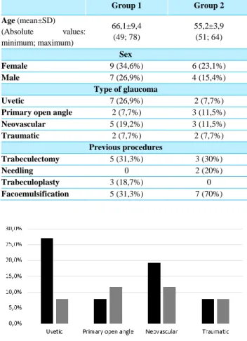
The study sample included 26 eyes of 26 patients (11 males and 15 females), with an average age of 61.7±10.1 years (minimum and maximum absolute values: 49 and 78, respectively). The results are expressed as mean ± standard deviation (SD), in absolute values (in decimal scale). The results in percentage (%) are also in decimal scale.
1. Description of the HTP (table 1): HTP was observed in 16 eyes (61.5%); 9 patients (34.6%) were female and the mean age was 66.1±9.4 years. The HTP occurred mainly at the 1st month (11 eyes, 68.7%; range: 1st –12th week). During the follow-up, it was observed an average IOP peak of 28.1±3.4 mmHg (absolute minimum and maximum pressure value of 22 and 34 mmHg, respectively) and an average number of hypotensive agents peak of 2.6±0.7 (absolute minimum and maximum number of hypotensive agents value of 1 and 4, respectively). The IOP stabilized after 3 months.
2. Comparison between groups (tables 2 and 3, and graphics 1-3):
2.1. The most prevalent type of glaucoma: in group 1 was uveitis (7 eyes, 26.9%) and in group 2, primary open-angle glaucoma and neovascular glaucoma (both 3 eyes, 11.5%).
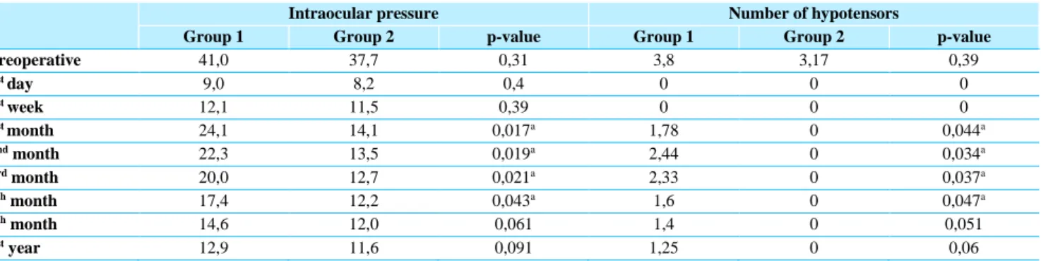
2.2. Evolution of the IOP throughout follow-up: mean preoperative IOP was 41.0±6.2 in group 1 and 37.7±3.7 in group 2. The mean IOP at the 1st day was 9.0±2.2 in group 1 and 8.2±0.32 in group 2; at the 1st week was 12.1±5.7 in group 1 and 11.7±1.2 in group 2; at the 1st month was 24.1±6.1 in group 1 and 14.1±1.5 in group 2; at the 2nd month was 22.3±5.2 in group 1 and 13.5±1.2 in group 2; at the 3rd month was 20.0±4.4 in group 1 and 12.7±0.9 in group 2; at the 6th month was 17.4±3.3 in group 1 and 12.2±0.4 in group 2; at the 8th month 14.6±1.7 in group 1 and 12.0±0.2 in group 2; and at the 1st year was 12.9±0.5 in group 1 and 11.6±0.3 in group 2. Group 1 had higher IOP values at all stages of the follow-up, with a statistically significant difference relative to group 2 between the 1st and 6th month after the surgery.
2.3. Evolution of the hypotensors’ number throughout follow-up: mean preoperative hypotensors’ number was 3.8±0.3 in group 1 and 3.2±0.5 in group 2. Both groups had any medication at the 1st day and the 1st week after the surgery. After that, only group 1 was medicated with hypotensors. The mean number at the 1st month was 1.8±0.9; at the 2nd month was 2.4±0.8; at the 3rd month was 2.3±0.8; at the 6th month was 1.6±0.7; at the 8th month 1.4±0.5; and at the 1st year was 1.2±0.4. Group 1 had higher number of hypotensors values at all stages of the follow-up after the 1st week following the surgery, with a statistically significant difference relative to group 2 between the 1st and 6th month after the surgery.
2.4. Surgical success in the first year: there was overall surgical success of 89% in group 1 (100% qualified sucess) and 100% in group 2 (67% qualified sucess and 33% complete sucess), with a statistically significant difference in complete surgical success (p = 0.042).
3. HTP management (table 1 and figure 1): only the HTP group was submitted to hypotensive medication and other surgical procedures.
3.1. Surgical procedures: were performed in 11 eyes (73.3%) during follow-up. 5 eyes (33.3%) performed injection of 5FU, 3 eyes needling (20%) and 3 eyes surgical revision (bleb excision, 20%).
3.2. Hypotensive agents (active principles): all eyes were medicated with at least 1 hypotensive agent during the follow-up. 2 hypotensors were prescribed for 47% of the eyes and 3 hypotensors were prescribed for 53% of the eyes. In the last clinical evaluation (in the first postoperative year), all eyes were medicated with hypotensive medication. 33.3% of the eyes were medicated with 1, 2 and 3 hypotensors.
Table 1 – Description and management of the hypertensive phase (HTP). IOP – intraocular pressure.
Hypertensive phase (HTP) Period of time to the HTP
1st month 11 eyes (68,7%)
2nd month 3 eyes (18,7%)
3rd month 2 eyes (12,5%)
Average IOP peak
(mmHg)
28,1±3,4
(absolute values: minimum; maximum – 22; 34)
Average number of hypotensors peak (in
follow-up)
2,6±0,7
(absolute values: minimum; maximum – 1; 4)
Management
Hypotensive medication 16 eyes (100%)
Surgical procedures
11 eyes (73.3%)
5-fluorouracil injection 5 eyes (33,3%)
Needling 3 eyes (20%)
Bleb revision
(surgical excision) 3 eyes (20%)
Table 2 – Comparison between groups: age, sex, type of glaucoma and previous procedures. SD – standard deviation.
Group 1 Group 2 Age (mean±SD) (Absolute values: minimum; maximum) 66,1±9,4 (49; 78) 55,2±3,9 (51; 64) Sex Female 9 (34,6%) 6 (23,1%) Male 7 (26,9%) 4 (15,4%) Type of glaucoma Uvetic 7 (26,9%) 2 (7,7%)
Primary open angle 2 (7,7%) 3 (11,5%)
Neovascular 5 (19,2%) 3 (11,5%) Traumatic 2 (7,7%) 2 (7,7%) Previous procedures Trabeculectomy 5 (31,3%) 3 (30%) Needling 0 2 (20%) Trabeculoplasty 3 (18,7%) 0 Facoemulsification 5 (31,3%) 7 (70%)
Table 3 – Evolution of intraocular pressure and number of hypotensors. Values express in mean. a Statistical significance: p-value<0.05.
Intraocular pressure Number of hypotensors
Group 1 Group 2 p-value Group 1 Group 2 p-value
Preoperative 41,0 37,7 0,31 3,8 3,17 0,39 1st day 9,0 8,2 0,4 0 0 0 1st week 12,1 11,5 0,39 0 0 0 1st month 24,1 14,1 0,017a 1,78 0 0,044a 2nd month 22,3 13,5 0,019a 2,44 0 0,034a 3rd month 20,0 12,7 0,021a 2,33 0 0,037a 6th month 17,4 12,2 0,043a 1,6 0 0,047a 8th month 14,6 12,0 0,061 1,4 0 0,051 1st year 12,9 11,6 0,091 1,25 0 0,06
Graphic 2 – Evolution of the intraocular pressure (IOP, mmHg) during the follow-up
Graphic 3 - Evolution of the number of hypotensive agents during the follow-up
Figure 1 – Bleb excisional revision: surgical excision of encapsulated cyst around the valve plate. A-E – sequence of the surgical technique
DISCUSSION
HTP is one of the early findings after AGV implantation and its occurrence has been associated with more uncontrolled IOP and an increased risk of surgical failure following AGV implantation due to the fibrosis surrounding the plate of the drainage device, causing an increase in the resistance to the flow of aqueous humor.3,5,10
Our results, related to the description of the HTP, are in agreement with the literature and other similar studies. In our study, an HTP occurred in 61.5% of the eyes over the first 3 months, with IOP peaking at 1 month (in 68.7%). Ayyala RS. et al. reported an HTP in 82% of the eyes over the first 6 months after surgery, with IOP peaking at 1 month and stabilizing at 6 months; Nouri-Mahdavi K. and Caprioli J. observed an HTP in 56% of the eyes in their study over the first 3 months, which occurred after a mean of 5 weeks with a mean peak IOP of 30.1±7.5 mmHg; Won H. et al. reported a prevalence of 31.1% over 3 months; Huang et al. reported the presence of a higher mean IOP 2 months after surgery; Jin Jeong et al. reported an HTP in 74% of the eyes over 3 months.1 The prevalence of HTP is variable in different studies according to different definition of HTP.2,9 HTP is by definition a transitory phase, that will spontaneously resolve after some time with or without treatment, but its duration is not clearly defined: some authors use a cut-off time of 2 or 3 months for the definition of HTP (instead of 6 months) because they believe that eyes showing an increase in IOP 6 months postoperatively may not be demonstrating temporary hypertension and might be indicating a poor IOP outcome rather than HTP.1,7 On the other hand, some authors reported that only a few eyes have HTP resolution, and most eyes will need continued
use of medication at least during the first postoperative year. Thus, the definition of “transitory phase” would not be completely correct and more active and timely treatment could be initiated to prevent surgical failure rather than waiting for the HTP to resolve. It is also noted that, due to the fact that many eyes remain on hypotensive medication after AGV implantation, it is difficult to estimate exactly the number of eyes in which the HTP is indeed transient.4,10
The results of our study related to the evolution of IOP and the number of hypotensors during follow-up are also consistent with previous studies, with the hypertensive group having a higher mean IOP and more uncontrolled IOP as compared with the non-hypertensive group. Consequently, eyes of the hypertensive group have a greater number of hypotensors and surgical procedures after valve implantation and a worse sucess rate. Some factors have been studied to identify an eventual association with HTP as risk factors for its development. Among the several factors analyzed by Won H. et al, only a higher preoperative IOP was associated with HTP development. However, in our study, the difference between groups in terms of preoperative IOP was not statistically significant. Other studies mention association with other factors. Jung KI. and Park C. observed that preoperative IOP were not different between the hypertensive and non-hypertensive groups (p=0.054 and 0.065, respectively), and axial length was significantly longer in the hypertensive group (25.2±2.6 mm), eyes with high myopia being at the greatest risk of developing hypertension following valve implantation (probably related to a collagen disorder in high myopia eyes).2
Among the following variables analyzed by Nouri-Mahdavi K. and Caprioli J., none was predictive of development of HTP after placement of the AGV: age, gender, eye laterality, race, diagnosis, lens status, number of previous glaucoma surgeries, preoperative IOP and number of medications.10
In our study, IOP and number of hypotensors were higher in the HTP group with a statistically significant difference only between the 1st and 6th month after surgery. In line with other studies, another aspect to highlight in our results is that, despite the absence of a statistically significant difference regarding these two parameters at the end of the follow-up, the hypertensive group obtained those values under hypotensive medication and, in some cases, after secondary surgical procedures, unlike the
non-HTP group.5 On the other hand, although Won H. et al. reported a significantly higher IOP at 1 year postoperatively in eyes that experienced an HTP, the IOP differences were no longer statistically significant at 2-year follow-up. They mention that, although long-term IOP outcomes might not be affected by the occurrence of HTP, they observed that significantly more eyes experiencing HTP required additional glaucoma surgeries (28.6%) compared with eyes that did not develop HTP (3.2%).4,6 As in this study and as in our study, other studies reported surgical procedures perfomed after the valve implantation to control IOP. Ayyala RS. et al. state that 20% of the eyes underwent bleb needling with 5FU injection, whereas 28% needed secondary surgical intervention to control the HTP.9 Eslami Y. et al. mention that needling or surgical removal of fibrotic tissue could theoretically restore outflow through the drainage device, and excisional bleb revision in failed AGV resulted in IOP control with or without hypotensive medication in 65.5% of the eyes in 1-year follow-up.3 Also, Eibschitz-Tsimhoni et al. reported that surgical excision of an encapsulated cyst around valve plate was effective in lowering IOP in 73% of eyes during 11 months follow-up, and Shah et al. reported a success rate of 42% in surgical revision group with median follow-up of 25 months.3
In our study all eyes in the hypertensive group showed a good outcome after hypotensive medication and/or secundary surgical procedures, with an IOP <17 mmHg at the 1st year of follow-up. However, on the one hand, the different studies used different success criteria, which makes it difficult to make any comparison; on the other hand, some authors reported that most eyes do not reach HTP resolution.4,7,10,13 Despite different sucess rate criteria, Kouros and Caprioli demonstrated in their study a success rate of 71% and 60% at 12 and 24 months, respectively, and also that there was a trend toward a higher failure rate in eyes with an HTP compared with those without, but the difference was not statistically significant.10 As in our study, in the study of Won H. et al. the overall success rate was not significantly different between groups (81.2% in the HTP group vs. 89.7% in the non-HTP group; p=0.111), with the complete success rate significantly higher in the non-HTP group (26.7% vs. 43.3%, p=0.003) at postoperative year 1.4 In relation to HTP resolution, Nouri-Mahdavi K. and Caprioli J. reported that only 28% of eyes with HTP resolved spontaneously and that many of the eyes that experienced
HTP continued to require the same number of IOP-lowering medications. However, the AGV in their study had a polypropylene plate, which seems to induce greater inflammation than silicone material (which appeared later) in similarly sized plates.10 Thus, in the study of Won H. et al., with silicone plate AGV, it is reported that approximately 76% of HTP cases had resolved by the end of postoperative year 1.4 Therefore, HTP occurrence was relatively less frequent and resolved more often following silicone plate AGV. However, as indicated above and as reported in previous studies, comparisons of surgical outcomes between different studies must be interpreted with caution; there are some differences in the studies including the criteria used for the various definitions, so HTP remains an issue as yet not clearly clarified.
Thus, the importance of this study is related to the fact that a HTP has clinical significance because of elevated IOP, even if it remains only for the short term, may cause substantial glaucomatous damage to the optic nerve, particularly in patients with advanced glaucoma. Consequently, patients should frequently be observed at the early postoperative stage following valve surgery, even after the hypotensive phase. On the other hand, the long-term prognosis for eyes that exhibited HTP is uncertain and has not been fully studied yet, with some authors reporting that HTP resolves only in a minority of eyes.4,7,10,13
In this context, some authors support the adoption of prophylactic procedures during the intraoperative period and/or in the early postoperative period, as well as intraoperative application of MMC, transplantation of amniotic membrane or subtenon triamcinolone injection; initiation of aqueous suppressant therapy after surgery and before the onset of HTP; topical NSAID medications such as ketorolac as an alternative to topical steroids after surgery; and 5FU injection after surgery. 3,12-15 Yazdani S. et al. reported that, in eyes submitted to surgery with application of MMC and transplantation of amniotic membrane, HTP was slightly but insignificantly more common with standard surgery (82% vs 60% and 70%, respectively); IOP was lower in the MMC group only 3 weeks postoperatively, but comparable at other time intervals; and the number of medications and overall success rates were comparable in the 3 groups at 12 months.14 MMC application was also included in the study of Cui QN. et al., as well as 5FU injection after AGV implantation, and the antimetabolites decreased HTP and
improved surgical outcomes without impacting complication rates at 1 year.15 Surgery with subtenon triamcinolone injection seems to have the same incidence of HTP, without a significant improvement in the success rate of the procedure.12 Some authors report that early initiation of aqueous suppressant therapy enabled a significantly lower postoperative IOP to be maintained at 3-4 weeks, when HTP usually occurs, without increasing the complication rate, and postoperative IOP was better controlled in the first 4 months if aqueous suppressant therapy was initiated before the onset of the HTP.5,6 Yuen D. et al. reported that the incidence of HTP was lower in the ketorolac group compared to the non-HTP group, although the difference between the 2 groups was not statistically significant.13
The limitations of our study are the retrospective profile, the sample size and the duration of the follow-up (despite being comparable to most studies). The strengths of our study are: given that it is still a controversial issue, the study of the HTP may contribute to an improvement in clinical practice; the comparison between groups; and the indication of possible strategies to manage the HTP.
To conclude, our study corroborates the idea that HTP is associated with greater difficulty in IOP control, requiring hypotensive treatment and other surgical procedures in some cases. It may have an impact on the final prognosis, and so regular follow-up is essential to allow its identification and early management.
BIBLIOGRAPHY
1. Jeong HJ, Park HYL, Park CK. Effects of Early Postoperative Intraocular Pressure after Ahmed Glaucoma Valve Implantation on Long-term Surgical Outcomes. Korean J Ophthalmol. 2018;32(5):391-9
2. Jung KI, Park C. Risk factors for the hypertensive phase after implantation of a glaucoma drainage device. Acta Ophthalmol. 2016;94:260-7
3. Eslami Y, Fakhraie G, Moghimi S, Zarei R, Mohammadi M, Nabavi A, et al. Excisional Bleb Revision for Management of Failed Ahmed Glaucoma Valve. J Glaucoma. 2017;26(12):1144-8
4. Won HJ, Sung KR. Hypertensive Phase Following Silicone Plate Ahmed Glaucoma Valve Implantation. J Glaucoma. 2016;25(4):313-7
5. Schimiti RB, Abe RY, Tavares CM, Vasconcellos JPC, Costa VP. Intraocular Pressure Control after Implantation of an Ahmed Glaucoma Valve in Eyes with a Failed Trabeculectomy. J Curr Glaucoma Pract. 2016;10(3):97-103
6. Law S, Kornmann H, Giaconi JA, Kwong A, Tran E, Caprioli J. Early Aqueous Suppressant Therapy on Hypertensive Phase Following Glaucoma Drainage Device Procedure: A Randomized Prospective Trial. J Glaucoma. 2016;25(3):248-57
7. Jung KI, Park H, Jung Y, Park CK. Serial changes in the bleb wall after glaucoma drainage implant surgery: characteristics during the hypertensive phase. Acta Ophthalmol. 2015;93(4):248-53
8. Hong CH, Arosemena A, Zurakowski D, Ayyala RS. Glaucoma drainage devices: a systematic literature review & current controversies. Surv Ophthalmol. 2005;50:48-60
9. Ayyala RS, Zurakowski D, Smith JA, Monshizadeh R, Netland PA, Richards DW, et al. A clinical study of the Ahmed glaucoma valve implant in advanced glaucoma. Ophthalmology. 1998;105(10):1968-76
10. Nouri-Mahdavi K, Caprioli J. Evaluation of the hypertensive phase after insertion of the Ahmed Glaucoma Valve. Am J Ophthalmol. 2003;136(6):1001-8.
11. Lieberman MF, Ewing RH. Drainage implant surgery for refractory glaucoma. Int Ophthalmol Clin. 1990;30:198– 208
12. Yazdani S, Doozandeh A, Pakravan M, Ownagh V, Yaseri M. Adjunctive triamcinolone acetonide for Ahmed glaucoma valve implantation: a randomized clinical trial. Eur J Ophthalmol. 2017;27 (4):411-6
13. Yuen D, Buys Y, Jin YP, Alasbali T, Smith M, Trope GE. Corticosteroids Versus NSAIDs on Intraocular Pressure and the Hypertensive Phase After Ahmed Glaucoma Valve Surgery. J Glaucoma. 2011; 20(7):439-44
14. Yazdani S, Mahboobipour H, Pakravan M, Doozandeh A, Ghahari E. Adjunctive Mitomycin C or Amniotic Membrane Transplantation for Ahmed Glaucoma Valve Implantation: A Randomized Clinical Trial. J Glaucoma. 2016;25(5):415-21
15. Cui QN, Hsia YC, Lin SC, Stamper RL, Rose-Nussbaumer J, Mehta N, et al. Effect of mitomycin c and 5-flurouracil adjuvant therapy on the outcomes of Ahmed glaucoma valve implantation. Clinical and Experimental Ophthalmology. 2017;45:128-34.
16. Heuer DK, Barton K, Grehn F, Shaarawy TM, Sherwood MB. Consensus on definitions of success – Guidelines on
Design and Reporting of Glaucoma Surgical Trials. 2 ed, pp.15–24. World Glaucoma Association. Kugler, Amsterdam: The Netherlands; 2009.
CONTACTO
Ana Sofia LopesRua Álvaro de Sousa, Nº6, 1A, Alfragide, 2610-313 Amadora, Lisboa
E-mail: [email protected] Conflicts of interest: none.
The manuscript has not been previously published and the authors assign the copyright to the SPO.