Identification of proteases as diagnostic and drug targets
in bovine babesiosis
Tiago Miguel Lopes Martins
III Dissertation presented to obtain a PhD degree in Biomedical Sciences, speciality Parasitology, by the Universidade Nova de Lisboa, Instituto de Higiene e Medicina Tropical
V The research described in this thesis was supervised by Doctor Ana Domingos and co-supervised by Doctor Vírgilio Estólio do Rosário. The work was performed at the Unidade de Tecnologias e Anticorpos Monoclonais, (formerly Instituto Nacional de Engenharia, Tecnologia e Inovação), Instituto de Higiene e Medicina Tropical, Universidade Nova de Lisboa, Campus do Lumiar, Lisboa, Portugal; Centro de Malária e Doenças Tropicais, Instituto de Higiene e Medicina Tropical, Universidade Nova de Lisboa, Rua da Junqueira, Lisboa, Portugal; and Faculdade de Veterinária, Universidade Eduardo Mondlane, Maputo, Mozambique.
VII The following thesis benefited from the insights and guidance of several people, to whom I am grateful.
I would like to express my gratitude to my supervisor, Dr. Ana Domingos, whose guidance, support, and patience, added considerably to my experience over the past years from a graduate student until the present time.
I am grateful to my co-supervisor, Professor Dr. Virgílio E. do Rosário for accepting me as student and for his unique guidance, planning, and whom I respect immensely.
I would like to thank the other member of my scientific committee, Dr. Sónia Centeno-Lima for the assistance she provided at all levels and especially in the initial development of this study.
To Dr. Luís Neves from the Faculty of Veterinary (Maputo, Mozambique), that received me in his laboratory, and for his endless support, advice and scientific enthusiasm that is an inspiration to whoever loves science.
To Dr. Varda Shkap from Kimron Veterinary Institute (Israel), for her support in this research project and her desire to contribute to a better knowledge on babesiosis.
To Dr. Carlos Novo, director of UTPAM, for the support, defense of his investigation group and constant instigation for us to acquire and improve our scientific abilities.
To Susana Teixeira for her work and dedication in the very beginning of the babesial adventure, when this research project was no more than a destiny.
VIII
friendship.
To my colleagues in Biotechnology Center of Faculty of Veterinary (Maputo, Mozambique) Olívia, Titus, and all the people that I have met there; that welcomed me to a second homeland, and for all the help, and friendship.
To my Mozambican “father”, Ibraímo, that received me in his home as a son, and for his friendship.
À Lucia pelo amor e apoio constante que sempre me ilumina quando mais preciso.
Aos meus pais, familia e amigos que me ajudaram a crescer com valores, responsabilidade, amor e alegria.
Esta tese é dedicada ao irmão de um “cientista maluco”, que sempre me incentivou a prosseguir este caminho depois de me ter guiado para um curso que adorei.
To the financial support provided by Fundação para a Ciência e Tecnologia (FCT) with a PhD fellowship grant (SFRH/BD/19059/2004) under POCI 2010 and the European Social Fund.
GOVERNMENT OF THE EUROPEAN UNION
IX Bovine babesiosis is a tick–borne disease with significant morbidity and mortality, and the economic losses associated to this disease can be considerable. Control measures of bovine babesiosis include the eradication or reduction of ticks, good diagnosis, use of vaccination and correct treatment. The aim of this work was to contribute to a better diagnosis of the infection aiming at the improvement of some of the control measures, as well as to identify and characterize protease genes for the development of a diagnostic method and studied as potential drug targets. For this study, collaborative work was carried out in Mozambique, from where blood samples from naturally infected cattle were collected in five farms located in the Maputo province, south of the country. A new molecular detection PCR method was then developed and tested using genomic DNA and random field samples collected from one farm. Primers were designed based in the babesial aspartic protease putative babesipsin gene identified in the genomes of Babesia bigemina and B. bovis. The new seminested hot-start PCR method was developed using the combination of 30 bp long primers and a hot start polymerase that theoretically allows the use of annealing temperatures above the melting temperatures of the primers and prevents the formation of unspecific amplifications and therefore increases the specificity.
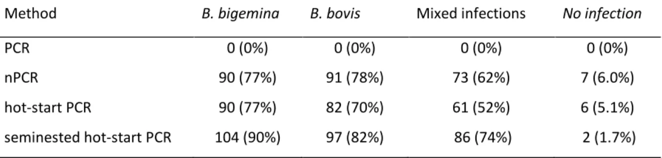
The new seminested hot-start PCR method was assayed using 117 field samples in parallel with the widely used nested PCR method. The babesipsin seminested hot-start PCR was in this study more sensitive than the nested PCR. With the seminested hot-start PCR, 90% of the samples were positive for B. bigemina and 82% were positive for B. bovis. The results suggested that bovine babesiosis is common and endemic in Mozambique and that the disease was in an endemically stable situation.
The status of bovine babesiosis in Mozambique was then further studied, by testing random field samples from four more farms using the seminested hot-start PCR. All the samples from the five farms were also analysed using the reverse line blot (RLB) assay, and the results were compared with the data obtained by the seminested hot-start PCR. The detection of Babesia spp. differed considerably
X
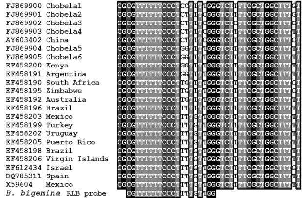
frequency of 53%. Using the RLB assay B. bigemina was not detected and detection of B. bovis ranged between 0% and 17% with a total frequency of 5.1%. Analysis of new sequences of the 18S rRNA gene revealed that the current B. bigemina RLB probe is unspecific for the detection of all the identified isolates from Mozambique. The seminested hot-start PCR was therefore more sensitive than the RLB assay. Nevertheless, ten different species of the four genera Anaplasma, Babesia, Ehrlichia and Theileria were detected by the RLB assay, and this illustrates that multiple infections are widespread in Mozambique.
The results of this study show that bovine babesiosis is common in Maputo province, but there are some locations with low prevalence of infections and therefore the results suggest that this disease is not in an endemically stable situation in Maputo province. Further epidemiological studies are now needed to corroborate these findings.
Proteases have been shown to have essential roles in parasitic protozoa and are under study as promising drug targets. Some cysteine proteases of protozoan parasites are now recognized drug targets and specific inhibitors are in validation for chemotherapy of leishmaniasis, malaria, and trypanosomiasis. In this study our focus on the identification and characterization of proteases as drug targets was therefore in this class of proteases.
Cysteine protease putative genes were identified by sequence similarity search in the ongoing B. bigemina genome sequencing project database and were compared with the annotated genes from the complete bovine piroplasms genomes of B.
bovis, Theileria annulata, and T. parva. Multiple genome alignments and sequence
analysis were used to evaluate the molecular evolution events that occurred in the C1 family of cysteine proteases in these piroplasms of veterinary importance. There are five distinct groups of cysteine protease genes of C1 family in B.
bigemina (5 genes), four groups in B. bovis (4 genes) and six groups in Theileria
spp. (13 genes). Molecular evolution in Theileria occurred through the duplication of genes and sequence diversity. These considerable differences observed in the
XI One of the identified cysteine proteases in the B. bigemina genome, babesipain-1, was expressed as a fusion protein with gluthatione S-transferase (GST) and the soluble protein was purified by affinity chromatography. The recombinant babesipain-1 showed activity against typical peptide substrates of cysteine proteases, and was inhibited by a general inhibitor of its class, but the low yield of the soluble purification prevented additional characterization.
Babesipain-1 was then purified from the insoluble fraction, and the denatured protein was refolded and activated to produce an active mature enzyme. Analysis of the activity of babesipain-1 revealed typical properties of a papain-family cysteine protease, including hydrolysis of typical papain-family peptide substrate, an acidic pH optimum (5.5-6.0), requirement for a reducing environment for maximum activity, and inhibition by standard cysteine protease inhibitors as E-64, leupeptin, ALLN and cystatin. The results suggest that babesipain-1 has a role in cytosol environment, since babesipain-1 retained high activity against peptide substrates at pH 7.5 (83% of maximum), an uncommon feature of cysteine proteases of parasitic protozoa.
Thus the results of this study demonstrate that bovine babesiosis is widespread in Maputo province in Mozambique, although the disease is not in an endemically stable situation. The results also suggest that cysteine proteases of Babesia spp. are promising drug targets for the development of an effective treatment of bovine babesiosis. In face of these results a plan for future work is associated. Some aspects and results from this work can be adjusted to other countries, including Portugal.
XIII A babesiose bovina é uma doença transmitida por carraças, que causa elevada morbidade e mortalidade, e provoca consideráveis perdas económicas devido aos esforços para controlar esta doença. As medidas de controlo da babesiose bovina incluem a erradicação ou redução de carraças, correcto diagnóstico, assim como tratamento e vacinação apropriados. Este trabalho tem como objectivo contribuir para um melhor diagnóstico da infecção e a consequente melhoria de algumas das medidas de controlo, bem como identificar e caracterizar genes de proteases utilizados para o desenvolvimento de um método de diagnóstico e estudados como potenciais alvos para fármacos.
Para este estudo, foi realizado um trabalho de colaboração em Moçambique, onde foram colhidas amostras de sangue de bovinos naturalmente infectados, em cinco explorações situadas na província de Maputo, no sul do país. Um novo método de detecção molecular por PCR foi desenvolvido e testado utilizando DNA genómico e amostras de campo aleatórias colhidas numa das explorações. Os iniciadores de PCR foram desenhados com base no gene putativo da protease aspártica babesipsina-1 identificado nos genomas de Babesia bigemina e de B. bovis. O novo
seminested hot-start PCR foi desenvolvido utilizando a combinação de iniciadores
longos de 30 pb de comprimento e uma hot-start polimerase, que permitem teoricamente a utilização de temperaturas de emparelhamento acima da temperatura de melting, impedindo assim a formação de amplificações não específicas, o que aumenta a especificidade do método.
O novo seminested hot-start PCR foi avaliado utilizando 117 amostras de campo, e em paralelo com um método amplamente utilizado, o nested PCR. O seminested
hot-start PCR neste estudo foi mais sensível que o nested PCR. Com o seminested hot-start PCR, 90% das amostras foram positivas para B. bigemina, e 82% foram
positivas para B. bovis. Os resultados sugeriram que a babesiose bovina é comum e endémica em Moçambique, e que a doença se encontra numa situação de estabilidade endémica.
O estudo do estado da babesiose bovina em Moçambique, foi então aprofundado, através da análise de amostras de campo aleatórias de mais quatro explorações
XIV
Babesia spp. diferiu significativamente entre os métodos utilizados e os locais de
recolha. Com o seminested hot-start PCR, a detecção de B. bigemina nas várias explorações, variou entre 30% e 89%, com uma detecção total de 61%, e a detecção de B. bovis variou entre 27% e 83% com uma frequência global de 53%. Utilizando o RLB, não foi detectado B. bigemina e a detecção de B. bovis variou entre 0% e 17% com uma frequência total de 5,1%. A análise de novas sequências do gene 18S rRNA, revelou que a actual sonda do RLB para B. bigemina não é adequada para a detecção de todos os isolados desta espécie identificados em Moçambique. O seminested hot-start PCR foi portanto mais sensível que o RLB. No entanto, dez espécies diferentes dos quatro Géneros Anaplasma, Babesia, Ehrlichia e Theileria foram detectadas pelo ensaio RLB, e isso demonstra que as infecções múltiplas são comuns em Moçambique.
Os resultados deste estudo mostram que a babesiose bovina é comum na província de Maputo, e também que existem alguns locais com baixa prevalência de infecções, e portanto, os resultados sugerem que esta doença não está numa situação de estabilidade endémica na província de Maputo. São agora necessários novos estudos epidemiológicos para confirmar estes resultados.
Tem sido demonstrado que as proteases têm papéis essenciais em parasitas protozoários e estão sob estudo como promissores alvos de fármacos. Algumas proteases cisteínicas de parasitas protozoários, são já reconhecidos alvos de fármacos, e encontram-se em validação inibidores específicos para a quimioterapia da leishmaniose, da malária e da tripanossomíase. Neste estudo, o nosso principal interesse na identificação e caracterização de proteases como alvos de fármacos foi portanto nesta classe de proteases.
Foram identificados no banco de dados do projecto em curso de sequenciação do genoma de B. bigemina, os genes putativos de proteases cisteínicas em pesquisas por similaridade de sequência, que posteriormente foram comparados com os genes anotados nos genomas completos das espécies de piroplasmas bovinos B.
XV proteases cisteínicas da família C1 em B. bigemina (5 genes), quatro grupos em B.
bovis (4 genes) e seis grupos em Theileria spp. (13 genes). No Género Theileria a
evolução molecular ocorreu através da duplicação de genes e da diversificação da sequência das proteínas codificadas por estes genes. Estas importantes diferenças observadas entre os Géneros Babesia e Theileria na família das proteases cisteínicas, podem parcialmente explicar os diferentes mecanismos de infecção destas espécies, em que parasitas Babesia não invadem linfócitos e parasitas
Theileria invadem primeiro os linfócitos no hospedeiro vertebrado.
A babesipaína-1, uma das proteases cisteínicas identificadas no genoma de B.
bigemina, foi expressa como uma proteína de fusão com a glutationa S-transferase
(GST) e a respectiva fracção solúvel foi purificada por cromatografia de afinidade. A babesipaína-1 recombinante apresentou actividade contra certos péptidos que são substratos típicos de proteases cisteínicas, e foi inibida por um inibidor geral da sua classe, mas o baixo rendimento da purificação da fracção solúvel impediu a sua caracterização adicional.
A babesipaína-1 foi então purificada a partir da fracção insolúvel, e a proteína desnaturada foi re-enrolada e activada para produzir uma enzima activa. A análise da actividade da babesipaína-1 revelou propriedades típicas de uma protease cisteínica da família da papaína, incluindo a hidrólise de alguns péptidos que são substratos típicos da família da papaína, um pH ácido óptimo (5.5-6.0), o requisito de um ambiente redutor para ter actividade máxima, e a inibição por inibidores de proteases cisteínicas como o E-64, a leupeptina, o ALLN e a cistatina. Os resultados sugerem que a babesipaína-1 tem um papel no citosol, já que a babesipaína-1 manteve elevada actividade contra substratos a pH 7,5 (83% do máximo), uma característica incomum das proteases cisteínicas de parasitas protozoários.
Assim, os resultados deste estudo demonstram que a babesiose bovina é uma infecção comum na província de Maputo em Moçambique, embora a doença não esteja numa situação de estabilidade endémica. Os resultados também sugerem que as proteases cisteínicas de Babesia spp. são alvos promissores para fármacos e
XVI
XVII Abs Absorbance
AEBSF 4-(2-Aminoethyl) benzenesulfonyl fluoride AIDS Acquired immunodeficiency syndrome ALLN N-acetyl-leucineleucine-norleucinal AMC 7-amino-4-methyl coumarin
bp Base pair
CP Cysteine protease
DCI 3,4-dichloroisocoumarin DNA Deoxyribonucleic acid
dNTP Deoxynucleotide triphosphate DTT Dithiothreitol
E-64 N-(trans-epoxysuccinyl)-l-leucine 4 guanidinobutylamide
E-64d (2S,3S)-trans-epoxysuccinyl-L-leucylamido-3-methylbutane ethyl ester
EDTA Ethylene diamine tetra acetic acid ELISA Enzyme-linked immunosorbent assay EST Expressed sequence tag
G+C Guanine-cytosine content GSH Glutathione
GSSG Glutathione disulfide GST Glutathione S-transferase HIV Human immunodeficiency virus
IFAT Indirect immunofluorescence antibody test IPTG isopropyl-β-D-thiogalactopyranoside LAMP Loop-mediated isothermal amplification LB-Lennox Lysogeny broth, Lennox formulation nPCR Nested PCR
ORF Open reading frame PBS Phosphate-buffered saline PCR Polymerase chain reaction
XVIII
rRNA Ribosomal RNA
RT-PCR Reverse transcription polymerase chain reaction SDS Sodium dodecyl sulfate
SDS-PAGE Sodium dodecyl sulfate polyacrylamide gel electrophoresis SSU rRNA small subunit ribosomal RNA
TBD Tick borne disease TBE Tris/Borate/EDTA buffer
TLCK Nα-Tosyl-L-lysine chloromethyl ketone hydrochloride Tm Melting temperature
TPCK N-p-Tosyl-L-phenylalanine chloromethyl ketone
XIX
ACKNOWLEDGMENTS VII
ABSTRACT IX
RESUMO XIII
ABBREVIATIONS XVII
TABLE OF CONTENTS XIX
INDEX OF FIGURES XXII
INDEX OF TABLES XXIII
CHAPTER 1. GENERAL INTRODUCTION 1
1.1. Introduction 3
1.2. The Apicomplexa 4
1.3. The Genus Babesia 4
1.3.1. Life Cycle 6
1.3.1.1. Events in the vertebrate host 8
1.3.1.2. Events in the tick 8
1.3.2. Epidemiology 9
1.3.3. Diagnosis and identification of parasites 10
1.3.4. Treatment and vaccination 11
1.4. Proteases as drug targets 13
1.4.1. Protease classification 15
1.4.2. Proteases of Babesia and other Apicomplexans 15
1.5. Outline of the study 16
CHAPTER 2. DEVELOPMENT OF A NEW MOLECULAR DIAGNOSTIC METHOD FOR THE DETECTION OF BOVINE BABESIOSIS, ITS APPLICATION IN
MOZAMBIQUE 19
XX
2.2.3. PCR reactions 23
2.2.3.1. Primers 23
2.2.3.2. Babesipsin hot-start PCR and seminested hot-start PCR 24
2.2.3.3. nPCR 25 2.2.4. Analysis of PCR products 25 2.2.5. Statistical analysis 25 2.3. Results 25 2.3.1. Hot-start PCR 25 2.3.2. nPCR 28 2.3.3. Statistical analysis 29 2.4. Discussion 29
CHAPTER 3. BOVINE BABESIOSIS AND OTHER CATTLE HAEMOPARASITES INFECTIONS IN MAPUTO PROVINCE, MOZAMBIQUE AS DIAGNOSED BY
MOLECULAR TECHNIQUES 33
3.1. Introduction 35
3.2. Materials and methods 36
3.2.1. Cattle 36
3.2.2. DNA extraction 36
3.2.3. Hot-start PCR and seminested hot-start PCR 36
3.2.4. Reverse line blot assay 37
3.2.5. Cloning of 18S ribosomal RNA genes 37
3.3. Results 38
3.3.1. Seminested hot-start PCR 38
3.3.2. Reverse line blot assay 38
3.3.3. 18S ribosomal RNA gene sequences analysis 39
3.4. Discussion 41
CHAPTER 4. IDENTIFICATION OF PAPAIN-LIKE CYSTEINE PROTEASES FROM
BABESIA BIGEMINA AS POTENTIAL DRUG TARGETS 47
4.1. Introduction 49
4.2. Materials and methods 50
XXI
4.3. Results 53
4.3.1. Database search for similarity, genome alignments and protein sequence
analysis 53
4.3.2. Recombinant expression and activity of babesipain-1 60
4.4. Discussion 63
CHAPTER 5. CHARACTERIZATION OF THE POTENTIAL DRUG TARGET
CYSTEINE PROTEASE BABESIPAIN-1 69
5.1. Introduction 71
5.2. Materials and methods 72
5.2.1. Materials 72
5.2.2. Expression and purification of recombinant babesipain-1 72
5.2.3. Refolding of recombinant babesipain-1 73
5.2.4. Activity assays 73
5.2.5. Enzyme Kinetics 74
5.3. Results 74
5.4. Discussion 79
CHAPTER 6. GENERAL DISCUSSION AND CONCLUSIONS 81
6.1. Discussion 83
6.1.1. Development of a new molecular diagnostic method 84 6.1.2. Survey on the status of babesiosis in Mozambique 85 6.1.3. Identification of proteases as potential drug targets 88 6.1.4. Biochemical characterization of babesipain-1 as a new drug target 90
6.2. Conclusions 91
6.3. Future work 91
REFERENCES 93
XXII
Rhipicephalus (Boophilus) microplus 7
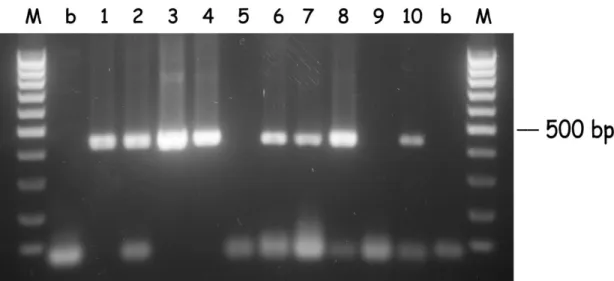
Figure 2.1. Hot-start PCR of B. bigemina DNA from random samples 26 Figure 2.2. Hot-start PCR of B. bovis DNA from random samples 27 Figure 2.3. Seminested hot-start PCR of B. bigemina DNA from random samples 27 Figure 2.4. Seminested hot-start PCR of B. bovis DNA from random samples 28 Figure 2.5. Nested PCR of B. bigemina DNA from random samples 29 Figure 3.1. Map of the locations in Maputo province, Mozambique where samples
were collected 36
Figure 3.2. Alignment of worldwide B. bigemina 18S rRNA gene sequences with
the respective species-specific RLB probe 40
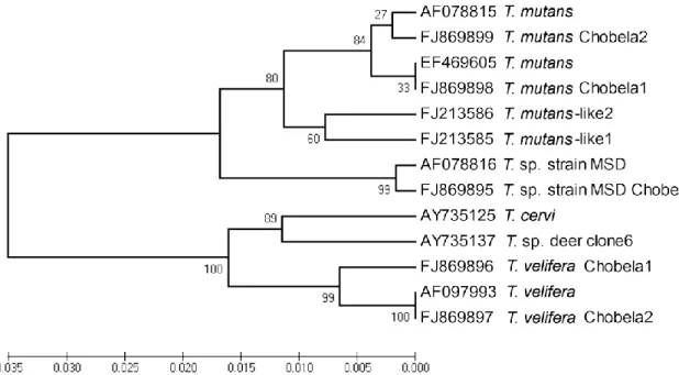
Figure 3.3. Phylogenetic tree inferred from Theileria spp. 18S rRNA gene
sequences 41
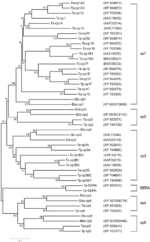
Figure 4.1. VISTA identity plots between homologous regions of Piroplasmida species genomic DNA displaying cysteine protease genes 54 Figure 4.2. Phylogenetic tree inferred from piroplasma cysteine proteases amino
acid sequence data 56
Figure 4.3. Multiple sequence alignment of the deduced amino acid sequences of
the cathepsin L-like CPs BbiCP1 to CP3 and papain 58
Figure 4.4. Sequence alignment spanning the active site cysteine and the predicted glutamine of the oxyanion hole of cysteine proteases 60 Figure 4.5. Expression, purification and gel substrate activity of recombinant
babesipain-1 61
Figure 4.6. Babesipain-1 activity assays 62
Figure 5.1. Expression, purification and gel substrate activity of recombinant
babesipain-1 76
Figure 5.2. Effect of pH, reductant concentration, and inhibitors on the activity of
XXIII
Index of Tables
Table 1.1. Known Babesia species of cattle, their ixodid tick vector genus and
geographical distribution 6
Table 2.1. Primers sequence and melting temperature 24
Table 2.2. Comparison of methods in the detection of cattle infected with B.
bigemina and/or B. bovis in Boane district, Mozambique 30 Table 3.1. Prevalence of cattle haemoparasites infections in Maputo
province, Mozambique 39
Table 4.1. Protein sequence similarity and identity matrix 57
Table 5.1. Optimization of refolding of babesipain-1 75
Table 5.2. Kinetic parameters for substrate hydrolysis by recombinant
Chapter 1. General Introduction
3
1.1. Introduction
Babesiosis caused by intraerythrocytic parasites of the Apicomplexan genus
Babesia, is a common infection of vertebrate animals worldwide (Uilenberg, 2006).
In 1893, Smith and Kilbourne discovered that Babesia bigemina, a causative agent of bovine babesiosis, was transmitted by the tick Boophilus annulatus. This revelation was a breakthrough in the history of parasitology, as it was the first proof that an arthropod was the vector of any disease agent (Uilenberg, 2006). Ticks from several genera are now known to be vectors and reservoirs of numerous Babesia spp. transmissible to reptiles, birds and mammals (Uilenberg, 2006). The major economic impact of babesiosis is on the cattle industry and the two most important species in cattle, B. bigemina and B. bovis threaten the health and safety of about 300 million cattle in tropical and subtropical regions of the world (Wright, 1990). The pathogenicity varies both between and within species but in many cases has a high rate of mortality in susceptible untreated animals (Bock et al., 2004). Many factors contribute to the emergence and re-emergence of babesiosis: parasite drug resistance, tick resistance to acaricides, genetic evolution, and the so-called global change (economic, social and environmental factors) (L’Hostis and Seegers, 2002). Climate change and increased climatic variability are particularly likely to affect vector borne diseases at the global scale in the near future. Control measures of bovine babesiosis include the eradication or reduction of ticks, better diagnosis, vaccination and treatment of clinical cases. The ongoing genome sequencing projects of Babesia spp., and more importantly, the vast knowledge acquired with the study of the closely related malaria pathogen may boost the study of Babesia diversity and drug resistance and the development of specific fast acting drugs, recombinant vaccines, and of more specific and sensitive diagnostic methods currently required for the control of babesiosis. This tick borne disease is also gaining increasing interest as an emerging zoonosis in humans, as the incidence of babesiosis has been rising steadily both in the U.S.A. and Europe (Homer et al., 2000; Hildebrandt et al., 2008). Human babesiosis may have previously been overlooked in many parts of the world due to its rarity or a lack of medical awareness and microbiological detection methods (Hildebrandt et al., 2008). On the other hand, the number of patients with risk factors is currently
4
increasing (e.g. immunocompromised individuals and organ transplant recipients) and asymptomatic but chronically infected blood donors may be a source of transfusion-transmitted babesiosis (Hildebrandt et al., 2008). New and old Babesia pathogens of veterinary and medical importance continue to emerge around the world and the substantial health impact of babesiosis on livestock and man is continuing.
1.2. The Apicomplexa
The eukaryotic phylum Apicomplexa comprises more than 5000 species of endoparasitic protozoa, including the Plasmodium parasites responsible for malaria, a major disease of poverty (Cavalier-Smith, 1993; Roos, 2005). Apicomplexan parasites infect virtually all animals (Levine, 1985), and while some parasite life cycles are relatively simple, involving only a single host, others require sexual recombination in a vector species for transmission. Sexual recombination is relevant in the production of diverse parasites within a species and this subject area is important in understanding drug resistance or immune mechanisms of defense. Some parasites are specialists, restricted to particular species and tissues, whereas others are generalists. For example, Plasmodium falciparum, which causes the most lethal form of malaria, infects only Hominids, and is transmitted only by anopheline mosquitoes. In contrast, Toxoplasma gondii can infect almost any tissue of warm-blooded animals. In the complex life cycle stages of Apicomplexan parasites, extracellular forms in the host include an apical complex that gives the phylum its name, including secretory organelles associated with host cell attachment, invasion, and establishment of an intracellular parasitophorous vacuole (Sibley, 2004). Different stages of development are also relevant for the study of drug targets and their selection.
1.3. The Genus Babesia
It was at the end of the 19th century that the Romanian biologist Victor Babeş discovered micro-organisms in erythrocytes of cattle in Rumania and associated them with bovine hemoglobinuria or red water fever (Babes, 1888). Currently it is know that members of the genus Babesia are one of the most ubiquitous and
5 widespread blood parasites in the world, second only to the trypanosomes (Homer et al., 2000). The genus Babesia belongs to the phylum Apicomplexa, class Sporozoasida, order Eucoccidiorida, suborder Piroplasmorina and family Babesiidae (Levine, 1971, 1985; Allsopp et al., 1994). The Babesia species sensu
stricto, or true Babesia, are characterized by transovarial transmission in the
vector tick and the limitation of infecting only erythrocytes in the host (Uilenberg, 2006). Babesia organisms are protozoan parasites that are all transmitted by ticks to their specific vertebrate host. Completion of a life cycle and therefore the maintenance of Babesia are completely dependent on both the tick and the vertebrate host (Mehlhorn and Shein, 1984) and therefore, the distribution of all the different Babesia species is governed by the geographical distribution of the tick vectors that transmit them. The Babesia spp. known to infect cattle, and their vectors, are listed in Table 1.1.
Babesia organisms can be visualized in a Giemsa stained infected blood smear.
They are pyriform (pear shaped) or seen as round or amoeboid forms (Levine, 1985). Depending on the Babesia species, a host erythrocyte may be parasitized by single, paired, or multiple organisms. The size of the organisms varies depending on the species and are therefore classified as either small (1.0-2.5 μm; B. bovis, B.
gibsoni, B. ovis and B. divergens) or large Babesia (2.5-5.0 μm; B. bigemina, B. caballi
and B. canis), accordingly to the size of the trophozoites (Levine, 1985). Electron microscopy of extracellular forms reveals the presence of an apical complex with micronemes, rhoptries, apicoplast and other organelles (Kawai et al., 1986; Kawai et al., 1999) characteristic of the phylum Apicomplexa. Babesia extracellular infective forms, the sporozoites, move by gliding or body flexion, and lack flagella or pseudopodia, characteristic of some Apicomplexans (Levine, 1985).
6
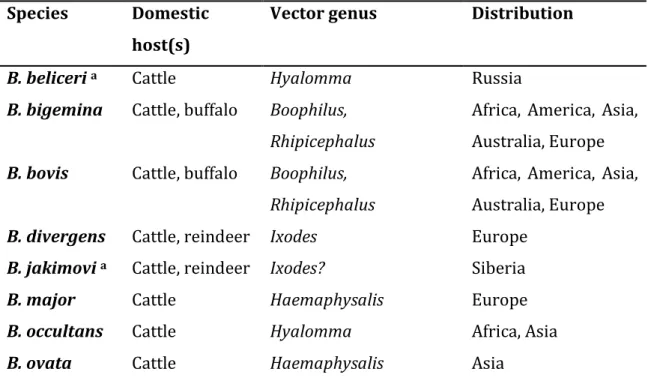
Table 1.1. Known Babesia species of cattle, their ixodid tick vector genus and geographical distribution.
Species Domestic host(s)
Vector genus Distribution
B. beliceri a Cattle Hyalomma Russia
B. bigemina Cattle, buffalo Boophilus, Rhipicephalus
Africa, America, Asia, Australia, Europe
B. bovis Cattle, buffalo Boophilus, Rhipicephalus
Africa, America, Asia, Australia, Europe
B. divergens Cattle, reindeer Ixodes Europe
B. jakimovi a Cattle, reindeer Ixodes? Siberia
B. major Cattle Haemaphysalis Europe
B. occultans Cattle Hyalomma Africa, Asia
B. ovata Cattle Haemaphysalis Asia
a These species are not recognized by all scientists and some may not be valid.
Adapted from Uilenberg (2006).
1.3.1. Life Cycle
The complex life cycle of Babesia spp. takes place in two hosts and sexual and asexual reproduction proceeds through at least three stages:
(i) gamogony – sexual development with formation and fusion of gametes inside the tick gut,
(ii) sporogony - asexual reproduction in tick salivary glands, (iii) merogony - asexual reproduction in the vertebrate host.
Babesia spp. are not characterized by a life cycle that is specific for the genus
(Mackenstedt et al., 1995). Therefore, we describe here the life cycle of B. bigemina (Fig. 1.1), which is similar to the life cycle of B. bovis.
7 Figure 1.1. The life cycle of Babesia bigemina in cattle and the ixodid tick vector Rhipicephalus (Boophilus) microplus. Adapted from Bock et al. (2004).
8
1.3.1.1. Events in the vertebrate host
Once in the vertebrate, the transmitted bovine Babesia spp. sporozoites do not parasitise any vertebrate host cell other than erythrocytes (Mackenstedt et al., 1995). Each sporozoite penetrates the cell membrane of an erythrocyte with the aid of a specialised apical complex, forming a parasitophorous vacuole (Sibley, 2004). The vacuole membrane gradually disintegrates, and the parasite is left with the defining piroplasm feature of a single membrane, in contrast to Plasmodium species, which invade by a similar mechanism but retain the host membrane in addition to its own. Once inside the erythrocyte, it transforms into a trophozoite from which two merozoites develop by a process of merogony (Levine, 1985). The merozoites lyse the cell and go on to infect additional erythrocytes. Rapid reproduction destroys the host cell and leads to hemoglobinuria in the host. Some trophozoites develop into a diploid ovoid type of merozoite, called a gamont precursor. These gamont precursors do not develop further until they are taken up by the tick in the blood meal (Mackenstedt et al., 1995).
1.3.1.2. Events in the tick
Environment changes in the passage from host blood to the midgut of the tick vector stimulate the development of two populations of gamonts called ray bodies (Gough et al., 1998). The ray bodies undergo further multiplication within the consumed erythrocyte and form large aggregations of multinucleated ray bodies. After the consumed erythrocyte is digested, and once gametogenesis is complete, single-nucleated and haploid gametes (Mackenstedt et al., 1995) emerge from the aggregates and then fuse together in pairs to form a zygote (Gough et al., 1998; Mosqueda et al., 2004). The zygote selectively infects the digestive cell of the tick gut where they probably multiply and then the basophilic cells where further multiplication occurs. At some stage in development in the gut the zygote undergoes one-step meiosis to form a haploid zygote (Mackenstedt et al., 1995). In the gut cells, schizogony occurs with the formation of polyploid kinetes, and these motile club-shaped kinetes then escape into the haemolymph and infect a variety of cell types and tissues, including the oocytes where successive cycles of secondary schizogony take place (Mackenstedt et al., 1995). Consequently,
9 transovarial transmission occurs with further development taking place in the larval stage. This is an important life cycle adaptation as the Boophilus vectors are one-host ticks. Kinetes enter the salivary glands and are transformed into multinucleated stages (sporogony) and these then break up to form sporozoites (Mackenstedt et al., 1995). In all species, sporozoite development usually begins when the infected tick attaches to the vertebrate host. In B. bigemina, infective sporozoites take about 9 days to appear and therefore transmission only occur in the nymphal and adult stages of the tick (Hoyte, 1961; Potgieter and Els, 1977). In the case of B. bovis, the formation of infective sporozoites usually occurs within 2 to 3 days of larval tick attachment (Riek, 1966), and transmission occurs only in this stage as infective sporozoites do not persist beyond the larval stage (Mahoney and Mirre, 1979).
1.3.2. Epidemiology
Different breeds of cattle are known to differ in susceptibility to infection and manifestation of clinical signs of babesiosis. Bos taurus breeds are more susceptible to both B. bigemina and B. bovis than Bos indicus cattle (Bock et al., 1997). Age resistance was also demonstrated in calves from non immune mothers and this is related to passively acquired resistance from colostrum and innate immunity. Calves exposed to babesiosis during the first 6 to 9 months rarely show clinical symptoms and develop a solid long-lasting immunity (de Waal and Combrink, 2006), and if the infection recurs repeatedly the immunity is permanent. Under conditions of endemic instability, some animals will not be challenged for some periods after birth and may therefore develop severe, life threatening disease if they are exposed later in life (Bock et al., 2004). If at least 75% of calves are exposed to B. bovis infection by 6 to 9 months of age the disease incidence would be very low and a state of natural endemic stability would exist (Mahoney, 1974). One infected tick is sufficient to transmit B. bovis, but tick infection rates can be low and the rate of transmission to cattle depending on the breed is therefore usually slow. The use of tick control with acaricides unbalances the natural conditions, and numbers of infected ticks may fall below those required to maintain endemic stability. In endemic areas the animals clinically affected are
10
mostly susceptible cattle introduced for breeding purposes, for slaughter, or in transit. Severe clinical cases which occur in these cattle are caused by exposure to stresses such as parturition and starvation (Bock et al., 2004). Animals that are aged, splenectomized or immunocompromised are more susceptible to babesiosis than young and healthy animals (Levine, 1985).
1.3.3. Diagnosis and identification of parasites
Diagnosis of clinical cases of babesiosis is most frequently made by examination of blood smears stained with Giemsa or acridine orange (Bӧse et al., 1995; Trees, 1974). Blood films are prepared from capillary blood, as blood of the general circulation may contain up to 20 times fewer B. bovis parasites due to the sequestration of infected erythrocytes in the capillaries of the brain and other organs (Bӧse et al., 1995). In B. bigemina infections, parasitized cells are evenly distributed throughout the blood circulation. Thick blood films are 10 times more sensitive and are therefore very useful for the detection of low level B. bovis infections (Bӧse et al. 1995). These films differ from thin ones in that the blood is not spread over a large area and is not fixed before staining, thus allowing lyses of the red blood cells and concentration of the parasites (Bӧse et al. 1995). These techniques are inexpensive and reasonably portable. However, the accuracy of diagnosis relies on the training and skill of the microscopist. Low parasitaemias and the presence of different and morphological similar parasites (e.g. other
Babesia spp. and also Theileria spp.) may adversely affect the proper classification
of infections. Diagnosis of babesial infections can be further confirmed by serologic evaluation and polymerase chain reaction (PCR) based assays. These tests also have improved sensitivity to diagnose chronic or asymptomatic infections, for the purposes of research, epidemiological studies, export certification or where vaccine breakdowns are suspected.
Different serodiagnostic tests have been described. The indirect immunofluorescence antibody test (IFAT) is the most widely used, while the enzyme-linked immunosorbent assay (ELISA) is the test system which holds the greatest promise for the future (Bӧse et al. 1995). The advantage that this test has over IFAT is that interpretation of results is less subjective and it is easily
11 automated for large numbers of samples, although it requires more antigen and very defined assay procedures. Thus far, improvements to the ELISA have been limited as the quality of crude antigen preparations made from infected blood is generally poor. Potentially, most of the problems associated with crude antigens can be overcome by the production of recombinant antigens. Some ELISAs based on highly defined recombinant antigens have been described and show promise (Goff et al., 2006; Goff et al., 2008).
In cases that are difficult to diagnose by smear or serology, or when the detection of carrier animals with very low parasitaemias is required, the direct detection of parasites by PCR based assays is used. With the evolution of more sensitive PCR based techniques, several methods have been described including nested PCR (Figueroa et al., 1993), reverse line blot (RLB) hybridization (Gubbels et al., 1999), LAMP (Iseki et al., 2007) and real time PCR (Buling et al., 2007) for detection and differentiation of bovine babesiosis infections. Currently, none of these methods is used globally since some of them have not been validated to worldwide use, some require complicated post-PCR detection methods to further enhance the sensitivity or differentiation, others require special equipment, and also some may be prone to amplicon contamination issues.
1.3.4. Treatment and vaccination
Chemotherapy is generally effective against bovine babesiosis, with essentially the same drugs used for B. bigemina and B. bovis. Successful treatment depends on early diagnosis and the prompt administration of effective drugs. There is less likelihood of success if treatment is delayed until the animal has been weakened by fever and anaemia. Current treatments provide protection from clinical diseases but usually allow a sufficient level of infection (low level parasitaemias) for immunity to develop which is interesting in areas where babesiosis is endemic. Only a few babesiacides are now available commercially and diminazene aceturate and imidocarb dipropionate are the most widely used:
1) Diminazene – used intramuscularly at a dose of 3.5 mg/kg for treatment (de Vos, 1979).
12
2) Imidocarb - used subcutaneously at a dose of 1.2 mg/kg for treatment; is also the only chemoprophylactic, at a dose of 3 mg/kg, which will prevent clinical infection from B. bovis for 4 weeks and B. bigemina for at least 2 months (Taylor and McHardy, 1979).
At the high dose of 3 mg/kg, imidocarb may also completely eliminate B. bovis and
B. bigemina leaving the animals susceptible to reinfection and for this reason
reduced drug levels are sometimes indicated (Vial and Gorenflot, 2006, Bock et al., 2004), especially in endemic areas where the development of protective immunity is desirable. But, the use of reduced drug levels increases the risk of development of resistance against the drug when used extensively (Rodriguez and Trees, 1996). When a treatment is carried out on infected animals, and at a later stage parasites are detected again in the blood stream, several hypotheses can be made, as:
a) parasites under treatment were resistant to the administered drugs and therefore we are in presence of recrudescent parasite resistance;
b) a new infection has taken place, at a time where the drugs were not any longer in the blood stream and had no effect on the new parasites;
c) there has been no proper metabolization of the drug and therefore parasites were not totally affected by the treatment.
In any case, these are examples related to curative treatment and not prophylaxis. Imidocarb has a 28-day withholding period for meat consumption and restrictions for lactating cattle (the Chemical Safety Information from Intergovernmental Organizations, http://www.inchem.org/). Imidocarb at either dose can interfere with the development of immunity following live vaccination (de Vos et al., 1986). Treatment with long-acting oxytetracycline following vaccination significantly reduces parasitaemia and red blood cell destruction without inhibiting the development of immunity (Pipano et al. 1987). Oxytetracyclines are not usually able to control virulent field infections. Supportive treatment for chemotherapy is sometimes desirable, particularly in valuable animals. Blood transfusions may be life-saving in very anaemic animals. Anti-inflammatory drugs, such as
13 phenylbutazone, help relieve the inflammatory processes that occur, particularly with B. bovis infections (Vial and Gorenflot, 2006).
In addition to chemotherapy, life attenuated vaccines for cattle are used with over 95% efficacy from a single vaccination (Bock et al., 2004). Attenuated Babesia spp. strains used in vaccines are produced by different methods according to the species. Avirulent B. bovis parasites are produced by multiple passages of selected isolates in splenectomised calves, and avirulent B. bigemina are produced by splenectomising latently infected calves and using the ensuing relapse parasites to repeat the procedure in other animals (Bock et al., 2004). In vitro methods have also been used to grow attenuated organisms for preparation of live vaccines (Shkap and Pipano, 2001). While attenuated live vaccines lead to significant decreases in mortality, they do not prevent infection or full protection against milder symptoms of the disease (de Waal and Combrink, 2006). Live vaccines have been used routinely or experimentally in several countries (de Waal and Combrink, 2006; Bock et al., 2004; Shkap and Pipano, 2001). The observed decreasing efficiency of live vaccines in some regions, the complicated production methods, reduced shelf life and transportation issues, the major risks involved by using blood-derived live vaccines like transmission of contaminating pathogens and the reported heterogeneity between and within natural isolates urges the search for efficient recombinant vaccines (Bock et al., 2004). However the lack of understanding of immune mechanisms to primary and secondary infection and the fact that many protozoa have developed elaborate mechanisms such as antigen variation for evading host immunity remain obstacles to developing effective vaccines using recombinant technology (Jenkins, 2001).
1.4. Proteases as drug targets
Proteases catalyse the cleavage of amide linkages in macromolecular proteins and oligomeric peptides. Proteases have been identified in all biological systems from viruses to vertebrates. Once thought as only nonspecific digestive enzymes, proteases are now also known to be involved in substrate cleavages that result in changes of protein function. Proteases are involved in a wide range of biological processes such as DNA replication, cell signalling, immunity and apoptosis. From
14
genome sequencing data it is know that more than 550 proteases are defined in the human genome accounting for 2% of the expressed genes and similar percentages are present with little variance in all organisms (Puente et al., 2003; Wu et al., 2003). In addition, about 77 mutated human proteases have been identified to date which often contribute to hereditary diseases and, therefore, represent target opportunities (Quesada et al., 2009). Therefore, proteases are recognized as one of the largest potential drug target enzyme families. In addition, there are many more proteases found in viruses, bacteria, and parasites, which are also potential drug targets, due to their lower homology to their mammalian orthologs, and offer target opportunities to identify selective inhibitors that have minimal cross-reactivity with mammalian proteases (McKerrow et al., 2008).The inhibition of HIV-1 protease and human angiotensin-converting enzyme (ACE) involved in hypertension and congestive heart failure, demonstrated the potential of protease inhibitors in the treatment of infections and other diseases (Lüthi, 2002). Currently, several other proteases are in advance stages of study. Clinical development of protease inhibitors for diabetes, cancer, thrombosis, and osteoporosis is advancing. Phase III clinical trials are ongoing for the inhibitor Telaprevir (VX-950) of Hepatitis C virus NS3 protease and the inhibitor Rupintrivir (AG7088) of common cold human Rhinovirus 3C protease (Lüthi, 2002). The cysteine protease of the protozoan parasite Trypanosoma cruzi, cruzipain, is a validated target of effective inhibitors, and the drug candidate K777, is in late preclinical trials for Chagas disease (Doyle et al., 2007). Some HIV antiretroviral drugs have also been found to have anti-malarial and anti-giardiasis properties (Dunn et al., 2007; Martins et al., 2006). The widespread use of protease inhibitors as effective therapy for hypertension and AIDS, confirmed that proteases are valuable drug targets and current research will likely increase the number of available chemotherapeutic strategies based on the inhibition of proteases.
Drugs for treatment of infections must be very inexpensive for widespread use in resource poor regions. Many protease inhibitors can be made cheaply. The chemistry of protease inhibitors is so diverse that inexpensive synthetic schemes using simple starting materials can be developed or selected. The cost of HIV protease inhibitors is dependent on market considerations. HIV protease
15 inhibitors are being distributed to economically poor regions of the world at increasingly lower cost (WHO, Towards universal access, Scaling up priority HIV/AIDS interventions in the health sector, progress report 2008). Also, many viral or parasite infections will require only short courses of treatment, leading to much cheaper therapeutic regiments compared to the chronic treatment required for hypertension, diabetes or chronic viral infections (McKerrow et al., 2008).
1.4.1. Protease classification
Proteases (or peptidases) are divided into seven classes on the basis of the catalytic mechanism used during the hydrolytic process: (1) serine proteases, (2) threonine proteases, (3) cysteine proteases, (4) glutamic proteases, (5) metalloproteases, (6) aspartic proteases and (7) unknown. In the first three classes, an amino acid residue (serine, threonine or cysteine respectively) acts as the catalytic nucleophile that binds to the target peptide bond, whereas in the case of the glutamic, metallo and aspartic proteases a water molecule performs that role. Each of the main classes is subdivided into clans, which are further divided into families according to sequence homology (Rawlings et al., 2008).
1.4.2. Proteases of Babesia and other Apicomplexans
Since members of the Apicomplexa are obligate intracellular parasites, their survival depends on their ability to invade host cells, avoid degradation by host cell machinery and propagate intracellularly. Thus, in these organisms, proteases carry out tasks common to many eukaryotes as well as functions highly specific to the parasite life cycles. The Apicomplexa are named for the unique set of secretory organelles that are closely related with these functions. Data from diverse pathogens like Plasmodium, Toxoplasma, Eimeria and Cryptosporidium place the proteolytic processing by cysteine and serine proteases of parasite surface proteins, required for attachment and penetration, in the centre of parasite rupture and subsequent invasion of host cells (Binder and Kim, 2004; Koussis et al., 2009; Wickham et al., 2003; Smith et al., 2005; Fetterer et al., 2007). In
Plasmodium, aspartic, cysteine and metallo- proteases are also important in the
16
vacuole (Francis et al., 1997). Proteolytic enzymes play key roles in the life cycle of Apicomplexa protozoan parasites or the pathogenesis of diseases they produce, and therefore, proteases have been validated as drug targets in a number of parasitic infections including malaria, toxoplasmosis, and cryptosporidiosis (McKerrow et al., 2008).
An initial analysis of the P. falciparum genome revealed over 90 genes with homology to well characterized protease families (Wu et al., 2003) and Toxoplasma
gondii is likely to encode as many protease genes (www.toxodb.org). The
distribution by protease classes of the putative genes of P. falciparum (11% aspartic, 36% cysteine, 22% metallo, 17% serine, and 14% threonine) resembles those in other model organisms, and is noteworthy that cysteine proteases predominate as in many other parasitic protozoa (North et al., 1990). Therefore, it is not surprising that the early studies on proteases from Piroplasms were made in the identification of cysteine protease genes from Theileria spp. (Baylis et al., 1992; He et al., 2005; Holman et al., 2002; Nene et al., 1990; Sako et al., 1999). However, the identification of such enzymes or other proteases has not been explored in detail in Babesia species. Recently a serine protease from B. divergens, BdSUB-1, was identified and localized in the apical complex dense granules (spherical bodies), and anti-BdSUB-1 antibodies had an inhibitory effect on the invasion process (Montero et al., 2006). Serine proteases were also shown to be important in the invasion process and growth of B. divergens by inhibition of in vitro cultures with the serine class inhibitors TPCK and TLCK (Montero et al., 2007). It was also showed recently, that the lipophilic cysteine protease class inhibitors E-64d and ALLN reduced in vitro the invasion of erythrocytes as well as the growth of B. bovis (Okubo et al., 2007). Therefore, the possibility of developing selective inhibitors of key proteases of Babesia parasites into novel chemotherapeutic strategies can be explored with probable success.
1.5. Outline of the study
In this study, the identification of protease genes from bovine Babesia spp. was prosecuted in order to develop a new diagnosis method (Chapter 2 and 3) and to characterize potential drug targets for the future development of new therapeutic
17 strategies (Chapter 4 and 5). Chapter 2 reports the development of a new molecular diagnostic method of bovine babesiosis denominated seminested hot-start PCR, using 117 field samples from Mozambican cattle, and based on the amplification of the aspartic protease babesipsin-1 gene. In Chapter 3, the current status of bovine babesiosis in Mozambique was further studied using the seminested hot-start PCR and the RLB assay to detect infections with B. bigemina and B. bovis in 477 samples from 5 different farms located in Maputo Province. Chapter 4, describes the identification of all the genes from the cysteine protease family C1 in the B. bigemina genome and the relationships with genes of the same family in the Piroplasms. For the first time, the activity of a cysteine protease from Piroplasms is reported. Recombinant babesipain-1 was expressed and purified as a soluble fusion protein with GST, and showed activity against peptide substrates. With this strategy, the amount of active enzyme produced was insufficient to proceed with further characterization. The production of sufficient amount was achieved with the purification of the insoluble GST-babesipain-1 fusion protein and the consequent refolding, activation and biochemical characterization as described in Chapter 5. In the final chapter, Chapter 6, the findings of the study described here are discussed and future potential studies are elaborated.
Chapter 2. Development of a new molecular diagnostic
method for the detection of bovine babesiosis, its
application in Mozambique
Development of a new molecular diagnostic method for
the detection of bovine babesiosis, its application in
21
2.1. Introduction
Babesiosis is a tick borne disease (TBD) caused by parasites of the genus Babesia, with considerable worldwide economic, medical, and veterinary impact. In recent years, efforts have been made to rebuild the livestock population in Mozambique. Cattle have been imported mostly from the neighbouring countries Zimbabwe and South Africa. The success of this approach is being impaired by high mortality among the imported cattle, which is estimated at around 50% within the first year (Alfredo et al., 2005). Tick Borne Diseases, particularly babesiosis, anaplasmosis and cowdriosis are arguably the major causes of mortality. Although the distribution and prevalence of TBDs in Mozambique is mostly unknown, seroprevalence of Babesia bovis (39%) and Anaplasma marginale (63%) was recently reported for Tete province (Alfredo et al., 2005).
Bovine babesiosis in Africa, and particularly in Mozambique, is caused by B. bovis and B. bigemina (Uilenberg, 2006). There are reports of B. occultans in South Africa (Gray and De Vos, 1981) and possibly Nigeria (Dipeolu and Amoo, 1984), but the prevalence and distribution of this benign form of cattle babesiosis is unknown. On the other hand, the tick vector responsible for the transmission of B. occultans,
Hyalomma marginatum rufipes, was identified in Mozambique, suggesting that B. occultans may therefore be present. In vitro cultivation of B. occultans was already
accomplished (VanNiekerk and Zweygarth, 1996), but unfortunately there are no published sequences of B. occultans in the public databanks, and there is also no Polymerase Chain Reaction (PCR) detection method available. Nevertheless, molecular studies should consider the presence of this and possibly other unidentified species. Detection methods based on the 18S rRNA gene sequence or other genus conserved sequences, can originate false positives as these sequences are expected to be more conserved with the sequences from unsequenced and unidentified Babesia spp..
B. bovis was detected in Mozambique by serologic tests (Alfredo et al., 2005), but
these methods are less sensitive and specific in the detection of the carrier state of animals and do not usually distinguish between past exposure and present infections. PCR based techniques constitute an alternative method for the direct detection of Babesia in carrier cattle. The carrier state occurs after acute or
22
primary infections, in which the animals are not clinically ill. Identification of carrier animals is important for the assessment of infection risk, given that they serve as reservoirs for infection of ticks and, ultimately, wider infection of the herd (Calder et al., 1996).
Several PCR based methods have been published that allow the detection of B.
bovis and B. bigemina. More specifically, two methods have been used by various
authors for the detection of Babesia from blood and ticks: the multiplex nested PCR (nPCR) for the detection of B. bovis and B. bigemina (Figueroa et al., 1993; Almeria et al., 2001; Gayo et al., 2003; Oliveira et al., 2005; Costa-Júnior et al., 2006) and the reverse line blot hybridization assay (RLB) (Gubbles et al., 1999; Georges et al., 2001; Brígido et al., 2004; Oura et al., 2004). Both methods appear to have similar sensitivities at 10-6 % parasitaemia (Gubbles et al., 1999; Costa-Júnior et al., 2006).
However, it is interesting that the non-multiplex nPCR has an increased detection of 10-7 % parasitaemia (Oliveira-Sequeira et al., 2005).
The nPCR is a modification of the PCR procedure designed to increase the sensitivity. This modification consists of two sets of primers directed against the same target, used in two consecutive runs of PCR. The first set of primers is selected in the usual manner, whereas the second set of primers are localized internally or nested to the product of the first PCR run. A variation of the nPCR is the seminested PCR, where the second set of primers incorporates one of the primers of the first set (Winn Jr., 2006).
In this chapter, we report the successful development of a new method for the detection of B. bovis and B. bigemina in field samples and in face of the results the state of bovine babesiosis in Mozambique will be briefly addressed.
2.2. Materials and methods
2.2.1. Blood samples from cattleA total of 117 blood samples were collected from cattle in the province of Maputo, Mozambique. The samples were collected in September near Umbelúzi in the Boane District, mainly from Friesian cattle. Approximately 2–4 ml of blood were collected from the coccygeal vein into ethylenediaminetetraacetic acid (EDTA)
23 buffered vacutainer tubes. Samples were kept at 4 °C while being transported to the laboratory at the Faculty of Veterinary of Maputo. The blood was stored at -20 °C until DNA extraction.
2.2.2. DNA extraction
DNA extraction was performed according to Centeno-Lima et al. (2003). Two hundred microlitres of EDTA buffered whole blood was added to 500 μl phosphate-buffered saline (PBS), vortexed for 10 s and then centrifuged at 16000 g for 5 min. The cells pellet was washed with PBS three more times or until the supernatant was clear. The pellet was then ressuspended with 100 μl of lysis buffer [50 mM KCl, 0.5% (v/v) Tween-20, 10 mM Tris-HCl (pH 8.0) and 10 μg of proteinase K added before use], incubated overnight in a water bath at 56 °C and heated for 10 min at 100 °C to inactivate proteinase K. Samples were stored at -20 °C.
2.2.3. PCR reactions 2.2.3.1. Primers
The primers used in hot-start PCR amplification (Table 2.1), are localized within the putative aspartic proteinase babesipsin genes from both B. bovis and B.
bigemina. The babesipsin putative gene sequences were identified in the Sanger
Institute databases: in the B. bovis EST Sequencing Project (de Vries et al., 2006) and in the B. bigemina genome project (http://www.sanger.ac.uk/Projects/). The oligonucleotide primers were designed using the online GeneFisher program (Giegerich et al., 1996) with the following parameters: G+C content from 40 to 60%, melting temperature between 60 and 80 °C, and primer size between 27 and 31 bp (Table 2.1). The expected size using babesipsin primers in hot-start PCR is 614 bp for B. bigemina and 426 bp for B. bovis. The amplification products span 2 partial exons and 1 intron. The seminested hot-start PCR products have the expected size of 469 bp and 275 bp respectively for B. bigemina and B. bovis.
The primer sequences described by Figueroa et al. (1993) were used in nPCR; BoF/R and BilA/B the outer primers, BoFN/RN and BilAN/BN the inner or nested primers (Table 2.1).
24
Melting temperatures were calculated using the Wallace–Itakura rule (Wallace et al., 1979).
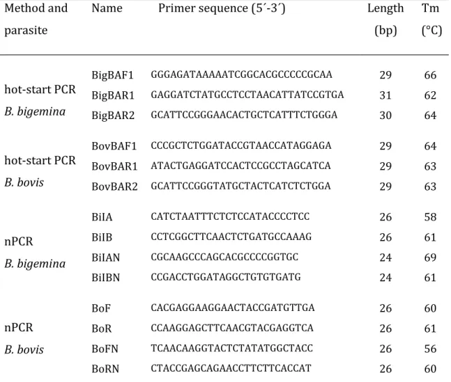
Table 2.1. Primers sequence and melting temperature (Tm). Method and
parasite
Name Primer sequence (5´-3´) Length
(bp) Tm (°C) hot-start PCR B. bigemina BigBAF1 GGGAGATAAAAATCGGCACGCCCCCGCAA 29 66 BigBAR1 GAGGATCTATGCCTCCTAACATTATCCGTGA 31 62 BigBAR2 GCATTCCGGGAACACTGCTCATTTCTGGGA 30 64 hot-start PCR B. bovis BovBAF1 CCCGCTCTGGATACCGTAACCATAGGAGA 29 64 BovBAR1 ATACTGAGGATCCACTCCGCCTAGCATCA 29 63 BovBAR2 GCATTCCGGGTATGCTACTCATCTCTGGA 29 63 nPCR B. bigemina BiIA CATCTAATTTCTCTCCATACCCCTCC 26 58 BiIB CCTCGGCTTCAACTCTGATGCCAAAG 26 61 BiIAN CGCAAGCCCAGCACGCCCCGGTGC 24 69 BiIBN CCGACCTGGATAGGCTGTGTGATG 24 61 nPCR B. bovis BoF CACGAGGAAGGAACTACCGATGTTGA 26 60 BoR CCAAGGAGCTTCAACGTACGAGGTCA 26 61 BoFN TCAACAAGGTACTCTATATGGCTACC 26 56 BoRN CTACCGAGCAGAACCTTCTTCACCAT 26 60
2.2.3.2. Babesipsin hot-start PCR and seminested hot-start PCR
The babesipsin hot-start PCR reaction mixtures (20 μl) contained 16 mM (NH4)2SO4, 67 mM Tris-HCl (pH 8.8), 0.01% (v/v) Tween-20, 2.5 mM MgCl2, 0.5 μM
each primer F1 and R1, 200 μM each dNTP, 1 U of Superhot Taq DNA polymerase (Bioron GmbH), and 1 μl total DNA. Hot-start PCR was carried out in a PTC-200 MJ Research thermocycler for 40 cycles. Each cycle consisted of 20 s of denaturation at 95 °C (1 min for the first cycle), 30 s of annealing at 69 °C, and 45 s of extension at 72 °C. The same conditions of the first PCR were used in seminested hot-start PCR apart from using the inner reverse primers R2 instead of R1, 1 μl of the first PCR products as template and an annealing temperature of 69 °C for B. bovis and
25 71 °C for B. bigemina. Only the samples that were negative in the first hot-start PCR were submitted to seminested hot-start PCR. Twenty random PCR products were gel purified and sequenced in outsourcing at STAB Vida, Portugal.
2.2.3.3. nPCR
The first PCR using the primers described by Figueroa et al. (1993), was carried out using the same buffer as described for babesipsin hot-start PCR, 1 U DFS-Taq DNA polymerase (Bioron GmbH), 0.5 μM outer primers, 1 μl total DNA and the thermocycler program described by Oliveira et al. (2005): 35 cycles (1 min at 95 °C, 1 min at 60 °C for B. bovis and 64 °C for B. bigemina, 1 min 30 s at 72 °C) and a final extension step at 72 °C for 5 min. The same conditions of the first PCR were used in nPCR apart from using inner primers, 2 μl of the first PCR products as template and an annealing temperature of 65 °C for B. bovis and 70 °C for B.
bigemina.
2.2.4. Analysis of PCR products
Ten microlitres of first PCR and 5 μl of second PCR products were separated by electrophoresis in 1.2% (w/v) agarose gel containing ethidium bromide in 0.5 x TBE buffer (44.5 mM Tris-HCl, 44.5 mM Boric Acid and 1 mM EDTA, pH 8.3). After electrophoresis, PCR products were visualized by transillumination with UV light.
2.2.5. Statistical analysis
The data obtained from DNA amplification (nPCR and seminested hot-start PCR) was used for comparisons among methods by Kappa coefficient analysis, a statistical measure of concordance for qualitative items, that takes into account the agreement occurring by chance.
2.3. Results
2.3.1. Hot-start PCR
The combinations using the babesipsin primers described in this study and genomic DNA (kindly provided by Dr. Varda Shkap from Kimron Veterinary Institute, Israel), allowed the amplification of single band products with the
26
expected size. There was no cross reaction between primers for one species and genomic DNA of the other species. And even if it occurs with other isolates, it is expected that the amplification products will have different sizes, since there is an intron between the locations of the primers that is 200 bp shorter for B. bigemina. Hot-start PCR using field samples also allowed the amplification of single band products with the expected size (see Fig. 2.1 and 2.2).
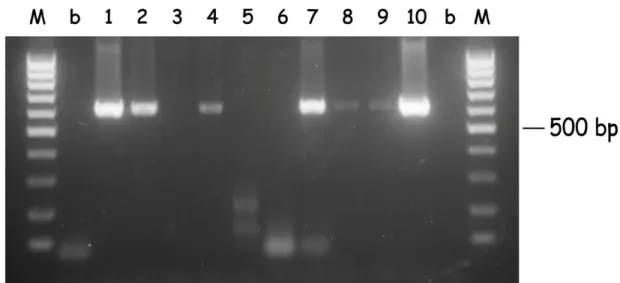
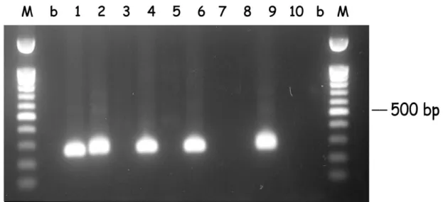
Figure 2.1. Hot-start PCR of B. bigemina DNA from random samples (1 to 10). DNA was subjected to hot-start PCR for the babesipsin 614 bp sequence amplification using the primers BigBAF1 and BigBAR1. Samples 3, 5 and 6 were negative and the remaining were positive. M: 100 bp DNA ladder marker; b: no DNA, negative control.
After the seminested hot-start PCR (see Fig. 2.3 and 2.4), 14 samples that were negative in the first PCR were positive for B. bigemina, and 15 were positive for B.
bovis. Thus, the detection of B. bigemina and B. bovis after the seminested hot-start
PCR increased and were respectively 90% and 80%. The mixed infections also increased to 86 samples (74%) and the number of negatives for both Babesia decreased to 2 (1.7%). Only one strong amplification with a different size was observed in the B. bigemina seminested hot-start PCR, and was considered as negative (Fig. 2.3, sample 2).