UNIVERSIDADE DE LISBOA
FACULDADE DE MEDICINA DE LISBOA
PREDICTION OF SMALL FOR GESTATION NEONATES
FROM BIOPHYSICAL AND BIOCHEMICAL MARKERS AT
35-37 GESTATIONAL WEEKS
Cristina Maria Patronilho Fadigas
Orientador: Prof. Luís Fernando Pacheco Mendes da Graça
Co-Orientador: Prof. Kypros Herodotou Nicolaides
Tese especialmente elaborada para obtenção do grau de Doutor
em Medicina, especialidade de Ginecologia e Obstetrícia
UNIVERSIDADE DE LISBOA
FACULDADE DE MEDICINA DE LISBOA
PREDICTION OF SMALL FOR GESTATION NEONATES FROM
BIOPHYSICAL AND BIOCHEMICAL MARKERS AT 35-37
GESTATIONAL WEEKS
Cristina Maria Patronilho Fadigas
Orientador: Prof. Luís Fernando Pacheco Mendes da Graça
Co-orientador: Prof. Kypros Herodotou Nicolaides
Tese especialmente elaborada para obtenção do grau de Doutor em
Medicina, especialidade de Ginecologia e Obstetrícia
Júri:
Presidente: Doutor José Augusto Gamito Melo Cristino, Professor Catedrático e Presidente do Conselho Científico da Faculdade de Medicina da Universidade de Lisboa
Vogais:
- Doutor José Paulo Achando Silva Moura, Professor Associado da Faculdade de Medicina da Universidade de Coimbra;
- Doutor João Francisco Montenegro de Andrade Lima Bernardes, Professor Catedrático da Faculdade de Medicina da Universidade do Porto;
- Doutor Luís Fernando Pacheco Mendes da Graça, Professor Catedrático Jubilado da Faculdade de Medicina da Universidade de Lisboa (Orientador);
- Doutor Carlos Calhaz Jorge, Professor Catedrático da Faculdade de Medicina da Universidade de Lisboa - Doutora Ana Isabel Gouveia Costa da Fonseca Lopes, Professora Catedrática da Faculdade de Medicina da Universidade de Lisboa;
- Doutor Diogo de Matos Graça Ayres de Campos, Professor Associado com Agregação da Faculdade de Medicina da Universidade de Lisboa.
Tese financiada por:
• The Fetal Medicine Foundation (Charity No: 1037116)
• European Union 7th Framework Programme - FP7-HEALTH-2013-INNOVATION-2 (ASPRE Project #601852)
• Roche Diagnostics Limited, que forneceu a máquina e os reagentes para os ensaios.
As opiniões expressas nesta publicação são da exclusiva responsabilidade da sua autora.
Contents
Preface 6 Abstract 8 Resumo 10 Acknowledgements 12 Abbreviations 13 Figure Legends 15 Table Legends 17 Chapter 1: Introduction 201.1. Definition and epidemiology of small for gestational age and fetal growth
restriction 21
1.2. Etiology and pathophysiology of growth restriction 23
1.2.1. Maternal factors 24
1.2.2. Environmental factors 29
1.2.3. Fetal factors 30
1.2.4. Placental factors 32
1.3. Adverse outcomes of small for gestational age 34
1.3.1. Impact of placental insufficiency on organ functions 34
1.3.2. Adverse perinatal outcomes 35
1.3.3. Long-term adverse outcomes 36
1.4. Screening for small for gestational age 38
1.4.1. Medical and obstetric history 39
1.4.2. Clinical examination 41
1.4.3. Mean arterial blood pressure 42
1.4.4. Ultrasound fetal biometry 42
1.4.5. Uterine artery dopplers 43
1.4.5. Fetal Doppler 44
1.4.6. Biochemical markers 45
1.4.7. Combination models 47
1.5. Objectives of the thesis 48
2. Patients and Methods 49
2.1 Study population 50
2.2. Ethical comittee approval 50
2.3. Data collection 50
2.3.1. Maternal characteristics and history 50
2.3.2. Mean arterial blood pressure 51
2.3.3. Estimated fetal weight 51
2.3.4. Measurement of uterine artery doppler 52
2.3.6. Outcome measures 52
2.4. Statistical analysis 53
Chapter 3: Screening by maternal charateristics and fetal biometry at 35-37
weeks 55 Abstract 56 3.1. Introduction 57 3.1.1. Objectives 57 3.2. Methods 58 3.3. Results 58
3.3.1. Normal pregnancy outcome 59
3.3.2. Small for gestational age 60
3.4. Discussion 67
3.4.1. Main findings of the study 67
3.4.2. Comparison with findings of previous studies 68
Chapter 4: Screening by maternal characteristics, fetal biometry, uterine artery
Doppler and mean arterial blood pressure at 35-37 weeks 70
Abstract 71
4.1. Introduction 72
4.1.1. Objectives 72
4.2. Methods 72
4.3. Results 73
4.3.1. Normal pregnancy outcome 74
4.3.2. Small for gestational age 74
4.4. Discussion 81
4.4.1. Main findings of the study 81
4.4.2. Comparison with findings from previous studies 82
Chapter 5: Screening by maternal characteristics, fetal biometry, placental growth
factor and soluble fms-like tyrosine kinase-1 at 35-37 weeks 83
Abstract 84
5.1. Introduction 85
5.1.1. Objectives 85
5.2. Methods 85
5.3. Results 86
5.3.1. Normal pregnancy outcome 87
5.3.2. Small for gestational age 87
5.4. Discussion 93
5.4.1. Main findings of the study 93
5.4.2. Comparison with findings from previous studies 94
Chapter 6: Conclusion 95
6.1. Summary of results 96
6.3. Implications for clinical practice 97
6.4. Future studies 98
References 100
Preface
The studies described in this thesis comprise work performed at the Fetal Medicine Units of King's College Hospital and Medway Maritime Hospital (United Kingdom). The project was guided by Professor Kypros Nicolaides and funded by The Fetal Medicine Foundation.
According to "Artigo 4º, Diário da República, 2ª série, N.º 111, 9th June 2015
(Regulamento n.º 320/2015)" and "Artigo 19º, Diário da República, 2ª série, nº 153, 7th August 2015 (Regulamento n.º 519/2015)", the results presented and discussed in this thesis were published in the following scientific peer-reviewed journals:
• Fadigas C, Saiid Y, Gonzalez R, Poon LC, Nicolaides KH. Prediction of small-for-gestational-age neonates: screening by fetal biometry at 35-37 weeks. Ultrasound Obstet Gynecol. 2015 May;45(5):559-65. doi: 10.1002/uog.14816. Epub 2015 Apr 9. Impact factor: 5.654. Cited by 29 (Google Scholar, 28 Jan 2019).
• Fadigas C, Guerra L, Garcia-Tizon Larroca S, Poon LC, Nicolaides KH. Prediction of small-for-gestational-age neonates: screening by uterine artery Doppler and mean arterial pressure at 35–37weeks. Ultrasound Obstet Gynecol. 2015 June;45(6):715–721. doi: 10.1002/uog.14847. Epub 2015 May 4. Impact factor: 5.654. Cited by 15 (Google Scholar, 28 Jan 2019).
• Fadigas C, Peeva G, Mendez O, Poon LC, Nicolaides KH. Prediction of small-for-gestational-age neonates: screening by placental growth factor and soluble fms-like tyrosine kinase-1 at 35-37 weeks. Ultrasound Obstet Gynecol. 2015 Aug;46(2):191-7. doi: 10.1002/uog.14862. Epub 2015 Jun 18. Impact factor: 5.654. Cited by 13 (Google Scholar, 28 Jan 2019).
Partial results of this thesis were presented at international congresses, as part of a wider project, also lead by Prof. Kypros Nicolaides and funded by the Fetal Medicine Foundation. The referred research project aims to predict appropriate timing for third trimester growth scan, contingent to the second trimester scan. The meetings were the following:
• Poon C, Lesmes C, Bakalis S, Fadigas C, Nicolaides KH. Prediction of Fetal Growth Restriction. Advances in Fetal Medicine Course 2014, London, United Kingdom, 6-7 Dec 2014. [Oral presentation]
• Poon C, Lesmes C, Bakalis S, Fadigas C, Nicolaides KH. Small for gestional age: Timing for 3rd trimester scan. 14th World Congress in Fetal Medicine, Crete,
Greece, 21-25 Jun 2015. [Oral presentation]
• Poon C, Gallo D, Bakalis S, Fadigas C, Nicolaides KH. Contingent model for the prediction of delivery of small-for-gestational-age neonates. 26th World Congress on Ultrasound in Obstetrics and Gynecology, Rome, Italy, 24-28 September 2016 [Oral presentation]. Abstract available in Ultrasound in Obstetrics and Gynecology, 48, Suppl 1:2, September 2016. DOI: 10.1002/uog.16029.
Abstract
Small for gestational age (SGA) is common in pregnancy and it has been associated with an increase in adverse perinatal outcomes, predisposition for neurological and cognitive delay in childhood and cardiovascular and endocrine diseases in adulthood.
The classification is not consensual, having been defined in different studies as estimated fetal weight, abdominal circumference or birthweight below the 10th, 5th or 3rd percentiles,
with the prevalence varying with the definition that is used.
The increased risk of perinatal mortality and morbidity can be substantially reduced in cases identified prenatally, as close monitoring, timely delivery and prompt neonatal care can be undertaken, in comparison to those cases detected after birth.
Over time, several screening methods have been introduced, in order to optimize the detection rate for SGA. These approaches range from abdominal palpation, symphyseal-fundal height measurement, fetal biometries, uterine artery doppler assessment and, more recently, biochemical markers.
The aim of this thesis is to develop a model for prediction of SGA neonates in the absence of pre-eclampsia, based on maternal characteristics, clinical history, fetal biometry, uterine pulsatility index (Ut PI), mean arterial blood pressure (MAP) and serum biochemical markers (serum placental growth factor: PlGF; Soluble fms-like tyrosine kinase-1: sFlt-1) at 35-37 gestational weeks.
This was a prospective screening project for detection of SGA neonates, in women attending for their third-trimester hospital visit in pregnancy at King's College Hospital (London) and Medway Maritime Hospital (Kent). The project comprised three studies.
The first study included biophysical measurements of 5515 pregnant women, including 278 that delivered SGA (<5th) neonates. A SGA predictive model was developed based on
the combination of maternal factors, clinical history and estimated fetal weight.
In the second study, a subset of 5121 pregnant women was evaluated, 245 of which had SGA (<5th) newborns. A model was developed based on the combination of maternal
dopplers. It was found that the additional use of mean arterial pressure and pulsatility index of the uterine arteries did not significantly improve the performance of screening for delivery of SGA neonates in comparison to the first study.
In the third study, a subset of 3859 pregnant women was evaluated, comprising 158 SGA newborns. The SGA prediction model combined the following parameters: maternal factors, estimated fetal weight and biochemical values (serum placental growth factor, PlGF; fms-like soluble tyrosine kinase-1, sFlt-1). It was found that sFlt-1, when combined with maternal factors and fetal biometries, did not remain an independent factor in this predictive model. Additionally, serum PlGF only marginally improved the SGA screening performance when compared to the model of the first study.
Hence, based on the findings, the best prediction was provided by the combination of maternal factors, estimated fetal weight and serum placental growth factor (PlGF). This combined screening predicted, at a 10% false positive rate, 88, 96 and 94% of SGA neonates with birth weight below the 10th, 5th and 3rd percentiles delivering at <2 weeks
following assessment. The respective values for SGA delivering ≥37 weeks were 64, 75 and 80%.
In conclusion, combined screening by maternal factors, biophysical and biochemical markers at 35-37 weeks can identify a high percentage of pregnancies that will deliver SGA neonates.
Keywords: Small-for-gestational age; Late third trimester screening; Fetal biometry; Placental growth factor; Soluble fms-like tyrosine kinase-1.
Resumo
Ser leve para a idade gestacional (LIG) é comum na gravidez e tem sido associado a um aumento nos resultados perinatais adversos, predisposição para défices neurológico e cognitivo na infância e doenças cardiovascular e endocrinológica na vida adulta.
A classificação não é consensual, tendo sido definida em diferentes estudos como peso fetal estimado, circunferência abdominal fetal ou peso à nascença abaixo dos percentis 10, 5 ou 3. Deste modo, a prevalência de LIG varia com a definição utilizada.
O aumento do risco de mortalidade e morbilidade perinatal pode ser substancialmente reduzido nos casos identificados no período pré-natal, uma vez que a vigilância apertada da gravidez, com programação do parto na altura adequada e um atendimento neonatal apropriado podem ser oferecidos, em comparação com os casos apenas detectados após o parto.
Ao longo do tempo, vários métodos de triagem foram introduzidos, a fim de optimizar a taxa de detecção de LIG. Essas abordagens incluem a palpação abdominal, a medição da altura uterina, a avaliação da biometria fetal, a medição dos dopplers das artérias uterinas e, mais recentemente, a utilização de marcadores bioquímicos.
O objectivo desta tese é desenvolver um modelo para previsão de recém-nascidos LIG na ausência de pré-eclâmpsia, baseado em factores maternos, história clínica, biometrias fetais, índice de pulsatilidade das artérias uterinas, pressão arterial média e marcadores bioquímicos (factor de crescimento placentário sérico: PlGF; FMS-like tirosina cinase solúvel-1: sFlt-1) às 35-37 semanas de gestação.
Este foi um trabalho prospectivo de rastreio de gestações simples às 35-37 semanas gestacionais, que decorreu no Reino Unido, no King's College Hospital em Londres e no Medway Maritime Hospital em Kent. O projecto foi organizado em 3 estudos distintos.
No primeiro estudo foram incluídas medidas biofísicas de 5515 gestantes, em que 278 grávidas tiveram recém-nascidos LIG (<p5) e foi desenvolvido um modelo de previsão de LIG com base na combinação de factores maternos, história clínica e peso fetal estimado.
No segundo estudo avaliou-se um subgrupo de 5121 gestantes, das quais 245 tiveram recém-nascidos LIG (<p5) e desenvolveu-se um modelo com base na combinação dos factores maternos, história clínica, peso fetal estimado, pressão arterial média e fluxometria das artérias uterinas. Constatou-se que a utilização adicional da pressão arterial média e do índice de pulsatilidade das artérias uterinas não melhorou significativamente a taxa de detecção de recém-mascidos LIG face ao primeiro estudo.
No terceiro estudo, avaliou-se um subconjunto de 3859 grávidas, incluindo 158 recém-nascidos LIG. O modelo de previsão de recém-nascido LIG combinou os seguintes parâmetros: factores maternos, peso fetal estimado e valores bioquímicos (factor de crescimento placentário sérico, PlGF; fms-like tirosina cinase solúvel-1, sFlt-1). Verificou-se que o sFlt-1, quando combinado com os factores maternos e biometrias fetais, não permaneceu como um factor independente neste modelo de previsão. Adicionalmente, observou-se que o PlGF sérico apenas melhorou marginalmente a taxa de detecção de LIG face ao modelo do primeiro estudo.
Assim, dos vários modelos avaliados, aquele com melhor taxa de detecção de recém-nascidos LIG foi fornecido pela combinação de factores maternos, peso fetal estimado e factor de crescimento placentário sérico (PlGF). Este modelo previu, com uma taxa de falsos positivos de 10%, 88, 96 e 94% dos recém-nascidos LIG com peso ao nascer inferior aos percentis 10, 5 e 3, respectivamente, e que nasceram <2 semanas após a avaliação em consulta. A taxa de detecção para LIG com nascimento ≥37 semanas foi de 64, 75 e 80%.
Em conclusão, o rastreio pelo modelo combinado de factores maternos, marcadores biofísicos e bioquímicos às 35-37 semanas pode identificar uma percentagem elevada de gestações com recém-nascidos LIG.
Acknowledgements
The studies described here in comprise work performed at the Fetal Medicine Units of King's College Hospital and Medway Maritime Hospital (United Kingdom).
Professor Kypros Nicolaides, to whom I am eternally grateful, guided the project. Without his supervision, support and expertise, this project would have not have been possible.
The study was funded by a grant from The Fetal Medicine Foundation (Charity No: 1037116) and by the European Union 7th Framework Programme - FP7-HEALTH-2013-INNOVATION-2 (ASPRE Project #601852). Roche Diagnostics Limited provided the machine and reagents for the assays.
I would like to thank Professor Luís Graça for his valuable suggestions and for believing in me and in this study since the beginning.
I am extremely thankful to Leona Poon for her invaluable help with the statistical analysis and to Argyro Syngelaki and Dahiana Gallo for their guidance and exhaustive cooperation in the collection of outcomes.
I would like to thank all the Research Fellows of the Fetal Medicine Foundation, who helped to recruit the pregnant women, perform the scans and collect the data for this study.
To Professor Jorge Lima, I thank the continuous support and motivation.
I would also like to thank my family and friends, whose unconditional support made me persist in adversity and complete this project.
I am thankful for my colleagues' encouragement and motivation.
Finally, and foremost, my acknowledgment goes to the women who participated in this research and contributed to the development of this model.
Abbreviations
Abbreviation
AC Abdominal Circumference
ACOG American College of Obstetricians and Gynecologists
ADAM12 Metalloprotease Domain-Containing Protein-12
AGA Appropriate for Gestational Age
aOR Adjusted Odds Ratio
APH Antepartum haemorrhage
APLS Antiphospholipid Syndrome
aRR Adjusted Relative Risk
ART Assisted Reproductive Technology
AUC Area Under ROC (Receiver Operating Characteristic)
β-hCG free β-human chorionic gonadotropin
BMI Body Mass Index
BPD Biparietal Diameter BW Birth Weight CI Confidence Intervals CPR Cerebro-Placental Ratio CS Cesarean section DR Detection Rate
EFW Estimated Fetal Weight
FGR Fetal Growth Restriction
FL Femur Length
FPR False Positive Rate
GH Gestational Hypertension
HC Head Circumference
IGF Insulin Like Growth Factor
IQR Interquartile Range
iSUA Isolated Single Umbilical Artery
IVF In Vitro Fertilization
LGA Large for Gestational Age
LR+ Positive Likelihood Ratio
MAP Mean Arterial Pressure
MCA Middle Cerebral Artery
MCI Marginal Cord Insertion
MoM Multiple of the Median
MRI Magnetic Resonance Imaging
NBAS Neonatal Behavioral Assessment Scale
NICE National Institute for Health and Clinical Excellence
OR Odds Ratio
PAPP-A Pregnancy Associated Plasma Protein-A
PE Pre-eclampsia
PI Pulsatility Index
PlGF Placental Growth Factor
PP-13 Placental Protein-13
RCOG Royal College of Obstetricians and Gynaecologists
RI Resistance Index
ROC Receiver Operating Characteristic
RR Relative Risk
SD Standard Deviation
SGA Small for Gestational Age
SFH Symphyseal-Fundal Height
sFlt-1 Soluble fms-like tyrosine kinase-1
SGA Small-for-Gestational Age
SLE Systemic Lupus Erythematosus
SUA Single Umbilical Artery
UA Umbilical Artery
UK United Kingdom
UtA Uterine Artery
VCI Velamentous Cord Insertion
VEGF Vascular Endothelial Growth Factor
Figure Legends
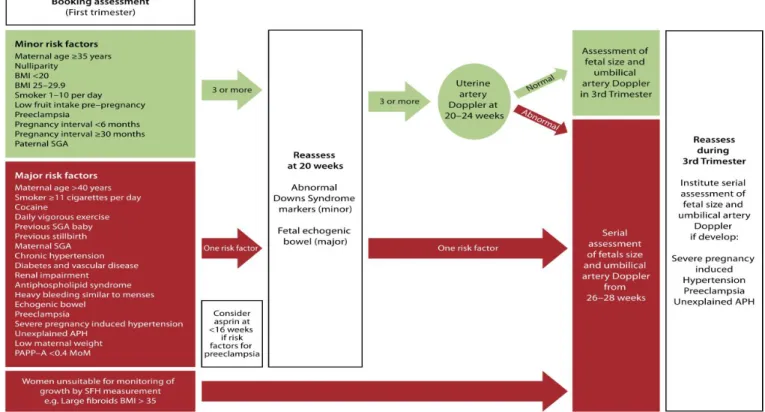
Figure 1.1 - Risk assessment for SGA as set out by the RCOG.
Figure 3.1. Z-scores for fetal head circumference (a), abdominal circumference (b), femur length (c) and estimated fetal weight (d) at 35-37 weeks.
Figure 3.2. Receiver-operating characteristic curves of maternal characteristics (black line), combination of maternal characteristics of HC, AC and FL z-score (blue line) and the combination of maternal characteristics with EFW z-score (red line) at 35-37 gestational weeks in the prediction of SGA with BW below the 10th (a), the 5th (b) and the 3rd (c)
percentile, delivering < 2 weeks following assessment (left) or ≥37 weeks' gestation.
Figure 4.1 Log10 uterine artery pulsatility index (UtA-PI) (a,b) and log10 mean arterial
pressure (MAP) (c,d) multiples of median according to assessment-to-delivery interval (a,c) and birth-weight Z‐score (b,d) in pregnancies delivering small-for-gestational-age neonates with birth weight < 5th percentile, plotted on the 50th (solid line), 90th and
95th (dashed line) percentile of the appropriate normal range.
Figure 4.2. Receiver–operating characteristics curves of maternal factors (black) and maternal factors with uterine artery pulsatility index (red), mean arterial pressure (blue), estimated fetal weight Z‐score (green) and their combination (purple), at 35–37 weeks'
gestation, in the prediction of small-for-gestational-age neonates with birth weight < 10th (a), < 5th (b) or < 3rd (c) percentile, delivering < 2 weeks following assessment
(top) or ≥ 37 weeks' gestation (bottom).
Figure 5.1. Log10 placental growth factor (a) and log10 soluble fms-like tyrosine kinase-1
(b) multiples of the median (MoM) according to assessment-to-delivery interval in pregnancies delivering small-for-gestational-age neonates with birth weight < 5th percentile, plotted on the 50th (solid line) and 10th (dashed line) percentile of
the normal range.
Figure 5.2. Receiver–operating characteristics curves of maternal factors (black line),
maternal factors with estimated fetal weight (EFW) (blue line), maternal factors with EFW and placental growth factor (red line) at 35–37 weeks' gestation, in the prediction of small-for-gestational-age neonates with birth weight < 10th (a), < 5th (b) and < 3rd (c) percentile,
Figure 5.3. Receiver–operating characteristics curves of maternal factors (black line), maternal factors with EFW (blue line), maternal factors with EFW and placental growth factor (red line) at 35–37 weeks' gestation, in the prediction of small-for-gestational-age neonates with birth weight < 10th (a), < 5th (b) and < 3rd (c) percentile, delivering
Table Legends
Table 1.1. Common causes of growth restriction (Sankaran et al45)
Table 1.2. Birthweight comparison (in grams) based on standard mother being defined as of European origin, height 163 cm, weight 64 kg, first pregnancy, with baby sex averaged between male and female (Gardosi et al31)
Table 1.3. RCOG risk factors for SGA fetus/neonate from history at booking(Robson et al, 2013).
Table 1.4. RCOG risk factors for SGA fetus/neonate from current pregnancy complications (Robson et al9)
Table 1.5. Studies showing the differences in PlGF in normal and pregnancies delivering a SGA neonate.
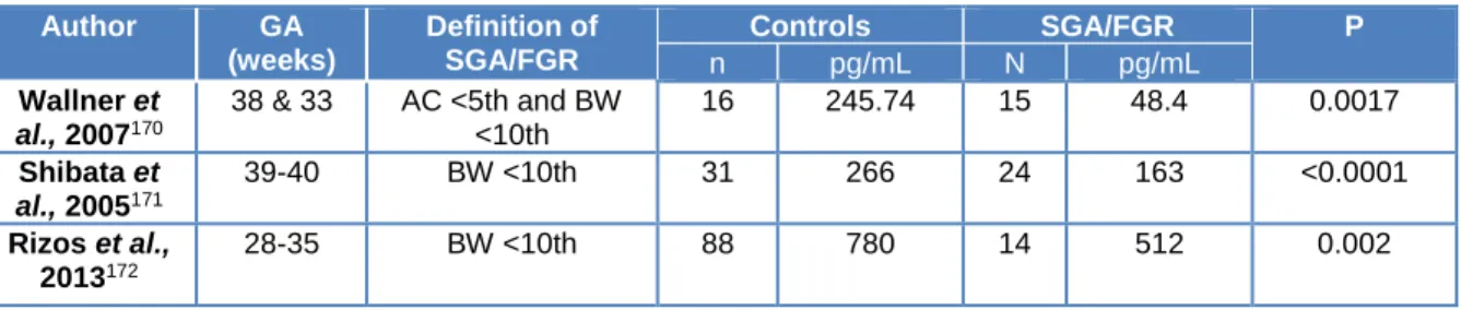
Table 1.6. Studies showing the differences in sFlt-1 in normal and pregnancies delivering a SGA neonate.
Table 1.7. Difference in PlGF/sFlt-1 ratio in normal and pregnancies delivering a SGA neonate
Table 3.1. Characteristics of the study population of women with a singleton pregnancy with normal outcome or with a small-for-gestational age (SGA) neonate, in the absence of pre-eclampsia (PE).
Table 3.2. Pearson correlation between Z-score values of head circumference, abdominal circumference, femur length and estimated fetal weights at 35-37 weeks’ gestation in the normal and small for gestational age groups.
Table 3.3. Fitted regression model with maternal characteristics and history for the prediction of small for gestational age with birth weight below the 5th percentile, in the
absence of preeclampsia.
Table 3.4. Fitted regression models with maternal characteristics and history, fetal head circumference (HC) score, abdominal circumference (AC) score, femur length (FL) Z-score or estimated fetal weight (EFW) Z-Z-score at 35–37 weeks’ gestation, for the
prediction of small-for-gestational age with birth weight < 5th percentile, in the absence of
pre-eclampsia
Table 3.5. Performance of screening for small for gestational age (SGA) neonates with birthweight <10th, <5th and <3rd percentiles, delivering within 2 weeks of assessment and ≥ 37 weeks' gestation, in the absence of pre-eclampsia, using maternal characteristics and history, fetal biometry or estimated fetal weight at 35-37 weeks' gestation
Table 3.6. Detection rates (DR) in screening for small-for-gestational-age neonates with birth weight < 10th, < 5th and < 3rd percentile, delivering within 2 weeks of assessment, in
the absence of pre-eclampsia, using maternal characteristics and history, fetal biometry or estimated fetal weight at 35–37 weeks’ gestation
Table 3.7. Detection rates (DR) in screening for small-for-gestational-age neonates with birth weight < 10th, < 5th and < 3rd percentile, delivering ≥ 37 weeks’ gestation, in the
absence of pre-eclampsia, using maternal characteristics and history, fetal biometry or estimated fetal weight at 35–37 weeks’ gestation
Table 4.1. Characteristics of the study population of women with a singleton pregnancy with normal outcome or with a small-for-gestational age (SGA) neonate, in the absence of pre-eclampsia (PE).
Table 4.2. Uterine artery pulsatility index (UtA-PI) and mean arterial pressure (MAP) at 35–37 weeks’ gestation in pregnancies that delivered small-for-gestational-age (SGA) neonates with birth weight < 5th percentile, in the absence of pre-eclampsia, and in
unaffected pregnancies
Table 4.3. Fitted regression models with maternal characteristics and history, estimated fetal weight (EFW) Z-score, uterine artery pulsatility index (UtA-PI) and mean arterial pressure (MAP) at 35–37 weeks’ gestation for the prediction of small-for-gestational-age neonates with birth weight < 5th percentile, in the absence of pre-eclampsia
Table 4.4. Performance of screening for small for gestational age neonates with birth weight <10th, <5th and <3rd percentile delivering within two weeks of assessment and at
>37 weeks’ gestation, in the absence of preeclampsia, with maternal factors, estimated fetal weight, uterine artery pulsatility index and mean arterial pressure at 35-37 weeks’ gestation.
Table 4.5. Detection rates (DR) in screening for small-for-gestational-age (SGA) neonates with birth weight < 10th, < 5th and < 3rd percentile, delivering within 2 weeks of assessment,
in the absence of pre-eclampsia, using maternal factors, estimated fetal weight (EFW), uterine artery pulsatility index (UtA-PI) and mean arterial pressure (MAP) at 35–37 weeks’ gestation
Table 4.6. Detection rates (DR) in screening for small-for-gestational-age (SGA) neonates with birth weight < 10th, < 5th and < 3rd percentile, delivering ≥ 37 weeks, in the absence of
pre-eclampsia, using maternal factors, estimated fetal weight (EFW), uterine artery pulsatility index (UtA-PI) and mean arterial pressure (MAP) at 35–37 weeks’ gestation Table 5.1. Characteristics of the study population of women with a singleton pregnancy with normal outcome or with a small-for-gestational-age (SGA) neonate, in the absence of pre-eclampsia (PE)
Table 5.2. Placental growth factor (PlGF) and soluble fms-like tyrosine kinase-1 (sFlt-1) at 35-37 weeks’ gestation in small for gestational age (SGA) neonates with birth weight below the 5th percentile, in the absence of preeclampsia and in the normal group.
Table 5.3. Fitted regression models with maternal characteristics and history (maternal factors), estimated fetal weight (EFW) Z-score, placental growth factor (PlGF) and soluble fms-like tyrosine kinase-1 (sFlt-1) at 35–37 weeks’ gestation for the prediction of small-for-gestational-age neonates with birth weight < 5th percentile, in the absence of
pre-eclampsia
Table 5.4. Performance of screening for small-for-gestational-age (SGA) neonates with birth weight < 10th, < 5th and < 3rd percentile delivering within 2 weeks of assessment, in
the absence of pre-eclampsia, using maternal characteristics and history, estimated fetal weight (EFW), placental growth factor (PlGF) and soluble fms-like tyrosine kinase-1 (sFlt-1) at 35–37 weeks’ gestation.
Table 5.5. Performance of screening for small for gestational age (SGA) neonates with birth weight <10th, <5th and <3rd percentile delivering at >37 weeks’ gestation in the
absence of preeclampsia, with maternal characteristics and history, estimated fetal weight, placental growth factor (PlGF) and soluble fms-like tyrosine kinase-1 (sFlt-1) at 35-37 weeks’ gestation.
C
HAPTER
1
Chapter 1. Introduction
1.1.DEFINITION AND EPIDEMIOLOGY OF SMALL FOR GESTATIONAL AGE AND FETAL GROWTH RESTRICTION
Small for gestational age (SGA) is usual in pregnancy1. It is associated with increased
adverse perinatal outcomes2, predisposition for neurological and cognitive delay in
childhood and cardiovascular and endocrine diseases in adulthood3, 4.The classification is
not consensual and the prevalence will vary with the definition that is used.
Historically, a birthweight (BW) below 2500g and, occasionally, under 1500g, was used as a cut-off for the definition of SGA. The World Health Organization still uses the definition of SGA as neonatal weight below 2500g at term5. This definition is especially useful in
developing countries, as it eliminates the impact of accurate pregnancy dating and since low birthweight has long been used as an important public health indicator.
However, with the introduction of population based gestational age dependent BW charts since Lubchenco6, there was a move to the use of percentiles, which has been widely
adopted in developed countries. The definition of SGA is not consensual and various cut-off limits have been proposed, including estimated fetal weight (EFW), BW, abdominal circumference (AC) or, more recently, BW charts comprising babies still in utero7 below
the 10th, 5th or 3rd percentiles or -2 standard deviations (SD) below the mean for
gestational age. The ideal cut-off remains uncertain.
The differentiation between constitutionally small and fetal growth restriction (FGR) and the subsequent obstetric management is challenging. The distinction is important, since SGA fetus have a good prognosis compared to FGR fetus. Some authors use the terms SGA and FGR interchangeably, although the majority of SGA are constitutionally small. FGR suggests an underlying pathology and refers to a fetus that has failed to achieve its optimal growth potential 8, 9. It affects up to 5-10% of all pregnancies10. Fetus with a BW
below the 10th percentile may not be growth restricted, but rather constitutionally small.
The incidence of FGR and SGA are approximately 10%11, 12. Although they are not the
same population, there is an overlap, as FGR concentrates in the SGA population. Using the 10th percentile as cut-off, it is estimated that in the SGA population, 40% neonates are
constitutionally small and 60% are growth restricted12, 13. The lower the weight percentile,
the higher the chances of pathology and, thus, growth restriction and problems after birth. Lowering the threshold for SGA, increases the likelihood of the fetus being fetal growth
restricted and lowers the false positive rate (FPR)14. Nevertheless, this definition fails to
recognize the fetus that have fallen across the percentiles, but still remain above the 10th
percentile14.
The challenge is to differentiate constitutionally SGA from FGR fetus and to define FGR fetuses in the group of appropriate for gestational age (AGA) or large for gestational age (LGA). The risks of inaccurate definition of FGR within these different growth groups are overtreatment of healthy SGA and undertreatment of FGR fetus with normal growth.
The traditional approach for identifying pregnancies with SGA fetuses by maternal abdominal palpation and serial measurements of symphisis-fundal height has a detection rate of 30-85%15-21, as obesity and leyomiomas limit its accuracy.
A few studies comprising low-risk singleton pregnancies have examined the potential value of sonographic fetal biometry during the third trimester in the prediction of SGA neonates 22-28.Of these, only one study27, up until the end of the studies of this thesis,
examined the value of EFW in a late third trimester-ultrasound examination in low risk pregnancies
The use of mathematical models, as well as customized charts10 adjusted for
physiological variables, might improve the classification of fetal growth. They would help to identify fetuses that are small because of constitutional reasons and not because of FGR, reducing unnecessary investigations and interventions 29-32.
The advantage of serial ultrasound examinations for longitudinal growth assessment has not been clearly demonstrated33, 34.
Doppler studies of maternal and fetal circulation have also shown to improve the diagnosis of SGA and FGR, as often SGA have normal doppler studies, whereas FGR show doppler abnormalities, due to placental insufficiency35.
Histological studies report that in preeclampsia (PE) and SGA without PE there is impaired placentation, with inadequate trophoblastic invasion of the maternal spiral arteries. This leads to an altered placental production and systemic release of antiangiogenic (soluble fms-like tyrosine kinase-1: sFlt-1) and proangiogenic (placental growth factor: PlGF) factors36. Several studies, mainly case-control, reported that, in
increased, both in the second and third trimesters of pregnancy. This translates into an increase in the sFlt-1/PlGF ratio37-39, which is not as high in late-onset FGR as in early
onset FGR37, 40. There has been a positive correlation in between sFlt-1/PlGF ratio and the
likelihood of complications41, 42.
1.2.ETIOLOGY AND PATHOPHYSIOLOGY OF GROWTH RESTRICTION
At a cellular level, fetal growth has three different stages 43:
• 0-16 weeks: Hyperplasia
• 16-30 weeks: Hyperplasia and hypertrophy • 30 week - term: Hypertrophy
An exponential curve translates fetal growth between the end of the first trimester and the last part of the third trimester, with only a slight tailing off around term. However, if we consider the percentage of mass gained per week rather than absolute mass gain, the greatest growth rate occurs in the early stages of pregnancy (in between conception and middle of second trimester), whilst organogesis occurs, in a period of very rapid cell division. From that point onwards, growth occurs mainly due to organs' maturation and hypertrophy44.
Normal fetal growth relies on the coordination of several components, namely, the genetic growth potential of the fetus, the ability of the placenta to transfer nutrients and oxygen to the fetus and the capacity of the maternal body to deliver these nutrients to the placenta. All of which, are influenced by the surrounding environment44.
Hence, the etiology of fetal FGR can be categorized into maternal, fetal, placental and environmental factors (Table 1.1)45. Even though the pathophysiology of the various
underlying conditions is different, the majority of the cases will lead to sub-optimal placental perfusion and fetal nutrition1, 9.
Table 1.1: Common causes of growth restriction (Sankaran et al 45)
Common causes for FGR
Maternal factors
• Undernutrition
• Low maternal weight gain
• Low maternal BW
• Extremes of maternal age • Low socio-economic status • Nulliparity
• Medical conditions (eg, pregestational diabetes, renal insufficiency, systemic lupus erythematosus, antiphospholid antibody syndrome, hypertensive disease pregestational or pregnancy-related, cyanotic cardiac disease)
Environmental factors
• Substance use and abuse (tobacco, alcohol, cocaine or narcotics)
• Teratogen exposure
• Daily vigorous exercise • High altitude (above 1500m) • Irradiation Fetal factors • Chromosomal abnormalities • Genetic diseases • Congenital malformations • Intrauterine infections • Multiple gestation Placental factors • Abnormal placentation
• Chronic abruption, infarcts and focal lesions • Chronic inflammatory conditions
• Chorioangioma
• Single umbilical artery, velamentous cord insertion • Confined placental mosaicism
1.2.1. Maternal factors
Nutrition
Animal studies have shown that both maternal undernutrition and overnutrition reduce placental-fetal blood flows and reduce fetal growth, by decreasing placental synthesis of nitric oxide (a major vasodilator and angiogenic factor) and polyamines (key regulators of DNA and protein synthesis). There is some evidence that maternal nutrition status can alter the epigenetic state of the fetal genome, which may provide a molecular mechanism for the impact of maternal nutrition on both fetal programming and genomic imprinting46.
Studies of pregnant women during famine times have shown an association between SGA and maternal undernutrition. However, the American College of Obstetricians and Gynecologists (ACOG)47-49 reports that there is no high-quality evidence to support that an
additional nutrient intake will improve the outcome of FGR, in the absence of true malnutrition.
Weight, height, body mass index and low maternal weight gain
The median weight and the median height of women delivering an SGA fetus is lower than those delivering an AGA fetus50, 51. More often, the studies have assessed maternal
characteristics on the basis of body mass index (BMI) and several of these have shown that the lower the BMI, the higher the risk of delivering a SGA neonate52, 53.
Low maternal weight gain has been shown to be associated with SGA, even when adjusted for confounding factors as height, BMI, parity, race, toxemia and diabetes. The relative risk (RR) was 1.8 (95% CI, 2.6) in the second trimester and 1.7 (95% CI 1.3-2.3) in the third trimester54. This increased risk was observed across the spectrum of
maternal BMI54. Regardless, the Royal College of Obstetricians and Gynaecologists
(RCOG)9 no longer recommends that women are routinely weighted during pregnancy as
a form of screening for SGA.
Low maternal birthweight
Parental contribution for fetal birthweight through inherited genes is estimated to be around 30-70%55. Potential interaction in between fetal genes and uterine environment
influences fetal size, with animal studies suggesting that growth is modified towards maternal size. In general, fetal growth tends to be restrained by maternal environment and this is more evident in the first pregnancy. This trace appears to be inherited through the maternal line, with several genes being potentially associated, namely mitochondrial DNA 16189 variant and common variants of maternally only expressed genes, such as H19. Several other genes have been reported to be in association with SGA and FGR (insulin-like growth factor IGF-1, IGF-2, G-protein beta 3 subunit, inducible cytochrome P450, genes encoding angiotensinogen, placental alkaline phosphatase and vitamin D receptor45, 56, 57. Paternally inherited genes seem to play a role when the maternal
component of restraining fetal growth is less evident. With paternal birthweight history of SGA, a study58 reports a 3.47 fold increase risk of the fetus being SGA (95% CI,
Maternal age
Extremes of maternal age have been associated with a higher risk of SGA.
Women<17 years: A study59 reports the highest incidence of SGA in mothers <17 years
of age (3.2%). Nevertheless, young age did not remain as an independent risk factor, when it was adjusted to other maternal factors, such as race, education, parity, marital status and prenatal care. These results indicate that the higher incidence of SGA in younger mothers apparently reflects their poor sociodemographic and prenatal care status.
Advanced maternal age: Women over 35 years are at higher risk of many pregnancy complications, mainly because they are more likely to have pre-existing medical conditions than their younger counterparts, predisposing them to develop pregnancy complications60. It was observed that women over 40 years of age were at increased risk
of pregnancy complications and this risk persisted even when the data was adjusted for preexisting maternal disease61. Hence, maternal age 40 years or older constitutes a major
risk factor for having a small-for-gestational-age neonate, with an odd ratio (OR) of 3.2 (95% CI 1.9-5.4) having been reported62.
Ethnicity
It has been referred that birthweight is affected by ethnicity. Gardosi's study31 to assess
the factors that affect fetal growth and birthweight in the population to derive the coefficients to obtain customized charts, shows that african babies can weight less than 218g less than European babies, whereas Chinese babies can weight 100g more (Table 1.2).
Table 1.2 - Birthweight comparison (in grams) based on standard mother being defined as of European origin, height
163 cm, weight 64 kg, first pregnancy, with baby sex averaged between male and female (Gardosi et al 31)
Ethnic origin United States England New Zealand Australia
African American -161.0 — — — African Caribbean — -127.5 — — African — -218.5 — -297.4 Hispanic -38.6 — — — Middle Eastern. — -89.9 — -110.0 Bangladeshi — -79.3 — — Indian/Pakistani — — — -162.0 Indian — -149.4 -149.5 — Pakistani — -187.3 — — Chinese — — 100.9 — Maori — — — -66.8 Samoan — — — 84.2 Tongan — — — 124.1 Other -140.8 — — —
This also impacts on the incidence of SGA neonates. In 2008, the Office of National Statistics63 released its data on all deliveries in England and Wales from 2005. The data
showed that babies born to white mothers were larger than those of south Asian or black mothers. Further dividing those two groups into Pakistani, Indian, Bangladeshi, African and Caribbean, showed that the lowest mean BW was in Bangladeshi mothers. However, the largest percentage of babies born either <2.5 kg or <1.5 kg was in the Caribbean group. Further United Kingdom (UK) studies, documented that Afro-Caribbean, South Asian, East Asian and mixed race women were statistically significantly more likely to deliver an SGA baby than white women50-51, 64.
Parity
Nulliparity - The biological mechanisms by which parity and SGA are correlated are not quite understood. Nulliparous women have significant associations with adverse outcomes, particularly, when women are also of young age (<18 years). A review65 has
shown an association with term SGA and nulliparity. For nulliparous aged under 18 years, the adjusted odds ratio (aOR) was 1.80 (95% CI: 1.62-2.01)65. For nulliparous aged 18-34,
the association was not as strong, but still significant, with aOR of 1.51 (95% CI: 1.39-1.64)65. Several studies have hypothesized that in young mothers, maternal-fetal
competition for nutrients and/or the mother’s incomplete physical growth might contribute to adverse neonatal outcomes65, 66.
Grand Multiparity - Multiple studies have shown an association in between grand multiparous and medical, obstetric and placental complications. Biological mechanisms have been used to explain this association (eg: chronic hypertension for abruption or atrophy of the endometrium for placenta previa)65. A study67 reports that grand multiparity
in an economically stable population is not a major risk factor and that previous studies reflect socio-economic factors and not parity itself. This was also shown in a meta-analysis, where a higher risk of SGA was identified in the subgroup of less developed countries, whereas the increase in uterine blood flow associated with increasing multiparity leads to an higher BW in subsequent newborns65.
Maternal medical conditions
Medical conditions that are associated with vascular disease and interfere with utero-placental circulation increase the risk of FGR. These include hypertensive disorders, pregestational diabetes, auto-immune diseases (eg: systemic lupus erythematosus - SLE), renal insufficiency and antiphospholid syndrome. As for hereditary thrombophilias (eg, factor V Leiden or prothrombin gene mutations), the association with FGR is not consistent1. The effects of placental underperfusion vary, whether the placental
insufficiency has early or late onset. Early onset insufficiency leads to the underdevelopment of terminal villi, with terminal villous hypoplasia. Late onset insufficiency leads to advanced maturation of the villi, with increased capillary branching, which only compensates the degree of hypoxia temporarily45, 68. Where the maternal
supply of the placenta has been completely occluded, there will be infarctation of the associated area.
The risks of SGA associated with pathology that affect placental perfusion are significant. The adjusted relative risk (aRR) for chronic hypertension is 2.5 (95% CI 2.1-2.9) 69, being
associated with the inadequate conversion of the spiral arteries in the decidua and myometrium. Diabetes with vascular disease has an OR of 6 (95% CI1.5-2.3)70, the
underlying mechanism involving small ischaemic villi, immature/dysmature villi and inconsistent glucose supply to the fetus. For renal impairment, the aOR is 5.3 (95% CI 2.8-10)71. SLE has an OR of SGA of 5.6 (95% CI 4.1-7-8)72. The presence of
anticardiolipin antibodies gives a RR of SGA 6.22 (95% CI 2.42-16.0)73.
Previous pregnancy history
Having a previous SGA neonate, increases up until three-fold the risk of having another SGA, using a cut-off of BW below the 10th percentile (OR 3.9; 95% CI 2.14-7.12)9, 74. This
risk is further increased, after two SGA deliveries75. The King’s College Group, in two
prospective studies51, 64, showed, not only that women with a previous SGA neonate were
more likely to deliver another SGA baby than those who had previous normal babies, but also that this risk remained significantly higher even if they had delivered a normal baby after an SGA baby.
Method of conception
In comparison with naturally conceived children, singletons born after assisted reproductive technology (ART) have a higher risk of FGR and other adverse outcomes, with studies reporting a risk of 40-60% of SGA in ART neonates61, 76. Regarding singleton
pregnancies after in vitro fertilization (IVF), a review76 reports an OR of 1.59 for SGA (OR
1.59; 95% CI 1.20-2.11). The results were in consistency with a meta-analysis77, which
reported an OR of 1.8 (95% CI 1.4, 2.2) for low birth weight (<2500g), an OR of 2.7 (95% CI 2.3, 3.1) for very low birth weight (<1500g) and an OR of 1.6 (95% CI 1.3, 2.0) for small for gestational age (<10th percentile). Nevertheless, according to a study78, the increased
risk of SGA observed among infertile couples, with or without infertility treatment, suggests that infertility may be a risk factor itself for FGR.
1.2.2. Environmental factors
Substance use and abuse
Smoking tobacco, both active and passive exposures, leads to SGA through its hypoxic effect. A variety of factors has been considered, including poor nutritional state of the mother, toxins and carbon monoxide disruption of oxygen binding. The use during pregnancy has been associated with a 3.5 fold increase of SGA79. Further studies have
shown that the risk is dose dependent, increasing with the number of cigarettes smoked. A study80 reported that women smoking up to 10 cigarettes per day have an OR of 1.54 of
having a SGA fetus (95% CI, 1.39-1.70) and those who smoke more than 10 cigarettes have an OR of 2.21 (95% CI, 2.03-2.40). A Cochrane review81 of fifty-six randomised
controlled trials (over 20,000 pregnant women) and nine cluster-randomised trials (over 5000 pregnant women) has shown that smoking cessation interventions reduced low birthweight (RR 0.83, 95% CI 0.73 to 0.95). Another study82 indicated that stopping
smoking prior to 15 weeks reversed the risk of SGA to that of a non smoker, showing aOR of SGA of 1.06 (95% CI 0.67-1.68) in women who stopped smoking by 15 weeks vs 1.76 (96% CI 1.03-3.02) in those who kept smoking.
Second and third trimester consumption of alcohol may result in SGA, with the impact being dose dependent83.A meta-analysis84 of 28 studies indicated an overall pooled RR of SGA of 1.12 (95% CI 1.04–1.20) for mothers drinking before or during pregnancy. However, this result was not significant when adjusted for confounders. The risk of SGA
only becomes apparent when the consuption of alcohol exceeds an average of one drink per day. The risk becomes two-fold for an average of four to five units per day, reaching a maximum RR of 7.48 (95% CI 4.46–12.55)84 for 12 units of alcohol per day. Conversely, consuming less than one drink per day has a minimal effect on intrauterine growth and birth weight84.
Cocaine acts on the central nervous system and, through its sympathomimetic vasoconstritive effects, can cause hypertension in the mother and fetus, leading to infarcts or hemorrhages in the placenta, at any time in gestation. Due to its high water content, lipid solubility, low molecular weight, and low ionization at physiologic pH, it is believed that it crosses the placenta by simple diffusion85. Cocaine use during pregnancy is
associated with SGA, with an OR of 3.66 (95% CI, 2.90-4.63)86.
Teratogen exposure
A variety of pharmacological substances has been implicated with FGR, with the teratogenicity being dependent not only on the substance itself, but also on the dosage, timing and duration of exposure and on the individual genetic predisposition1.
Vigorous exercise
Exercise is recommended in pregnancy. However, high intensity exercise is associated SGA, with an aOR of 3.3 (95% CI, 1.5-7.2%)87. The mechanism in uncertain, but it can be
related with flow reduction in the uterine arteries during vigorous exercise88.
1.2.3. Fetal factors
Chromosomal abnormalities and genetic diseases
Growth potential is adversely influenced by genetic disorders and chromosomal abnormalities. Genetic diseases affect the rate of cellular division, leading to poor growth early in pregnancy. This growth restriction will be enhanced in later stages of pregnancy. On the other hand, in fetus with chromosomal abnormalities, not only the rate of cell
division is reduced, but also placental development is affected, resulting in an additional factor of poor nutrient supply later in pregnancy44.
Congenital malformations
Fetus with congenital malformations (with a normal karyotype), are at an increased risk of being SGA. Gastroschisis is frequently associated with FGR, being up to 25% of these fetuses growth restricted1, 89. Congenital heart problems also are correlated to SGA,
having being hypothesized that fetal hemodynamics impact on suboptimal fetal growth1.
Intrauterine infections
Intrauterine infections are responsible for around 5-10% of FGR cases1. All the common
bacterial, viral and protozoal infections have been associated with FGR. Cytomegalovirus, toxoplasmosis, rubeolla, varicella, syphilis and malaria are the infections most commonly involved with FGR, with the latter accounting for most of the cases of infection related FGR worldwide. Infections in pregnancy can affect fetal growth not only at a fetal cellular level, but also at a placental level, as inflammation and scarring of the placenta can interfere with nutrient supply1, 44.
Multiple gestation
The risk of SGA in multiple gestations is increased, having been reported as high as 25% in twin pregancies and reaching 60% for triplets and quadruplets1. The prevalence will
vary, not only according to the number of fetus, but also in regard to chorionicity, as complications as twin-to-twin transfusion and selective fetal growth restriction tend to arise due to the uneven share of the placenta in monochorionic fetus.
Fetal gender
It has been shown that male fetuses and neonates have both an EFW and a BW larger than females. Thus, the risk of a female being considered SGA is higher90.
1.2.4. Placental factors
Placenta is the interface between maternal and fetal circulation. The development of placental transport systems and the activation of endocrine and paracrine signaling pathways between the mother, the placenta, and the fetus, will eventually coordinate fetal growth.
The blastocyst implantation triggers the development of the placental vasculature. The migration of the cytotrophoblast forms anchoring villi among the decidua and the uterus. At the same time, hypoxia-stimulated angiogenesis forms vascular connections between the maternal circulation and the intervillous space43, 68, 91. Fetal villous budding and
trophoblastic invasion of the maternal spiral artery, promotes further nutrient, waste and gas exchange.
Trophoblast-induced vascular adaptation induces an increase in the diameter of the spiral arteries from 15–20 to 300–500 mm during the second trimester92. This process is
designated 'physiological changes of pregnancy', decreases the resistance to, and increases the volume of blood flow within the placenta. Hence, it optimizes fetal-maternal exchange in the intervillous space68, 91.
Further growth of the placenta results in a term placental exhange area of 12 m2, with
around 600 ml/min flow of maternal blood to be matched by 400 ml/kg/min of fetal flow. Once all the placental transport systems have been established, growth is determined by substrate availability, placental perfusion from the maternal circulation, transplacental paracrine and endocrine signaling, and the perfusion of the fetal placental compartment45.
Abnormal placentation
Abnormal placentation, with impaired placental vessels development, can lead to a reduction in fetal growth. The reduction in utero-placental blood flow can occur by a reduction in the number the following structures: normal villi at the fetal-maternal interface, arterioles in the tertiary stem villi, terminal capillary loops and villous tree elaboration44.
The rate of DNA synthesis is decreased in the trophoblasts in FGR and the placental cotyledon's cross-sectional area is reduced. These findings are suggestive of alterations in placental development45.
A small placenta has been associated with a small neonate93. However, this evidence is
not consistent, as the placenta can hold up unto 30-40% functional inactivation of the villous population without affecting fetal growth94. Clinically, the severity of placental
dysfuntion is assessed by Doppler ultrasound. Uterine artery (UtA) dopplers assess the maternal blood flow to the uterus and umbilical artery (UA) doppler assesses the response of the fetus to placental function. UA doppler resistance will only be raised when approximately 30% of the villous are affected43. That occurs due to the placenta's
potential for compensatory growth43,45.
Placental disorders
Also, SGA can be caused by any factor that leads to a decrease in utero-placental transfer of nutrients, such as placental abruption, infarcts, haematomas or abnormalities (eg: chorioangioma)45.
Umbilical cord abnormalities
Single umbilical artery (SUA) is a common finding, which is found in nearly 1% of liveborn fetuses. Still, the association with SGA with isolated SUA (iSUA) is not consensual. Regardless, it is reported an almost two fold increased risk of FGR for iSUA, even after adusting for smoking, gestational diabetes, African-American race and pre-eclampsia (aOR 1.9, 95% CI 1.4-2.5)95.
Regarding abnormal cord insertion (velamentous cord insertion - VCI; marginal cord insertion - MCI), in singleton pregnancies, it is stated that FGR neonates (BW<3rd
percentile) are at higher risk for poor neurological outcomes. Namely, for cerebral palsy the OR is of 10.1 (95% CI 2.4-41.5) for VCI and 4.3 (95% CI 1.6-11.9) for MCI96. As for
developmental disorders, the OR is of 6.7 (95% CI 1.7-26) for VCI96. Also, for SUA it is
reported an increased risk for development problems, with an OR of 3.9 (95% CI 1.1-14.2)96.
Confined placental mosaicism
Confined placental mosaicism occurs in up to 2% of pregnancies57 and was found to be
three times more common in SGA rather than in AGA fetus97.
1.3.ADVERSE OUTCOMES OF SMALL FOR GESTATIONAL AGE
In case of placental insufficiency, the fetus adjusts and adapts to the inadequate supply of nutrients, in order to optimize its chances of postnatal survival. The adaptive mechanism consists of several strategies, which lead to adjustment in fetal circulation, to spare the brain and the axial skeleton45.
A catabolic state, with consumption of subtracts to provide energy, is the immediate response to malnutrition. If insufficient supply of nutrients persists, alterations in metabolism occur, which are mediated by changes in hormonal synthesis, such as a decrease the production of IGF-1 and the sensitivity of the tissues to it98. The initial
response to late fetal growth restriction, does not necessarily translate into a weight change, but typically there is an increase in the brain-to-liver weight. This is followed by fetal adrenal hypertrophy, with increased glucocorticoid activity and a decrease in thymus weight. Further ahead, there will be a reduction in fetal growth and amniotic fluid, as well as myocardium hypertrophy99. If the compensatory mechanisms reach their limits, fetal
distress occurs and, ultimately, there may be intrauterine demise45, 100.
1.3.1. Impact of placental insufficiency on organ functions
Brain
Sparing mechanisms aim to compensate oxygen brain supply during hypotensive episodes. These mechanisms include an increased cerebral blood flow and a decreased metabolic rate, by electrophysiological and behavioral states changes101. However, in
case of chronic placental insufficiency, it is uncertain whether these mechanisms of protection are enough to ensure enough oxygen supply45.
Cardiovascular
In FGR, there is cardiac hypertrophy as a result of increased cardiac afterload. Also, there is a decreased cardiac output to the placenta. Both result in an increased recirculation of deoxygenated umbilical flow within the fetus. The shunting through the ductus-venosus is higher, with a reduction of the fraction of blood directed to the fetal liver102.
Lungs
In response to the increased levels of adrenocorticotrophic, there is accelerated lung maturation, as an adaptative mechanism, for increasing the chances of extra-uterine survival103.
Skeletal muscle
The DNA synthesis is reduced in skeletal muscle. Hence, growth-restricted fetus have a reduction in muscle mass, with a reduction in muscle fiber number, when compared with their appropriate grown counterparts. Muscle hypertrophy can only partially compensate for this limitation in fiber number104, 105.
Gastrointestinal tract
Reduction of the mesenteric blood flow can be associated with the poor nutrient absorption and postnatal intestinal motility syndrome, more frequently seen in FGR106.
1.3.2. Adverse perinatal outcomes
FGR is associated with stillbirth, neonatal death, cesarean section (CS) delivery for fetal distress, neonatal acidosis and neonatal unit admission1, 100, 107-110.
The risk of stillbirth for SGA (EFW <10th) is of 1.5%, which is twice as high as reported for
incidence of neonatal death (1.1 vs 0.4/1000 births) in a cohort of uncomplicated term pregnancies with SGA (BW <10th percentile), with an aOR of 2.56 (95% CI, 1.83–3.57).
The more severely affected the fetuses are, the higher the risk is1.
It is also reported an association between SGA (BW<10th percentile) and hypoxic
composite neonatal morbidity (5-minute Apgar score, hypoxic-ischemic encephalopathy, seizures and neonatal death)107. After adjusting for potential confounders, hypoxic
composite neonatal morbidity was significantly higher in SGA (1.1%) compared with normally grown babies (0.7%), with an aRR of 1.44 (95% CI, 1.07–1.93)107.
Another study showed that SGA (EFW<5th) had a higher risk of long neonatal hospital
stay (RR 2.7; 95%, CI 2.3-7.8), neonatal unit admission (RR 3.2; 95% CI, 2.2-4.8) and stillbirth (RR 7.7; 9% CI 2.6-23)108.
The higher risk for having a CS for SGA fetus (EFW<10th percentile) with normal dopplers
was shown by Savchev et al109. The risk for CS due to fetal distress was higher (22.0 vs
15.9%; p=0.21), but not the risk for intrapartum CS109. However, for fetus with EFW<3rd
percentile, the risk was both higher for CS for fetal distress (25.0 vs 8.3%; p<0.01) and for intrapartum CS (30.0 vs 15.3%; p 0.04)109.
In order to prevent potential adverse outcomes, it is important to identify SGA, as there is a four-fold increase of adverse fetal outcome (OR 4.1; 95% CI, 2.5-6.8) in SGA fetuses not recognised antenatally110. Breaking down the outcomes, considering SGA not
identified antenatally vs SGA identified antenatally, the risks are higher for the first, with an aOR of 2.3 (95% CI, 0.8-6.6; not statistically significant) for cerebral damage, 4.5 (95% CI, 2.1-8.5) for severe fetal distress and 4.2 (95% CI, 2.1-8.5) for fetal/infant death110. In
the same study, by comparing the SGA group with the AGA group, it was observed that the risk for umbilical pH<7 (OR 2.3; 95% CI 1.5-9.8) and Apgar score<4 at 5 minutes (OR 3.1; 95% CI, 1.8-5.4) was higher for the SGA fetuses110.
1.3.3. Long-term adverse outcomes
Intrauterine remodeling is a process that alters gene expression due to an intrauterine insult, leading to tissue hyperplasia, abnormal cell type balance or incorrect timing of gene induction. These changes, that are part of a survival strategy, not only have short-term impact, but also have long-term consequences45, 111.