Extraction and Characterisation of Cellulose Nanocrystals from
Pineapple Peel
Ana Raquel Madureira
a*, Tu˘
gba Atatoprak
a, Duygu C
¸ abuk
a, Fl´
avia Sousa
b,
Robert C. Pullar
c, and Manuela Pintado
aa Centro de Biotecnologia e Qu´ımica Fina, Escola Superior de Biotecnologia, Universidade Cat´olica
Portuguesa, Rua Dr. Ant´onio Bernardino de Almeida, 4200-072 Porto, Portugal b
CICS - Department of Pharmaceutical Sciences, Institute of Health Sciences-North, CESPU, Rua Central de Gandra, 1317, 4585-116 Gandra, Portugal
c
Dept. Engenharia de Materiais e Cerˆamica / CICECO - Aveiro Institute of Materials, Universidade de Aveiro, Campus Universit´ario de Santiago, 3810-193 Aveiro, Portugal
*Corresponding author
Tel: +351-225580044 Fax: +351-225580000
Received: 26 October 2016; Published online: 18 April 2018
Invited paper from the 4thInternational ISEKI Food Conference - ISEKI Food 2016 - Bridging Training and Research for Industry and the Wider Community - Responsible Research and Innovation in the Food Value
Abstract
The potential of pineapple peel as a source of cellulose nanocrystals was evaluated. Peels skin from fresh-cut fruit was used as raw material. These residues were purified to remove pigments, lipids and hemicellulose, and a bleaching process for delignification was carried out for 4-6 h. All resulting products were characterised for their lignin, hemicellulose, cellulose and ash contents using standard techniques. Dry matter at the end was low (ca. 50%) compared with the raw material (ca. 90%). The process applied resulted in ca. 20% (m/m) of purified cellulose (ca. 80% purity), with ineligible levels of lignin and hemicellulose present, especially when using 6h of bleaching. The purified cellulose was subject to acid hydrolysis for nanocrystal extraction with two testing times, 30 and 60 minutes. These cellulose nanocrystals had small sizes (< 1000 nm), with high variability and negative zeta potential values. The time of extraction did not affect the nanocrystals’ chemical and physical properties. The use of 6 h of bleaching treatment during purification was shown to be more effective than 4 h. Pineapple peel was demonstrated to be a good source of cellulose for the production of cellulose nanocrystals. Keywords: Cellulose; CNC; Pineapple peels
1
Introduction
In the last decade, there is a great deal of research focusing on the use of cellulosic-rich wastes. Cel-lulose fibre is the most abundant renewable ma-terial, constitutes the major component of plant fibres, and is a natural hydrophilic polymer. Plant/vegetable fibres consist of cellulose, hemi-cellulose and lignin. Hemihemi-cellulose is a branched
multiple polysaccharide polymer, composed of different types of sugars including glucose, xy-lose, galactose, arabinose and mannose. Lignin is a highly cross-linked phenolic polymer. Both hemicellulose and lignin are amorphous mers, whereas cellulose is a semi-crystalline poly-mer. There are two types of linkages between hemicellulose groups and lignin. One is an
ester-Nomenclature
CNC Cellulose Nanocrystal PP Pineapple Peel
TPP Treated Pineapple Peel
TPP4 Treated Pineapple Peel with a bleach-ing treatment of 4 hours
TPP6 Treated Pineapple Peel with a
bleach-ing treatment of 6 hours PS Particle Size
PI Polydispersity Index ZP Zeta Potential NP Nanoparticles
type bond between the hydroxyl of lignin and the carboxyl of uronic acid in hemicellulose. This linkage is sensitive to alkali solutions. Hence, starting from raw fibres, an alkali treatment can be applied to hydrolyse and remove the hemi-cellulose, soluble mineral salts and other compo-nents (Deepa et al., 2011). The second linkage is of an ether-type, and is formed between the hydroxyls of lignin and those of carbohydrates. Other linkage types are insensitive to alkali so-lutions, but an acid hydrolysis can be applied to disrupt these amorphous domains. This treat-ment releases individual rod-like rigid nanocrys-tals, named cellulose nanocrystals (CNCs) (As-pler et al.,2013), and introduces negative charges to their surface due to the formation of sul-phate ester groups. Cellulose nanocrystals pos-sess high mechanical strength, high surface area and aspect ratio, non-toxicity, biocompatibility and biodegradability, making them an excellent candidate for pharmaceutical applications (Lin & Dufresne, 2014). Because of their properties, CNCs could be used for numerous applications, such as the preparation of composite materials, regenerative medicine and drug delivery (Qiu & Hu,2013).
The majority of the studies of extraction of CNCs from food industry wastes and natural sources are very recent, owing to the inter-est generated in such systems. Studies were made on the extraction from wood, sisal, co-conut husks, agave fibres, bananas, rice husks, soy hulls, mango seeds, sweet potato residue, garlic skins, pineapple leaves, mengkuang and
capim momba¸ca (Cherian et al., 2010; Deepa et al., 2011; Flauzino Neto, Silverio, Dantas, & Pasquini, 2013; Henrique, Silv´erio, Neto, & Pasquini, 2013; Johar, Ahmad, & Dufresne, 2012; Prozil, Evtuguin, Silva, & Lopes,2014). However, pineapple processing residues have not been explored until now, and this fruit is one of the most important commercial fruits of the world. It is a typical tropical fruit, with a total global annual production between 16 and 19 mil-lion tonnes. Generally, pineapple is consumed as fresh fruit, or is processed into salads, fruit cocktail and jam/conserves, or canned. During pineapple fruit processing, the wastes (peel and stems mainly), ca. 50% (w/w) of total pineap-ple weight, are removed and discarded. In fact, pineapple peel consists of cellulose, hemicellu-lose, lignin and pectin, and represents the largest portion of pineapple wastes (30–42%, w/w) (Hu, Wang, & Huang, 2013). The composition is ca. 42 % fibre, ca. 9% protein and 1.5% lipids. Hence, the aim of this study was to evaluate the potential of using pineapple peel residues as a source of CNCs, and to characterise them in terms of physical properties.
2
Materials and Methods
2.1
Purification of pineapple
residues
Pineapple residues were provided from a fresh-cut fruit processing company. The peel was immediately frozen after processing procedures
tion line. Initially, the pineapple peels (PP) were dried and milled (Fig. 1b). They were treated with a 2% (w/w) NaOH solution for 4 h at 100 °C under mechanical stirring, and then washed several times with deionised water until the al-kali was completely removed, and finally dried at 50°C for 12 h in an air-circulating oven (Fig. 1c). After this treatment, the resulting fibres were bleached with a solution made up of equal parts (v:v) of acetate buffer (27 g NaOH and 75 mL glacial acetic acid, diluted to 1 L with dis-tilled water) and aqueous sodium chlorite (1.7 wt% NaClO2 in water). This bleaching
treat-ment was performed at 80°C for 4 h or 6 h (Fig. 1d & e). The bleached fibres were washed re-peatedly in distilled water, until their pH became neutral, and subsequently dried at 50 °C for 12 h in an air-circulating oven. The material which resulted after this purification was the treated pineapple peel (TPP), and this was subsequently bleached for 4 h (TPP4) and 6 h (TPP6).
2.2
Extraction of cellulose
nanocrystals (CNCs)
After the chemical treatment described previ-ously, TPP4 and TTP6 dried material was milled with a blender, and the nanocrystals extracted by acid hydrolysis. The hydrolysis was per-formed at 50 °C, for either 30 min or 60 min, under vigorous and constant stirring. For each gram of TPP, 20 mL of a solution of H2SO464%
(w/w) was used. Immediately following the hy-drolysis, the suspension was diluted 10-fold with cold water to stop the hydrolysis reaction, and centrifuged twice for 10 min at 7000 rpm to re-move the excess acid. The precipitate was then dialysed with tap water to remove non-reactive sulphate groups, salts and soluble sugars, until a neutral pH was achieved (5–7 days). Subse-quently, the resulting suspension of the dialysis process was sonicated for 5 min at 70% intensity in a VCX 130 ultrasonicator (Sonics & Materials, Newtown, USA), with sample tubes immersed in an ice bath to prevent heating. The colloidal sus-pension was stored in a refrigerator at 4°C, with the addition of some drops of chloroform to avoid any bacterial growth until the freeze-drying
pro-Figure 1: Images of fresh pineapple peel residues (a), dried and milled pineapple peel PP (b), after alkali drying and milling treatments (c), TPP4 (after 4h bleaching), drying and milling treat-ments (d), TPP6 (after 6h bleaching), drying and milling treatments (e), CNC30 colloidal and dial-ysed suspension after 30 min acid hydrolysis (f), and lyophilised CNC30 (g).
cess. The freeze-drying process was performed using a Vacuum Freeze Drier (Model FT33, Arm-field, UK), under a vacuum pressure of 100 mil-litorr; the temperature in the freezing chamber was -46 °C, and the temperature in the sample chamber was 15 °C. The cellulose nanocrystals were labelled as CNC30 or CNC60, depending on the time of extraction by hydrolysis (30 and 60 minutes, respectively).
2.3
Product chemical
characterisation
The chemical composition of the dried PP and TPP was measured as follows. The lignin con-tent was determined according to the standard method of the Technical Association of Pulp and Paper Industry TAPPI T222 om-88. The holocellulose (α-cellulose + hemicellulose) con-tent was estimated by the acid chlorite method (Browning, 1967). The α-cellulose content was determined by treating the holocellulose with a potassium hydroxide solution (Browning, 1967). The hemicellulose % was determined by sub-tracting the α-cellulose content from the holo-cellulose. An average of three replicates was cal-culated for each sample.
2.4
Fourier transform infrared
spectroscopy (FTIR)
An IRPrestige-21 infrared spectrophotometer (Shimadzu, Japan) was used to obtain spectra for PP, TPP and CNC30. The KBr disk (ultra-thin pellets) method was used to measure the IR spectra. Samples were ground and mixed with KBr (sample/KBr ratio = 1/100) to prepare discs. The experiments were carried out using the wavenumber range of 500-4000 cm−1, with a resolution of 4 cm−1 and a total of 32 scans for each sample.
2.5
Particle size and charge
measurements by dynamic
light scattering (DLS)
Particle size (PS), polidispersity index (PI) and zeta potential (ZP) were measured using
dy-namic light scattering (DLS) with a ZetaSizer NanoZSP (Malvern, UK). Particle sizes were measured by taking into account the first order result from a DLS experiment as an intensity dis-tribution of PS. The intensity disdis-tribution was weighted according to the scattering intensity of each particle fraction or family. Data was vali-dated only if the cumulants fit error was <0.005. Zeta potential was measured using Laser Doppler Anemometry (LDA). All analyses were carried out with an angle of 90° at 25 °C.
2.6
X-ray diffraction (XRD)
X-ray diffraction (XRD) was carried out using a Rigaku Geigerflex D/max Series diffractometer with Cu Kα radiation, between 20-70°, with a 2θ step size of 0.02° and a time of 10 s per step. The crystallite size was estimated from the FWHM (full width at half maximum) value of the 100 % cellulose peak at 22.3°, using the Scherrer equa-tion:
D = Kλ
h1/2cosθ
(1)
where D = average size of the crystallites, K = Scherrer constant (0.94 for spherical crystals), λ= wavelength of radiation (1.54056 ˚A ), h1/2=
FWHM, and θ = Bragg angle (the peak position = 2θ).
2.7
Scanning Electron Microscopy
(SEM)
Morphology of PP, TPP and CNC was evalu-ated by Scanning Electron Microscopy (SEM) us-ing a JEOL-5600 Lv microscope (Tokyo, Japan). Briefly, a small amount of freeze-dried sample was placed on a metallic stub with carbon tape and coated with gold/palladium using a Sput-ter CoaSput-ter (Polaron, Bad Schwalbach, Germany). SEM was operated at the high vacuum mode, with a potential of 15-20 kV. All analyses were performed at room temperature (20 °C).
3.1
Extraction process and
physical properties of CNC
Pineapple peel was obtained from a local fresh-cut fruit production company which is dedicated to the processing of ready-to-eat fruit, such as melon, mango and pineapple. The dried and milled pineapple peel (PP) contains several com-ponents (Fig. 1b), such as pigments, proteins and lipids. In Table1, the chemical composition of each product generated is depicted. In terms of dry matter, a decrease of ca. 90% to 50% occurred after the bleaching process. This is a consequence of the occurrence of the lost of lulose during the process, since no increase on cel-lulose content occurs after bleaching treatment. These results can be related to the breakdown of cellulose chains during bleaching and the loss of material during the filtering processes and mass transfer.
Lignin is the major component of the PP at ca. 29%, followed by cellulose (ca. 17%) and hemi-cellulose (ca. 16%) (Table 1). These values ac-cord with those found by a few other authors that characterized pineapple peel (Raji, Jibril, Misau, & Danjuma,2012; Thirawan, Karnnasuta, & Sri-norakutara,2017). In the samples purified by the alkali process, a bleaching was performed dur-ing two times, 4 and 6 h (TPP4 and TPP6). With the bleaching process, the delignification of the samples is supposed to occur (Cherian et al., 2010). For the two times of study, a high decrease in lignin was detected, which proved the efficiency of delignification treatment. The hemicellulose % was determined by subtracting the α-cellulose content from the holocellulose. Hemicelullose % decreased, as is supposed to happen with the first alkaline treatment. Holo-cellulose present in the bleached samples only contained cellulose and very low hemicelluloses’ content, ca. 54% of α-cellulose and increased to more than 80% of cellulose after bleaching (data not shown). When calculating the % of cellu-lose in the initial samples’ masses, the values were shown to be maintained or slightly increased (Table1). Nevertheless, the TPP products were used to extract the cellulose nanocrystals. At
a gelly and alkaline suspension was obtained as shown in Fig. 1f.
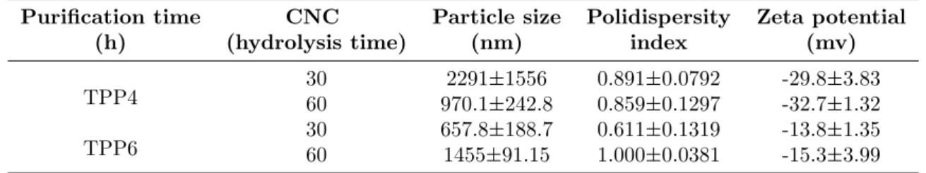
The colloidal suspensions of TPP4 and TPP6 were analysed by DLS to determine the CNC par-ticle size in suspension and charge (Table2). The sizes obtained were high since crystals’ length is also measured, not only the width, using a DLS scatter that is dynamic, and agglomeration may occur in solution. Nevertheless, smaller CNCs were obtained for TPP6 than for TPP4.
The polydispersity index (PI) indicates the vari-ation in the distribution of the particle size. Aa high polydispersity shows the existence of particle families with different sizes, which may mean the occurrence of aggregation (Hanaor, Michelazzi, Leonelli, & Sorrell, 2012). In gen-eral, all samples showed PI values much higher than 0.3, which indicates a polydisperse distribu-tion of CNCs. On the other hand, zeta potential (ZP) can give us an indication of whether re-pulsion between adjacent, similarly charged par-ticles in dispersion will occur or not. When ZP is high (whether they are positive or nega-tive values) means stability between the cles, whereas when the potential is low, parti-cles tend to coagulate/flocculate as attraction ex-ceeds repulsion in the dispersion. The ZP values where ca. 30 mV in the CNCs extracted from TPP4, which means a moderate stability. In the case of the CNCs extracted from TPP6, this value decreased but is still moderate. In both cases, the values are negative, which is a result of the acid hydrolysis, in which sulphuric acid removed the amorphous regions in the cellulose fibres, leaving only the highly ordered crystalline regions intact, resulting in negatively charged, sulphonated nanoparticles. Due to the relatively low surface charge, this method required ultra-sonication to disperse and stabilize the CNC sus-pension.
The colloidal suspensions with the lowest sizes, i.e. those originating from TPP6, were freeze-dried and a white and light powder was obtained as shown in Fig. 1g.
Table 1: Chemical characterisation in % (w/w) (means±SD) of the products obtained during samples processing
Samples Lignin Cellulose Hemicellulose Ash PP 28.9±0.390 16.9±2.02 15.8±2.02 3.92±0.721 TPP4 3.40±0.103 21.8±0.793 3.91±0.983 2.48±0.201 TPP6 2.48±0.041 21.0±1.23 2.79±1.03 2.00±0.510 PP-Pineapple peels residues; TPP-Treated and bleached residues 4 h (TPP4) and 6 h (TPP6)
Table 2: Chemical characterisation in % (w/w) (means±SD) of the products obtained during samples processing.
Purification time CNC Particle size Polidispersity Zeta potential
(h) (hydrolysis time) (nm) index (mv)
TPP4 3060 970.12291±1556±242.8 0.8910.859±0.0792±0.1297 -29.8-32.7±3.83±1.32 TPP6 3060 657.81455±91.15±188.7 0.6111.000±0.1319±0.0381 -13.8-15.3±1.35±3.99 TPP4-Treated pineapple residues after 4h bleaching; TPP6-Treated pineapple residues after 6h bleaching
3.2
FTIR spectra
The powders of the PP, TPP4, TPP6, and CNCs obtained from TPP6, were analysed by FTIR and compared (Fig. 2). The presence of peaks 1 and 4 at 3310 and 1640 cm−1, respectively, in all samples shows that during the purification, cellulose is present and was not removed during the purification and CNC extraction processes (Sheltami, Abdullah, Ahmad, Dufresne, & Kar-garzadeh, 2012; Sun, Xu, Sun, Fowler, & Baird, 2005). Peak 2 at 2900 cm−1 is from the C-H stretching vibration, is also present in all samples as expected, and was observed in other similar samples (Alemdar & Sain,2008; Sheltami et al., 2012). Peak 3 at 1700 cm−1originates from the acetyl and ester groups in hemicellulose, or car-boxylic acid groups in the ferulic and p-coumeric components of lignin. The existence of this peak shoulder was also reported in other works with wheat straw, rice husks and soy hulls (Alemdar & Sain,2008; Flauzino Neto et al.,2013; Sun et al., 2005). It should disappear with extraction, since hemicellulose is removed from PP with the pu-rification process. Indeed, this peak is more pro-nounced in PP than TPP4 and TPP6, and dis-appears in the CNC spectra. Peaks 6 and 7 at
1060 and 897 cm−1, respectively, correspond to the cellulose C-O stretching and C-H vibrations of cellulose, and appeared in all of the spectra, growing with extraction (Alemdar & Sain, 2008; Sheltami et al.,2012). Peak 5 at 1205 cm−1 is only present in the CNC spectra, and it is not common to see in other CNCs extracted from lignocellulosic materials. It can be attributed to the S-O vibration, due to the esterification reac-tion, as reported in soy hulls (Flauzino Neto et al., 2013) (Lu, Gui, Zheng, & Liu, 2013; Lu & Hsieh, 2012).
3.3
X-ray diffraction
measurements
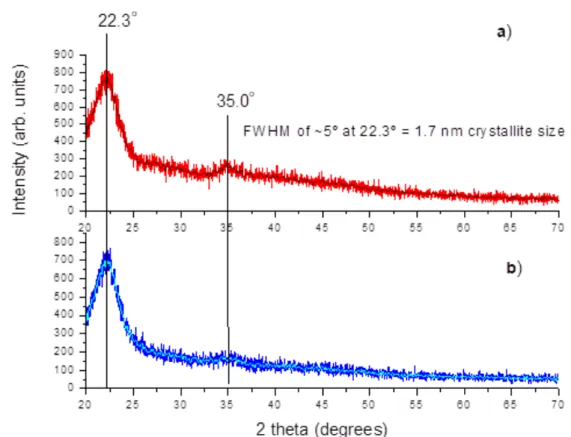
The CNCs obtained from the two purification treatments, TPP4 and TPP6, were evaluated re-garding their degree of crystallinity. Both intra-and intermolecular hydrogen bonding occurs in cellulose via hydroxyl groups, which results in various ordered crystalline arrangements. The x-ray diffractograms obtained (Fig. 3) are typi-cal of cellulose I, the more widespread crystalline form of the 4 existent cellulose polymorphs, with well-defined crystalline peaks around 22° and 35° (Klemm, Heublein, Fink, & Bohn, 2005). The
Figure 2: FTIR spectra of dried and milled pineapple peel (PP), after 4h bleaching, drying and milling treatments (TPP4) or after 6h bleaching (TPP6) and cellulose nanocrystals (CNCs) obtained from 6 h of purification (TPP6) and after 30 min (CNC30) and 60 min (CNC60) of acid hydrolysis. 1- 3310 cm−1; 2 – 2900 cm−1; 3 – 1742 cm−1; 4 – 1640 cm−1; 5 – 1307 cm−1; 6 – 1060 cm−1; 7 – 897 cm−1.
Figure 3: X-ray diffraction patterns of CNCs obtained from hydrolysis for 30 min of TPP6 (a) and TPP4 (b)
major peak, which is related to the crystalline structure of cellulose I, was seen for all samples at 22.3°, with a much smaller peak at 35.0°, and both indicate a poorly crystalline material, made of nanoscale crystals. The background “hump” around 20-30° also indicates the presence of some amorphous material. The peaks are slightly more pronounced in the CNC obtained from TPP6, which again shows the importance of using a higher bleaching time during purification to bet-ter remove lignin and hemicellulose. The average cross-sectional dimension of the elementary cel-lulose crystallites was estimated from their X-ray diffractograms by applying the Scherrer equa-tion, as described in Section 2. The Scherrer equation is unreliable for dimensions lower than 100 nm, as the broadening becomes excessive, but it can still be used to get a very approximate estimate of the average crystallite size. For both
samples in Fig. 3, with a FWHM of ∼5°, the value for the crystallite size was found to be only 1.7 nm.
3.4
Morphology
In Fig. 4a and b, the raw fibres have become narrow fibrils, with a reticular structure, after the chemical-purified treatment. This indicates that the purification process and bleaching did not provoke the breaking of the cellulose chains. In Fig. 4b, the release of cellulose microfibers can be clearly seen. Fig. 4c and d show the CNC samples. In Fig 4c, a drop of the colloidal suspension was put on the carbon tape and left to dry at room temperature. After covering with gold, the samples were observed and aggregations of small entities were identified. CNCs which are freeze dried appear as an aggregation of irregular
Figure 4: Micrographs of pineapple peels residues PP 1000x (a), TPP6 1000x (b), col-loidal suspension CNC30 4000x (c) and freeze-dried CNC30 3000x (d)
rod-like assemblies of CNCs. However, individ-ual NPs cannot be distinguished, as they are far too small for the resolution of the SEM method, which has a limit of around 30 nm. This was also seen with CNCs extracted from sweet pota-toes residues (Lu et al.,2013).
4
Conclusions
The purification process is important in the treatment of wastes for cellulose extraction, since pigments, waxes, hemicellulose and lignin are ef-fectively extracted from the residues, while cel-lulose is not removed. The best conditions for extraction include a 6 h bleaching process. The time of bleaching was shown to be important for the purification of cellulose, and this was re-flected by the composition, the physical proper-ties and FTIR spectra. Indeed, the pineapple peel that was bleached for 6 hours had lower hemicellulose and lignin contents, and the CNCs produced from TPP6 (6 h) were marginally more crystalline than those produced with TPP4 (4 h). In terms of hydrolysis of the extracted cellulose, the time used was not shown to be important, and 30 minutes was shown to be effective in the hydrolysis process. Even with low yields, the cel-lulose extracted had ca. 80% purity. Pineapple
for the extraction of CNC.
Acknowledgements
Authors acknowledge financing support by the European Regional Development Fund (ERDF) through the Programa Operacional Factores de Competitividade – COMPETE, by Portuguese funds through FCT, in the frame-work of the project PEst-C/SAU/LA0002/2013 and Multirefinery POCI-01-0145-FEDER-016403. Ana Raquel Madureira acknowl-edges FCT for the postdoctoral scholarship SFRH/BPD/71391/2010. Robert Pullar acknowledges the support of FCT grant SFRH/BPD/97115/2013.
References
Alemdar, A. & Sain, M. (2008). Isolation and characterization of nanofibers from agricul-tural residues - wheat straw and soy hulls. Bioresource Technology, 99 (6), 1664–1671. doi:10.1016/j.biortech.2007.04.029
Aspler, J., Bouchard, J., Hamad, W., Berry, R., Beck, S., Drolet, F., & Zou, X. (2013). Re-view of nanocellulosic products and their applications. In Biopolymer nanocompos-ites (pp. 461–508). John Wiley & Sons, Inc. doi:10.1002/9781118609958.ch20
Browning, B. L. (1967). Methods of wood chem-istry. volumes i & ii. John Wiley & Sons. Cherian, B. M., Leao, A. L., de Souza, S. F.,
Thomas, S., Pothan, L. A., & Kottaisamy, M. (2010). Isolation of nanocellulose from pineapple leaf fibres by steam explosion. Carbohydrate Polymers, 81 (3), 720–725. doi:10.1016/j.carbpol.2010.03.046
Deepa, B., Abraham, E., Cherian, B. M., Bis-marck, A., Blaker, J. J., Pothan, L. A., . . . Kottaisamy, M. (2011). Structure, mor-phology and thermal characteristics of ba-nana nano fibers obtained by steam ex-plosion. Bioresource Technology, 102 (2), 1988–1997. doi:10 . 1016 / j . biortech . 2010 . 09.030
Flauzino Neto, W. P., Silverio, H. A., Dantas, N. O., & Pasquini, D. (2013). Extraction and characterization of cellulose nanocrys-tals from agro-industrial residue - soy hulls. Industrial Crops and Products, 42, 480– 488. doi:10.1016/j.indcrop.2012.06.041 Hanaor, D., Michelazzi, M., Leonelli, C., &
Sor-rell, C. C. (2012). The effects of carboxylic acids on the aqueous dispersion and elec-trophoretic deposition of zro2. Journal of the European Ceramic Society, 32 (1), 235– 244. doi:10.1016/j.jeurceramsoc.2011.08. 015
Henrique, M. A., Silv´erio, H. A., Neto, W. P. F., & Pasquini, D. (2013). Valorization of an agro-industrial waste, mango seed, by the extraction and characterization of its cellu-lose nanocrystals. Journal of Environmen-tal Management, 121 (Supplement C), 202– 209. doi:https : / / doi . org / 10 . 1016 / j . jenvman.2013.02.054
Hu, X., Wang, J., & Huang, H. (2013). Impacts of some macromolecules on the character-istics of hydrogels prepared from pineapple peel cellulose using ionic liquid. Cellulose, 20 (6), 2923–2933. doi: 10.1007/s10570-013-0075-4
Johar, N., Ahmad, I., & Dufresne, A. (2012). Ex-traction, preparation and characterization of cellulose fibres and nanocrystals from rice husk. Industrial Crops and Products, 37 (1), 93–99. doi:10.1016/j.indcrop.2011. 12.016
Klemm, D., Heublein, B., Fink, H.-P., & Bohn, A. (2005). Cellulose: fascinating biopolymer and sustainable raw material. Angewandte Chemie International Edition, 44 (22), 3358–3393. doi:10 . 1002 / anie . 200460587
Lin, N. & Dufresne, A. (2014). Nanocellulose in biomedicine: current status and future prospect. European Polymer Journal, 59, 302–325. doi:10 . 1016 / j . eurpolymj . 2014 . 07.025
Lu, H., Gui, Y., Zheng, L., & Liu, X. (2013). Mor-phological, crystalline, thermal and physic-ochemical properties of cellulose nanocrys-tals obtained from sweet potato residue. Food Research International, 50 (1), 121– 128. doi:10.1016/j.foodres.2012.10.013
Lu, P. & Hsieh, Y.-L. (2012). Preparation and characterization of cellulose nanocrystals from rice straw. Carbohydrate Polymers, 87 (1), 564–573. doi:10 . 1016 / j . carbpol . 2011.08.022
Prozil, S. O., Evtuguin, D. V., Silva, A. M. S., & Lopes, L. P. C. (2014). Structural char-acterization of lignin from grape stalks (vi-tis vinifera l.) Journal of Agricultural and Food Chemistry, 62 (24), 5420–5428. doi:10. 1021/jf502267s
Qiu, X. & Hu, S. (2013). “Smart” Mate-rials Based on Cellulose: A Review of the Preparations, Properties, and Appli-cations. Materials, 6 (3), 738–781. doi:10 . 3390/ma6030738
Raji, Y. O., Jibril, M., Misau, I. M., & Dan-juma, B. Y. (2012). Production of vine-gar from pineapple peel. International Journal of Advanced Scientific Research and Technology, 3 (2), 656–666. Retrieved from http : / / www . academia . edu / 2080752 / PRODUCTION OF VINEGAR FROM PINEAPPLE PEEL
Sheltami, R. M., Abdullah, I., Ahmad, I., Dufresne, A., & Kargarzadeh, H. (2012). Extraction of cellulose nanocrystals from mengkuang leaves (pandanus tectorius). Carbohydrate Polymers, 88 (2), 772–779. doi:10.1016/j.carbpol.2012.01.062
Sun, X. F., Xu, F., Sun, R. C., Fowler, P., & Baird, M. S. (2005). Characteristics of degraded cellulose obtained from steam-exploded wheat straw. Carbohydrate Re-search, 340 (1), 97–106. doi:10 . 1016 / j . carres.2004.10.022
Thirawan, B., Karnnasuta, S., & Srinorakutara, T. (2017). Agricultural wastes potential (pineapple crown, durian peel and sugar-cane leaves) on reducing sugar production by using sulfuric acid pretreatment fol-lowing enzymatic hydrolysis. Asia-Pacific Journal of Science and Technology, 19 (3), 361–370.