UNIVERSIDADE DE LISBOA
FACULDADE DE CIÊNCIAS
DEPARTAMENTO DE QUÍMICA E BIOQUÍMICA
Supported lipid bilayers with
micro/nanodomains in the study of membrane
lipid organization and interactions
Doutoramento em Bioquímica
Especialidade: Biofísica Molecular
Joaquim Trigo Marquês
Tese orientada pelo Doutor Rodrigo F. M. de Almeida e
pela Doutora Ana S. Viana
2015
Documento especialmente elaborado para a obtenção
do grau de Doutor
Preface
i
“Life, as science, is a journey in the pursuit of the unknown.
Make sure you make the most of it and share all the personal accomplishments
with whom matters. But try not to do it through facebook.”
Membrane interactions in supported lipid bilayers with nano/microdomains
Preface
iii
Preface
Biomembranes are characterized by a great heterogeneity in their composition, in terms of
both lipids and proteins, and by a unique lateral organization that leads to the generation of
compartmentalized regions. These regions have been in focus in membrane biophysics and their
formation, function and properties are currently hot topics in membrane studies. To answer these
fundamental questions is crucial so that in a as short as possible time-window, we will be able to
use and manipulate such regions to our benefit, finding feasible applications in disease therapy and
diagnostic.
This dissertation aims to provide effective contributions for the following questions:
- How do lipid structure and composition determine membrane lateral organization?
- How can lipid composition and/or membrane lateral organization modulate membrane
function, in particular its interaction with other molecules?
- How to take advantage of gold as a substrate for the application of supported lipid
bilayers in the study of membrane-related phenomena using optical and electrochemical
techniques?
To that end, the attainment of more specific goals is required.
- Can bulk techniques be conjugated with atomic force microscopy images in order to
unravel complex phase diagrams for biologically relevant lipid mixtures?
- Is it atomic force microscopy a suitable technique to follow the effects promoted by
small molecules in lipid membranes?
- Is it possible to observe nanoscale heterogeneities in gold-supported lipid bilayers?
- How to quantify changes in lipid nanodomains and their properties using high resolution
label-free techniques?
- How to establish if a supported lipid bilayer has the required properties, such as
planarity, continuity, compactness and stability?
- How sensitive can be the detection of electroactive molecules adsorbed onto SLB
prepared on gold substrates?
- How to relate the information retrieved from cyclic voltammetry with the one from
fluorescence spectroscopy for the characterization of the interaction of electroactive
molecules with lipid membranes?
Membrane interactions in supported lipid bilayers with nano/microdomains
iv
These are the goals that hopefully were achieved through the execution of the doctoral work that
is the subject of this book.
The work presented in this thesis, which is included in the field of Biochemistry – Molecular
Biophysics, was developed as a strict collaboration between the Molecular Biophysics and
Interfacial Electrochemistry groups. The expertise from each group allowed to apply an unusual
approach in the characterization of the lipid systems studied throughout this thesis by the use of a
wide collection of techniques. All experimental results shown were obtained at Centro de Química
e Bioquímica from Faculdade de Ciências da Universidade de Lisboa. Most of the data obtained
during this thesis has already been published or is under consideration in peer view scientific
journals, hence the main text of this thesis comprises the compilation of those published works.
In chapter I the general properties and lateral organization of lipid membranes (first
subsection, which corresponds to a book chapter in the series Advances in Planar Lipid Bilayers
and Liposomes, Vol. 22) as well as the formation of supported lipid bilayers in gold substrates
(second subsection, review article in Electrochimica Acta) are discussed. In chapter II the working
principles of the techniques used throughout this work along with their major applications in the
context of lipid membrane domains is briefly described. Chapters III through VI describe the main
results obtained during this thesis. In chapter III (under consideration in Langmuir) the phase
behavior
of
the
pseudo-binary
lipid
system
N-stearoyl-phytoceramide
and
palmitoyloleoylphosphatidylcholine in its full hydration regime is thoroughly characterized and a
phase diagram for this system proposed. Chapter IV (published in BBA-Biomembranes) describes
the interplay between the effects of ethanol on lipid bilayers with their composition (from
single-component to three-single-component) and phase behavior. Chapter V (published in Soft Matter)
discusses the formation of multicomponent phase-coexistence lipid bilayers directly on bare gold,
whereas chapter VI (published in Langmuir) describes the formation of supported lipid bilayers on
modified gold surfaces and also the interaction of a biological relevant compound, epinephrine,
with lipid membranes, whether liposomes or supported, in this case using the newly developed
platform.
In the final section of this thesis the overall conclusions of this work are presented. There,
it is intended to highlight the common threads along the work presented throughout this thesis, and
to establish more general relationship between this work and selected reports from literature
Preface
v
considered as important marks within the topics of the thesis t. Finally, to conclude this manuscript
future directions are suggested.
At this point I feel it is necessary to acknowledge all the people or institution that, directly or not,
have contributed for the success of this thesis. During the course of my PhD I had the privilege of
being surrounded by many generous, friendly and kind people who had made my days, or at least
the great majority of them, a pleasure for the last years.
My first acknowledgement goes, inevitably, to my supervisors, Doctor Ana Viana and Doctor
Rodrigo de Almeida, (the order is just to respect the etiquette that says ladies first because I like
them both equally) that over the last years have given me their guidance, support, teaching and
mostly their fully commitment and friendship.
To Professor Jorge Correia and Doctor Pedro Lima whom, despite of their senior age, are playful
I would like to thank for their good advices and entertaining moments.
My friends and colleagues from the Molecular Biophysics (André Bastos, André Cordeiro,
Francisto Branco, Ana Margarida, António Soure, Alena Khmelinskaia, Natércia Rodrigues, Eva
Zupancic, Catarina Antunes, Telma Santos, Andreia Santos, Filipa Santos, Ana Carreira, Andreia
Sousa, Telmo Paiva) and Interfacial Electrochemistry (Inês Almeida, Ana Matos, Isabel Ornelas,
Joana Cabrita, Ana Melato, Ana Mourato, Virgínia Ferreira, Yu Niu, Wei Liu, Kang) groups that
contributed to a fantastic work environment, I am grateful for their friendship, companionship and,
even sometimes, fruitful scientific discussions.
I would also like to express my gratitude to the people that, one way or another, had contributed to
this thesis:
Enzymology group who let me use their power sonicator for the preparation of lipid vesicles
throughout the thesis;
Membrane interactions in supported lipid bilayers with nano/microdomains
vi
I thank Centro de Química e Bioquímica for receiving me and put at my disposal everything I
needed, whether reagents or equipment, for carrying out the experimental work that has culminate
in this thesis.
I thank the Fundação para a Ciência e Tecnologia for my PhD fellowship SFRH/BD/64442/2009
and the Byophysical Society for sponsoring my participation in two congresses.
I would also like to present my gratitude to Professor Gang Jin who have kindly receive me in
China, under the 6
thSino-Portugal Scientific and Technological Cooperation of 2013-2015, for
about a month, giving me the opportunity to broaden my horizons and work with a very power
optical technique – total internal reflection imaging ellipsometry.
To my good friends Bastos and Catarina (my little apprentice) I want make a special
acknowledgment cause with them hard working hours seemed more like hard laughing hours
without compromising working efficiency.
I would also like to present my gratitude to my good friends Cordel and my two walking
liposomes – Inna and Pinie, for the lovely moments we have enjoyed since we know each other.
Lastly but not the least… Margarida I thank you! For your love, constant motivation, support,
patience (a lot, I believe) and especially for making every day much more shining. Finally, I thank
my family, my parents and brother (who have bugged me with many questions about what I do),
who have gave me their love and their unequivocal support, who have settled the example and
encouraged me to pursuit my objectives, and to whom a simple expression of thanks does not
suffice.
Contents
vii
Contents
Preface
iii
List of abbreviations and symbols
xi
Resumo
xv
Palavras-chave
xix
Abstract
xxi
Keywords
xxii
1. Chapter I – Introduction
1
1.1. Biomembrane organization and function: the
decisive role of ordered lipid domains
3
1.2. Lipid bilayers supported on bare and modified
gold – Formation, characterization and relevance of lipid rafts
37
2. Chapter II – Characterization techniques
51
2.1. Atomic Force Microscopy
53
2.1.1. Modes of operation
55
2.1.2. Instrumentation
57
2.1.3. Biological Applications
60
2.2. Ellipsometry
62
2.2.1. Principles
62
2.2.2. Instrumentation and Biological Applications
64
2.3. Cyclic Voltammetry
68
2.4. Quartz Crystal Microbalance
72
2.5. Surface Plasmon Resonance
74
2.6. Fluorescence Spectroscopy
76
2.6.1. The Perrin-Jablonski diagram
76
2.6.2. Steady-state emission and excitation spectra
77
Membrane interactions in supported lipid bilayers with nano/microdomains
viii
2.6.4. Steady-state fluorescence anisotropy
79
2.6.5. Instrumentation and Biological Applications
81
2.7. References
82
3. Chapter III – Phytoceramide interactions with a fluid phospholipid
87
3.1. Formation and properties of membrane ordered
domains by phytoceramide: role of sphingoid base hydroxylation
89
3.2. Supporting Information
115
4. Chapter IV –Membrane lateral organization on ethanol effects
119
4.1. Ethanol effects on binary and ternary supported lipid bilayers
with gel/fluid domains and lipid rafts
121
4.2. Supporting Information
133
5. Chapter V – Building lipid rafts on bare gold
139
5.1. Biomimetic membrane rafts stably supported on unmodified gold
141
5.2. Supporting Information
153
6. Chapter VI – Lipid bilayers on gold to study electroactive molecules
159
6.1. A Biomimetic Platform to Study the Interactions of
Bioelectroactive Molecules with Lipid Nanodomains
161
6.2. Supporting Information
173
7. Chapter VII – Concluding remarks
181
My contribution to the articles included in the thesis:
1.1. Marquês, J.T., Antunes, C.A.C., Santos, F. C., de Almeida, R. F. M. (2015) Biomembrane
Organization and Function: The Decisive Role of Ordered Lipid Domains, Advances in
Planar Lipid Bilayers and Liposomes, Elsevier Inc. 2015, Vol 22.
Took an active part in the planning of the paper and the writing.
1.2. Marquês, J.T., de Almeida, R.F.M., Viana, A.S. (2014) Lipid bilayers supported on bare
and modified gold - formation, characterization and relevance of lipid rafts. Electrochim.
Acta 126, 139-150. DOI: 10.1016/j.electacta.2013.07.117.
Contents
ix
Writing of the paper
3. Marquês, J.T.; Cordeiro, A.M.; Viana, A.S.; Herrmann, A.; Marinho, H.S.; de Almeida,
R.F.M. (2015) Formation and properties of membrane-ordered domains by
phytoceramide: role of sphingoid base hydroxylation, Langmuir, 31, 9410-9421. DOI:
10.1021/acs.langmuir.5b02550.
Atomic force microscopy experiments, fluorescence spectroscopy and X-ray scattering data
analysis and interpretation. Contribution to the definition of the phase diagram and to the
writing of the paper.
4. Marquês, J.T., Viana, A.S., and de Almeida, R.F.M. (2011) Ethanol effects on binary
and ternary supported lipid bilayers with gel/fluid domains and lipid rafts. Biochim.
Biophys. Acta - Biomembr. 1808: 405-414. doi:10.1016/j.bbamem.2010.10.006.
Took an active part in the planning of and performed all the experiments. Took an active part
in the writing of the paper.
5. Marquês, J.T., de Almeida, R.F.M., Viana, A.S. (2012) Biomimetic membrane rafts
stably supported on unmodified gold. Soft Matter 8:2007-2016. DOI:
10.1039/C2SM06738B.
Took an active part in the planning of and performed all the experiments. Took an active part
in the writing of the paper.
6. Marquês, J.T., Viana, A.S., de Almeida, R.F.M. (2014) A Biomimetic Platform to Study
the Interactions of Bioelectroactive Molecules with Lipid Nanodomains. Langmuir, 30,
12627-12637. DOI: 10.1021/la503086a.
Took an active part in the planning of and performed all the experiments. Took an active part
in the writing of the paper.
Papers to which I have contributed that are not included in the thesis:
- Almeida, I., Marquês, J., Liu, W., Yu, N., de Almeida, R.F.M., Jin, G., Viana, A.S.,
Lipid/decanethiol mixtures for simple assembly of efficient immunosensing interfaces.
Accepted in Colloids and Surfaces B: Biointerfaces.
Membrane interactions in supported lipid bilayers with nano/microdomains
x
- R.F.M. de Almeida, J.T. Marquês, L.C. Silva (2014) Chapter 2. Biophysics of Lipid Rafts
and their Interplay with Ceramide: Studies in Model Systems and Biological Insights In:
Lipid Rafts: Properties, Controversies and Roles in Signal Transduction Ed. Dan Sillence,
Nova Publishers, NY, pp 21-52.
- Marquês, J.T. and de Almeida, R.F.M. (2013) Application of Ratiometric Measurements
and Microplate Fluorimetry to Protein Denaturation: An Experiment for Analytical and
Biochemistry
Students.
J.
Chem
Educ.
90,
1522-1527.
List of abbreviations and symbols
xi
List of abbreviations and symbols
Abbreviations
2H-NMR
Deuterium Nuclear Magnetic Resonance
2OHOA
2-hydroxyoleic acid
AFM
Atomic Force Microscopy
Chol
Cholesterol
Cer
Ceramide
CV
Cyclic Voltammetry
Cys
L-Cysteine
di-8-ANEPPS
4-(2-[6-(Dioctylamino)-2-naphthalenyl]ethenyl)-1-(3-sulfopropyl)pyridinium inner salt
DMPC
1,2-dimyristoyl-sn-glycero-3-phosphocholine
DOPC
1,2-dioleoyl-sn-glycero-3-phosphocholine
DPH
Diphenylhexatriene
DPPC
1,2-dipalmitoyl-sn-glycero-3-phosphocholine
DPTL
2,3-di-O-phytanoyl-sn-glycerin-1-tetraethyleneglycol-lipoic acid ester
DSC
Differential Scanning Calorimetry
EIS
Electrochemical Impedance Spectroscopy
EPR
Electron Paramagnetic Resonance
FCS
Fluorescence Correlation Spectroscopy
FRAP
Fluorescence Recovery After Photobleaching
FRET
Förster’s Resonance Energy Transfer
GM1
Monosialotetrahexosylganglioside (Ganglioside G
M1)
GPI
Glycosylphosphatidylinositol
GPMV
Giant Plasma Membrane Vesicles
Membrane interactions in supported lipid bilayers with nano/microdomains
xii
IR
Infrared
LB
Langmuir-Blodgett
LS
Langmuir-Schaffer
LUV
Large Unilamellar Vesicles
MLV
Multilamellar Vesicles
MUA
11-Mercaptoundecanoic acid
NMR
Nuclear Magnetic Resonance
PC
Phosphatidylcholine
PCer
N-palmitoyl-D-erythro-sphingosine
PE
Phosphatidylethanolamine
PLAP
Placental Alkaline Phosphatase
PM-IRRAS Polarization Modulation-Infrared Reflection-Absorption Spectroscopy
POPC
1-palmitoyl-2-oleoyl-sn-glicero-3-phosphocholine
PSM
N-palmitoyl-D-erythro-sphingosylphosphorylcholine
QCM
Quatz-Crystal Microbalance
SAM
Self-assembled Monolayer
SL
Sphingolipid
SLB
Supported Lipid Bilayer
SM
Sphingomyelin
SMase
Sphingomyelinase
SPR
Surface Plasmon Resonance
STED
Stimulated Emission Depletion
STM
Scanning Tunneling Microscopy
SUV
Small Unilamellar Vesicles
tBLM
tethered Bilayer Lipid Membranes
TIRF
Total Internal Reflectance Fluorescence
t-PnA
trans-parinaric acid
List of abbreviations and symbols xiii
UQ10
Ubiquinone-10
UV
Ultraviolet
X
Molar Fraction
Symbols
Absorption coefficient
d
Tthickness
Δ
Ellipsometric phase parameter
ΔE
pRedox peak separation, V
E
Potential of an electro versus a reference, V
E
1/2Measured half-wave potential, V
F
Faraday constant, 9.648523415(39)
10
4C mol
-1𝒇
𝟎Resonant frequency, Hz
h
Plank constant. 6.62606876(52)
10
-34J s
k
Extinction coefficient
l
dLiquid disorded
l
oLiquid ordered
ν
Potential rate, V s
-1n
Refractive index or number of electrons
𝑵
̃
Complex refractive index
Angle of incidence or propagation
Ψ
Ellipsometric amplitude parameter
Q
Charge, C
s
oSolid ordered
t
Time, s
R
qRoot mean square roughness
Membrane interactions in supported lipid bilayers with nano/microdomains
xiv
𝑹𝒑
𝑹𝒔
Fresnel coefficients in p and s directions
T
Absolute temperature, K
T
mMain gel-to-fluid phase transition temperature
𝝉̅
Lifetime weighted quantum yield or amplitude averaged lifetime
Γ
Surface coverage, mol cm
-2Resumo
xv
Resumo
As membranas biológicas apresentam na sua composição, além de proteínas, um vasto
número de espécies lipídicas e, por conseguinte, são estruturas bastante complexas. Atualmente, o
estudo da sua organização lateral em domínios assim como as suas propriedades e funções a elas
associadas são um dos campos de investigação mais ativos em biofísica. A forma como a
composição ou organização lateral da membrana podem influenciar a interação de pequenas
moléculas com esta é, igualmente, outro dos focos da investigação em biomembranas. O trabalho
descrito ao longo desta dissertação teve como principais objetivos a caracterização, por diversas
técnicas, de diversos sistemas modelo de membranas biológicas, ora lipossomas ora bicamadas
lipídicas suportadas (SLB) de variadas composições e, consequentemente, com comportamento de
fases distinto; o desenvolvimento de uma plataforma de deteção de espécies electroativas
adsorvidas ou incorporadas em SLB usando ouro como substrato sólido para a deposição da
bicamada; o estudo da interação de pequenas moléculas, como o caso do etanol e da epinefrina,
uma molécula electroativa, com bicamadas lipídicas exibindo um comportamento de fases bastante
variado – fluido, gel, coexistência gel/fluido e coexistência líquido ordenado (l
o) / líquido
desordenado (l
d), que mimetiza os domínios lipídicos conhecidos como jangadas lipídicas.
Neste trabalho estudou-se o comportamento de fases da mistura binária
1-palmitoil-2-oleoil-sn-glicero-3-fosfocolina (POPC) / N-octadecanoil(2S,3S,4R)-2-amino-1,3,4-octadecanetriol
(Fitoceramida (PhyCer)). O POPC é um dos fosfolípidos mais abundante nas membranas celulares
de organismos eucariotas enquanto a PhyCer é a principal ceramida, esqueleto dos esfingolípidos
complexos, em importantes grupos taxonómicos como plantas e fungos. Através da informação
recolhida a partir de diversas técnicas propõe-se o diagrama de fases para esta mistura. Neste estudo
recorreu-se tanto a bicamadas lipídicas em suspensão (lipossomas) como a SLB. Usou-se a
espectroscopia de fluorescência com recurso a sondas fluorescentes de membrana como o ácido
trans-parinárico (tPnA) e o 1,6-difenil-1,3,5-hexatrieno (DPH), as quais são extremamente
sensíveis ao ambiente em seu redor e através de cujas propriedades biofísicas é possível inferir
acerca da composição da membrana em termos de domínios. Enquanto o tPnA se distribui
preferencialmente nas fases mais rígidas, em particular o gel, o DPH é uma sonda que se distribui
equitativamente ao longo de toda a membrana e, portanto, reporta a ordem global desta.
Recorreu-se, igualmente, à análise por microscopia de força atómica (AFM), a qual devido à sua extrema
Membrane interactions in supported lipid bilayers with nano/microdomains
xvi
resolução lateral (na ordem dos nm) e vertical (na ordem dos Å) permite distinguir detalhes, como
diferenças de alturas entre diferentes domínios de membrana, na topografia de SLB. A
espectroscopia de difração de raio-X também foi usada na medida em fornece informação
relacionada com o empacotamento lateral dos lípidos numa bicamada. Os resultados obtidos
mostram que o comportamento de fases desta mistura difere daquele já publicado para outras
ceramidas quando também misturadas com POPC. O diagrama de fases proposto prevê a existência
de diferentes regiões de coexistência de fases, e, principalmente, antecipa a existência de
complexos POPC:PhyCer para determinadas proporções destes lípidos. As estequiometrias são
conforme se segue: POPC 3 : 1 PhyCer e POPC 1 : 2 PhyCer. Estes complexos são vistos como
entidades moleculares com características próprias e apresentam propriedades biofísicas muito
semelhantes àquelas já descritas para os domínios gel identificados in vivo em levedura
Saccharomyces cerevisiae.
No decurso desta tese investigou-se a forma como a organização lateral da membrana pode
modular a interação de pequenas moléculas com esta. Estudaram-se os casos do etanol, da
quercetina e da epinefrina. Os efeitos do etanol foram analisados para SLB formadas em silício e
mica, que é um substrato atomicamente liso e carregado negativamente a pH neutro e, portanto,
adequado à deposição de lípidos. Neste estudo usaram-se os lípidos
1,2-dipalmitoil-sn-glicero-3-fosfocolina (DPPC), 1,2-dioleoil-sn-glicero-3- 1,2-dipalmitoil-sn-glicero-3-fosfocolina (DOPC),
N-palmitoil-D-eritro-esfingosilfosforilcolina (PSM) e colesterol (Chol). Foram, igualmente, estudadas bicamadas
exibindo diferentes comportamentos de fase: apenas fluido (DOPC), apenas gel (DPPC e
DPPC:Chol 98:2), coexistência gel/fluido (DPPC:DOPC 1:1 e 92:8) e, finalmente, coexistência
l
o/l
d(DPPC:DOPC:Chol 2:2:1 e PSM:DOPC:Chol 2:2:1). Os efeitos do etanol sobre as diferentes
bicamadas foram seguidos em tempo real por AFM. Para todos os sistemas estudados observou-se
o adelgaçamento da bicamada seguido de expansão em consequência da interação com o etanol.
Relativamente aos sistemas com separação de fases, enquanto para o sistema gel/fluido, a baixas
concentrações de etanol, o adelgaçamento verifica-se apenas para a fase fluida, para os sistemas
l
o/l
do adelgaçamento ocorre em simultâneo nas duas fases. Contudo, em ambas as situações apenas
se observa a expansão da fase mais fluida, o que leva a um aumento da fração de domínios
desordenados cujas principais causas serão uma variação na tensão superficial na interface
água/lípido e diminuição do ponto de fusão que, em conjunto, levarão a um aumento da desordem
das cadeias acilo, aumento do ângulo de inclinação das cadeias acilo e, ainda, interdigitação. De
Resumo
xvii
referir ainda que o uso de duas misturas ternárias permitiu perceber se os efeitos induzidos pelo
etanol seriam dependentes da composição lipídicas ou, por outro lado, do tipo de domínios
presentes. Tendo em conta que as alterações observadas foram idênticas para ambos os sistemas
ternários DPPC:DOPC:Chol 2:2:1 e PSM:DOPC:Chol 2:2:1 concluiu-se que o tipo de fase
influencia mais os efeitos provocados pelo etanol que o tipo de lípido presente na composição das
bicamadas.
Um dos trabalhos fulcrais no decorrer desta tese foi o desenvolvimento de SLB em
superfícies de ouro. Como até à data da realização dos estudos descritos nesta tese eram escassos
os trabalhos encontrados na literatura a descrever a formação de bicamadas em ouro com separação
de fases ou mais do que um componente, pretendeu-se investigar quais as condições experimentais
que favorecem a formação de SLB compostas por 3 lípidos distintos e com separação de fases l
o/l
d.
O ouro é um substrato metálico condutor e com propriedades que permitem a aplicação de inúmeras
técnicas de superfície de elevada sensibilidade. No entanto, tem sido descrito que a hidrofobicidade
deste metal é representa um grande constrangimento à formação de SLB nesta superfície.
Bicamadas planas e contínuas com jangadas lipídicas, diferindo em espessura da membrana
circundante em cerca de 1,5 nm, conforme comprovado por AFM, foram depositadas diretamente
em ouro nu através da fusão de vesículas unilamelares pequenas ou grandes e na ausência total de
qualquer sal. De facto, observou-se que a presença de NaCl promove a formação de túbulos ao
invés de uma bicamada plana, conforme revelado pelas imagens obtidas AFM e as espessuras dos
filmes lipídicos estimadas por elipsometria. As SLB preparadas diretamente em ouro são estáveis
numa larga gama de potenciais o que torna o seu uso adequado ao estudo da interação da epinefrina
com membranas lipídicas. No entanto, ficou também provada a influência do substrato metálico
nas características da bicamada uma vez que foi obtida uma proporção domínios
ordenados/domínios desordenados diferente da prevista pelo diagrama de fases para essa mistura
obtido em suspensões de lipossomas e, por outro lado, observam-se corrugações na fase mais
desordenada que são consequência do ambiente assimétrico dos dois lados da bicamada. Enquanto
as cabeças dos fosfolípidos do folheto virado para a superfície de ouro encontram-se num ambiente
praticamente desprovido de água, o mesmo não se verifica relativamente ao folheto do topo da
bicamada. Adicionalmente, os domínios identificados por AFM apresentavam formas muito
angulares e, portanto, diferentes das já descritas para lipossomas em suspensão. Este aspeto
deve-se ao facto de o filme lipídico deve-seguir a topologia do ouro que é caracterizada por terraços
Membrane interactions in supported lipid bilayers with nano/microdomains
xviii
monoatómicos. Para ultrapassar estas limitações, inclusivamente o elevado grau de hidrofobicidade
do ouro que dificulta a formação de SLB contínuas, modificou-se a superfície previamente à
formação da SLB. Esta modificação foi feita com uma monocamada automontada de L-cisteína, a
qual apresenta um grupo tiol que estabelece uma ligação muito forte ao ouro e por outro lado torna
a superfície deste metal mais hidrofíla e, portanto, mais propícia à adsorção de lípidos.
Adicionalmente, pelo facto da monocamada de cisteína ser curta e pouco compacta não impede
trocas eletrónicas entre o substrato metálico e espécies electroativas, tornando-se, desta forma, uma
modificação adequada aos objetivos pretendidos. Dados de AFM, elipsometria e voltametria cíclica
mostram que as SLB formadas sobre ouro modificado com cisteína são contínuas, compactas e
estáveis.
Outra molécula cuja interação com a membrana foi também avaliada é a epinefrina. A
epinefrina apresenta duas propriedades, fluorescência e eletroatividade, que permitem o uso de um
leque de técnicas mais vasto na caracterização da sua interação com a membrana, incluindo a
espectroscopia de fluorescência e técnicas eletroquímicas. A avaliação da interação da epinefrina
foi, então, concretizada tanto por intermédio de espectroscopia de fluorescência usando
lipossomas, como através do uso da plataforma acima descrita com recurso a voltametria cíclica.
De referir ainda que uma grande variedade de sistemas lipídicos foram empregues conforme se
descreve: fluido (DOPC), gel (DPPC), gel/fluido (DPPC:DOPC 1:1), l
o/l
d(DPPC:DOPC:Chol
2:2:1 e DPPC:DOPC:Chol 2:2:1 com 10
% de gangliósido G
M1) e l
o(DPPC:DOPC:Chol 11:1:8).
Fazendo uso da fluorescência intrínseca da epinefrina concluiu-se que esta interage fracamente
com membranas lipídicas. No entanto, apesar desta interação ser fraca, a epinefrina foi claramente
detetada por voltametria cíclica quando adsorvida em SLB formadas sobre ouro modificado com
cisteína. Foi, ainda, possível aferir que na bicamada fluida ficou adsorvida uma maior quantidade
de epinefrina. É de realçar que os dados provenientes de voltametria cíclica permitiram estimar um
coeficiente de partição membrana/água (de cerca de 1.13×10
4para a membrana em fase fluida)
para a epinefrina. Finalmente, uma das principais observações deste estudo é a capacidade das
diferentes bicamadas preparadas, independentemente da sua composição ou comportamento de
fases, estabilizarem a epinefrina do ponto de vista estrutural, impedindo a sua oxidação, a qual
ocorre espontaneamente a pH neutro em soluções aquosas.
Este trabalho mostra como a combinação de várias técnicas experimentais pode ser
importante e como permite chegar a conclusões que de outra forma não seria possível. Ao longo
Resumo
xix
desta tese são descritas múltiplas evidências que demonstram, não só como a composição da
membrana influencia as suas propriedades biofísicas, mas também a capacidade que a organização
lateral da membrana tem de influenciar os mecanismos de interação de pequenas moléculas com
esta.
A plataforma desenvolvida no decurso desta tese abre perspetivas não só na condução de
estudos que visam investigar as propriedades gerais das membranas mas também como também
no potencial desenvolvimento de interfaces para biossensores com transdução de sinal ótico ou
eletroquímico.
Palavras-chave
- Microscopia de força atómica;
- Modificação da superfície de ouro;
- Diagramas de fase de lípidos;
- Técnicas óticas e eletroquímicas;
- Domínios de esfingolípidos e jangadas.
Membrane interactions in supported lipid bilayers with nano/microdomains
Abstract
xxi
Abstract
The lateral organization of lipid bilayers into domains, such as lipid rafts or gel domains,
and how they influence the properties and function of membranes is a current topic in biophysics.
In this work, the properties of single and multicomponent lipid bilayers (whether supported on a
solid substrate or free in solution as liposomes) exhibiting different phase behavior were studied by
a wide range of characterization techniques – atomic force microscopy, ellipsometry, cyclic
voltammetry, quartz crystal microbalance, surface plasmon resonance and fluorescence
spectroscopy.
The phase diagram for the binary mixture
1-palmitoyl-2-oleoyl-sn-glycero-3-phosphocholine
(POPC)
/
N-octadecanoyl(2S,3S,4R)-2-amino-1,3,4-octadecanetriol
(Phytoceramide (PhyCer)) is proposed by combining data from different techniques. The formation
of complexes between POPC and PhyCer with properties similar to those found for gel domains in
vivo is anticipated. These POPC/PhyCer complexes occur at two distinct stoichiometries, 3:1 and
1:2.
The influence of membrane lateral organization was investigated in the context of the
interaction of small molecules with lipid bilayers, namely ethanol and epinephrine. The effects of
ethanol were studied in supported lipid bilayers (SLB) formed on mica spanning a phase behavior
from single fluid to liquid ordered (l
o) / liquid disordered (l
d) phase coexistence. It was concluded
that the lateral organization of lipids into domains, but not the specific lipid composition, plays a
determinant role on the effects induced by ethanol.
Another purpose of this work was to study the properties of supported lipid bilayers (SLB)
formed on gold surfaces. For the first time raft-containing SLB were formed directly on bare gold
and the substrate seems to influence the properties of the lipid bilayer since an unexpected
proportion of lipid domains was obtained and corrugations were observed in the liquid disordered
phase. A lipid-based biosensing interface consisting on a SLB formed on top of a
L-cysteine-modified gold was also developed for the detection of membrane-interacting electroactive
molecules.The interaction of epinephrine, an electroactive molecule, was evaluated using both
liposomes and SLB formed on Lcysteine-modified gold. Despite the weak interaction between
epinephrine and liposomes as determined by fluorescence spectroscopy, its redox signal could be
clearly detected by cyclic voltammetry when adsorbed on SLB of various compositions. Moreover,
voltammetric data allowed to estimate a membrane/water partition coefficient for epinephrine. The
Membrane interactions in supported lipid bilayers with nano/microdomains
xxii
results presented show how lateral membrane organization and composition may influence its
function and properties.
Keywords
- Atomic force microscopy;
- Gold surface modification
- Lipid phase diagrams
- Optical and electrochemical techniques
- Sphingolipid domains and rafts
Abstract
1
Chapter I
Biomembrane organization and function: the decisive role of ordered lipid domains
3
1.1. Biomembrane organization and function: the decisive role of ordered lipid domains
Chapter I – Introduction
Biomembrane organization and function: the decisive role of ordered lipid domains
Chapter I – Introduction
Biomembrane organization and function: the decisive role of ordered lipid domains
Chapter I – Introduction
Biomembrane organization and function: the decisive role of ordered lipid domains
Chapter I – Introduction
Biomembrane organization and function: the decisive role of ordered lipid domains
Chapter I – Introduction
Biomembrane organization and function: the decisive role of ordered lipid domains
Chapter I – Introduction
Biomembrane organization and function: the decisive role of ordered lipid domains
Chapter I – Introduction
Biomembrane organization and function: the decisive role of ordered lipid domains
Chapter I – Introduction
Biomembrane organization and function: the decisive role of ordered lipid domains
Chapter I – Introduction
Biomembrane organization and function: the decisive role of ordered lipid domains
Chapter I – Introduction
Biomembrane organization and function: the decisive role of ordered lipid domains
Chapter I – Introduction
Biomembrane organization and function: the decisive role of ordered lipid domains
Chapter I – Introduction
Biomembrane organization and function: the decisive role of ordered lipid domains
Chapter I – Introduction
Biomembrane organization and function: the decisive role of ordered lipid domains
Chapter I – Introduction
Biomembrane organization and function: the decisive role of ordered lipid domains
Chapter I – Introduction
Biomembrane organization and function: the decisive role of ordered lipid domains
Chapter I – Introduction
Biomembrane organization and function: the decisive role of ordered lipid domains
Chapter I – Introduction
Lipid bilayers supported on bare and modified gold – formation, characterization and relevance of lipid rafts
37
1.2. Lipid bilayers supported on bare and modified gold – Formation, characterization
Chapter I – Introduction
Lipid bilayers supported on bare and modified gold – formation, characterization and relevance of lipid rafts
Chapter I – Introduction
Lipid bilayers supported on bare and modified gold – formation, characterization and relevance of lipid rafts
Chapter I – Introduction
Lipid bilayers supported on bare and modified gold – formation, characterization and relevance of lipid rafts
Chapter I – Introduction
Lipid bilayers supported on bare and modified gold – formation, characterization and relevance of lipid rafts
Chapter I – Introduction
Lipid bilayers supported on bare and modified gold – formation, characterization and relevance of lipid rafts
Chapter I – Introduction
Lipid bilayers supported on bare and modified gold – formation, characterization and relevance of lipid rafts
Chapter I – Introduction
51
Chapter II
Atomic Force Microscopy
53
In this chapter, the principles of the main characterization techniques used throughout this
work will be briefly described. The surface characterization (AFM, ellipsometry, QCM and SPR)
and electrochemical (cyclic voltammetry) techniques will be mentioned first, followed by a general
overview of fluorescence spectroscopy, as a tool to assess the biophysical properties of free
standing lipid bilayers (liposomes) and interaction of bioactive molecules with those liposomes.
Both surface and electrochemical techniques were employed in the characterization of SLB, the
solid substrates (mica, silicon and bare and modified gold) used for the preparation of those SLB
and in the study of the interaction of small molecules with lipid membranes. Since there is a great
complementarity regarding the techniques employed throughout this thesis, their combined use
allows for a comprehensive characterization of the systems under investigation. For example, for
SLB prepared on gold, electrochemical, optical and scanning probe techniques can be employed in
their characterization and, thus, it is possible to conclude about the thickness, compactness, stability
and continuity of SLB as well as assess the presence of lipid domains.
2.1. Atomic Force Microscopy
AFM is an exceptional technique for obtaining morphological information on any type of
surface, providing it is sufficiently rigid, and also structural details for very flat materials, with
lateral resolution in the nanometer scale and a vertical resolution in the Ånsgström range. The
sample can be probed whether exposed to air or immersed into a liquid and under different
conditions, such as at different temperatures. In this work AFM was used, at a first instance, to
gather information concerning the organization and morphological arrangement of SLB and lipid
films formed on different solid substrates, namely mica, silicon and gold. During this thesis, AFM
was also employed to follow the effects that small molecules promote in the organization of lipid
bilayers displaying a wide range of compositions and consequently exhibiting distinct phase
behavior. AFM can have indeed a broad spectrum of biological applications, spanning from the
study of DNA and lipid bilayers to cells, as it will be discussed ahead under section 2.1.3.
This scanning probe microscopy can analyze areas that may go from less than 1 nm
2up to
10000 μm
2or higher. Additionally, samples with distinct degrees of roughness can be analyzed
since the vertical scale may range up to 8 to 10 μm. This technique is also suitable to investigate
attractive and repulsive, short- and long-range atomic and molecular forces in the order of 10
-12Chapter II – Characterization techniques
54
Newton. Since its operational principle is the variation of forces established between the surface
and an interacting probe, it is not required that analyzed samples are electrically conductive, and
therefore there is no need for metallic coating or even the use of dyes or other contrast agent in
opposition to other microscopic techniques. In addition, AFM is non-destructive allowing the same
sample to be imaged over time, before and after different treatments, and enables the sample to be
reused e.g., for characterization with other techniques.
1-3In fact, during this thesis, the same sample
prepared on gold substrate was, frequently, analyzed by AFM, ellipsometry and cyclic
voltammetry.
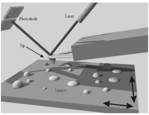
Figure 1 shows a schematic representation of one of the main components of an atomic
force microscope. Its operation is based on scanning and probing a sample by an extremely sharp
tip that is mounted at the end of a cantilever. As the tip interacts with the surface, the cantilever
undergoes a vertical deflection (up or down) acting as a spring sensitive to slight variations in force.
The most common approach to measure these variations is the use of a laser beam focused on the
cantilever end, which is reflected into a position sensitive photodetector. Consequently,
topographic variations in the sample will induce alterations in the cantilever angle, which, in turn,
generates different voltages in the detector. These voltages are recorded and sent to a computer for
processing and formation of the topographic image.
Figure 1 – Schematic representation of operation of an atomic force microscope. The sample is
moved by a piezoelectric scanner. As the sample is scanned, the movement of the tip is detected by the photodiode through a laser beam reflected from the back of the cantilever. Adapted from ref.1
Atomic Force Microscopy
55
2.1.1. Modes of operation
This section will focus on the modes of operation related to the acquisition of topographic
images. According to the purpose of an experiment and considering the general properties of a
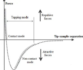
sample three main modes of operation can be employed. In Figure 2 the relation between the forces
(attractive or repulsive) generated upon tip-sample interaction and the different modes of operation
is illustrated.
Contact Mode
In this mode of operation, which can be also designated by constant force mode, the forces
ruling the tip-sample interaction are the interatomic repulsive ones. The tip is kept in contact with
the surface of the sample and the cantilever deflection is held constant during the scanning through
a feedback loop. The feedback acts in order to maintain the tip-sample separation constant along
the scan independently on the topographic features of the sample. In this sense, a topographic image
is formed and image contrast depends on the applied force, which in turn, depends on the spring
force of the cantilever. The major advantage of this mode is its high resolution even for samples
with large variations in height. However, because the tip is kept in close contact with the sample
large lateral forces are generated, which can damage soft biological samples or polymer networks.
In order to minimize the influence of the tip on the sample surface, this mode can be performed in
liquids allowing for a significant reduction of the capillary forces generated between the tip and
the sample during the scanning.
1-3Figure 2 – Expected behavior of the idealized forces between tip and sample according to their separation.
Chapter II – Characterization techniques
56
Non-Contact Mode
In this mode of operation the forces governing the tip-sample interaction are the attractive
van der Walls or long-range forces. Concretely, a cantilever oscillating at a frequency close to its
resonance frequency is brought into proximity of the sample surface without touching it. As a
consequence of the interaction a frequency shift in the oscillation of the cantilever is induced.
Through a feedback loop this oscillation is kept at a fixed frequency or amplitude and a topographic
map of the sample surface is generated. Though a good vertical resolution can be obtained, its
lateral resolution is not as good as the one attained with other scanning modes since the tip-sample
interaction is not very strong in this mode. Consequently, the sample suffers no damage by virtue
of the complete absence of any contact between the tip and the samples. The fact that this mode
cannot be performed in liquids constitutes its main drawback. The presence of a single layer of
water might be sufficient to trap the tip onto the surface of (supposedly) dried samples since the
low oscillating amplitude does not drive the required energy to promote the detachment of the tip.
1-3
Tapping Mode
This is perhaps the most employed mode, especially for the analysis of soft surfaces as in
the case of biological samples. In this mode, also designated as intermittent contact mode, the
forces in play are both attractive and repulsive depending on the tip-sample separation (Figure 2).
An oscillating cantilever is brought into close contact with the sample which causes a dampening
of the oscillation amplitude. Similarly to other modes, a feedback loop is used. It maintains the
oscillation amplitude at a fixed value by either lifting or lowering the sample (or the tip, depending
on the equipment used). In contact mode, the axial and lateral resolutions are better than for the
non-contact, and relatively to the contact mode, the tapping involves minimal tip-sample
interaction, which leads to a great reduction of the lateral forces that may damage soft samples.
1-3In the oscillating modes besides topography further information can be retrieved. In the
case of tapping mode by comparing the applied oscillation and the actual one, a phase signal is
generated, which is intimately related to the local mechanical and chemical properties of the
sample. In that sense, regions of the sample with the same height, although displaying distinct
chemical properties will interact differently with the tip and thus generate unique phase signals
allowing for their differentiation.
1-3Atomic Force Microscopy
57
This mode of operation in solution was the one used throughout this work. Although it is
common to find in the literature concerning the analysis of SLB results obtained in the contact
mode, in the present thesis, tapping mode was used. This was done to ensure that the information
contained in the topographic image had minimal influence of the tip in the sample. In this way,
lateral forces were greatly reduce and thus any damage promoted by imaging in contact could be
avoided, which enabled a clear observation of membrane domains, e.g. lipid rafts, which could not
be detected in this work with a similar resolution using contact mode. This can be especially
relevant in the case of studying the effect of small molecules on lipid bilayers where a change in
the shape and thickness of lipid domains occurs over time. Also, for the characterization of lipid
tubular structures which can be highly deformable when compared to a planar lipid bilayer, tapping
mode can be advantageous over contact mode.
2.1.2. Instrumentation
The atomic force microscope is comprised by innumerous components. The key
components of an atomic force microscopic are described next.
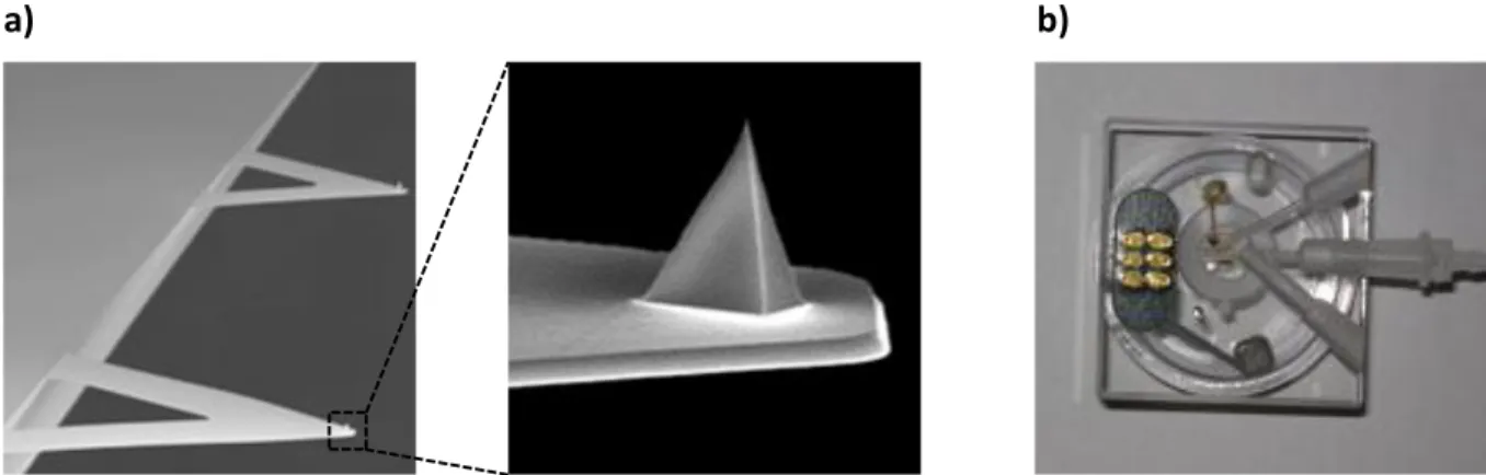
Tip and Cantilever
As mentioned above the tip is mounted at the end of a cantilever and is one of the crucial
components of the microscope since it is the element that interacts with the sample generating the
topographical images. Usually, the assembly tip/cantilever is fabricated in silicon, silicon nitride
or diamond. The most important parameters that define a good tip are the sharpness of its apex,
measured by the radius of curvature, and the ratio between the height of the tip and the width of its
base. Generally, the sharper the tip the most detailed (highest resolution) will be the final AFM
image.
1-3Cantilevers are constructed with two distinct geometries, rectangular and triangular, which,
essentially, respond differently to torsional/lateral forces that can affect the resolution of
topographic images. The most important characteristic of a cantilever are its dimension, resonance
frequency and spring constant, which is a measure of the cantilever flexibility. Shorter cantilevers
allow for a greater resolution. The resonance frequency should be high in order to avoid
interferences from the inherent vibrations of the building and the acoustical noise. Soft cantilevers
Chapter II – Characterization techniques
58
(with low spring constant) and of low resonance frequency are suitable for the analysis of samples
immersed in a liquid both in contact and resonance mode, whereas stiff cantilevers (higher spring
constant) with high resonance frequency are indicated for the study of samples in resonance mode
in air.
1-3In the present work soft triangular cantilevers (Figure 3a) were employed since lipid
bilayers are a soft and compressible biological sample. Moreover, in order to confer a more
biomimetic environment the samples were imaged in buffer solutions at physiological pH.
Figure 3 – Triangular cantilevers used throughout this thesis for imaging in liquids with a detail of the tip
(a) and the liquid cell used for experiments carried out with the sample immerse in buffer (b). In (a) tip height may go from 2.5 μm to 8.0 μm, whereas its radius is close to 2 nm.
Piezoelectric Scanner
The AFM scanner is made of piezoelectric materials which are characterized by a singular
property. They undergo an alteration of their dimensions upon the application of an electric field.
Considering that the applied voltages are strictly controlled, extremely exact movements in x, y
and z directions can be made. Instruments may display one of two configurations; either the sample
is mounted directly on top of the scanner or the assembly tip-cantilever is mounted on the scanner,
above the sample that, in this case, is fixed. The first condition allows for a higher scanning rate
and the visualization of larger areas, whereas the latter permits the use of samples with larger
dimensions.
1-3The first configuration is the one used thourghout this thesis. Although this design
imposes a restriction in sample size it was not a limiting factor for the AFM analysis carried out.
However, this configuration demands extra caution when working in liquids in order to avoid the
contact of the liquid with the scanner.
Atomic Force Microscopy
59
Photodetector
As mentioned above the method of detection is based on the reflection of a laser beam from
the top of the cantilever into a position sensitive photodetector. This photodetector consists of two
(or four) photodiodes side by side. With this configuration any minimal deflection of the cantilever
will promote a variation in the angle of the reflected laser and consequently the spot at which the
laser hits the detector. Thus, the difference between the signals coming from the two (or four)
photodiodes will indicate the position of the laser on the detector and therefore the angular
deflection of the cantilever. Since the distance between the cantilever and the detector is three
orders of magnitude higher than the length of the cantilever, this method of detection enables to
significantly magnify the movements of the tip with great sensitivity. The configuration of the
atomic force microscope allows detecting with high accuracy the movements of the cantilever and
thus providing a detailed topographic image of the surface.
1-3All AFM experiments described and presented throughout this work were performed with
the sample immersed in liquid and at room temperature using a liquid cell (Figure 3b). A
Multimode Nanoscope IIIa (Digital Instruments, Veeco) was used. Topographic images were
obtained in tapping mode and keeping the force exerted on the sample as low as possible by
continuously adjusting the “set point” parameter. Scan rate was close to 1.9 Hz. Before each
experiment the glass support where the cantilever is mounted was washed several times with water
and ethanol. The cantilevers used were fabricated in silicon nitride with a resonance frequency
close to 9 kHz in liquid.
2.1.3. Biological Applications
AFM imaging can be used to study a great diversity of biologically-related mechanisms.
One of the biological applications of AFM is in the study of nucleic acids, where it can be used to
evaluate the structural damage in chromosomes upon exposure to radiation
4,5, to study the
conformation of the double helix
6, to measure the mechanical properties of DNA and its
modulation by additions of small molecules
7-8. The study of protein-protein or protein-membrane
interactions at the molecular level has also benefited from the use of AFM
9-11. For example it has
been possible to study the dependence of amyloid fibril formation with lipid composition, charge
and phase as well as the aggregation of amyloid-beta peptide in various lipid systems
12,13or even
Chapter II – Characterization techniques
60
the inibhition of islet amyloid polypeptide fibril formation by compounds with therapeutic
potential
14. Recently, with the introduction of high-speed AFM a new field of applications emerges
and it has been possible, for example, to follow the motion of membrane protein assemblies in
SLB
15. The imaging of living cells has also been reported
16,17with real time visualization of
supramolecular assemblies of proteins in the surface of cell membrane of living cells
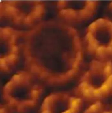
18-21(Figure
4). AFM can also find application in following cellular processes such as endocytosis
22. A
consequent derivation of using AFM in the inspection of living cells is the use of functionalized
tips in order to chemically map the surface of the membrane and in this way determine the precise
location of target proteins
23.
The first reports concerning the study of SLB by AFM date from more than 20 years ago
24.
Due to its unique vertical resolution, the heterogeneity of supported lipid bilayers can be probed
with extremely high spatial resolution and because of that it has been widely used in the
characterization of membrane domains, namely lipid rafts. Concretely, the presence of such lipid
domains differing in height, from the surrounding bilayer, by ~1 nm can be clearly depicted with
this technique
25,26(as showns in chapters III, IV and V). The remarkable resolution of AFM has
also helped in the characterization of corrugated lipid bilayers formed on gold substrates
27(see also
chapter V). Furthermore, it is possible to follow, in real time, the changes induced by small
molecules, such as anesthetics, in the structure of the lipid bilayer
28,29(discussed in chapter IV).
Figure 4 – Example of supramolecular structures observed by AFM. Here, the structure of photosynthetic
complexes of Rhodospirillum photometricum is illustrated. Adapted from reference 19
Recently, super-resolution optical techniques have witnessed an unprecedented growth
30.
Still, AFM emerges as one of the main techniques regarding the characterization of lipid bilayers.
Due to its versatility it can gather a considerable amount of information concerning different
properties of the lipid bilayers. It is possible to assess the topography of the lipid films and thus not
Atomic Force Microscopy
61
only quantitatively evaluate the phase behavior of the lipid system under study but also to
determine the thickness of each lipid phase. Furthermore, AFM enables to study the mechanical
properties of lipid bilayers. As mentioned above, in this work, AFM was employed to assess the
topography (nano/mirco-domains, thickness changes) and evaluate the quality and organization
(phase area fractions, domains size, shape) of multicomponent biomimetic lipid films formed on
diverse solid substrates (mica, silicon, gold), whether modified or not in the case of gold electrodes.
Moreover, AFM phase data has also been used to more easily assign gel and fluid phases as a
consequence of their distinct mechanical properties, as shown in chapter IV.
Chapter II – Characterization techniques
62
2.2. Ellipsometry
2.2.1. Principles
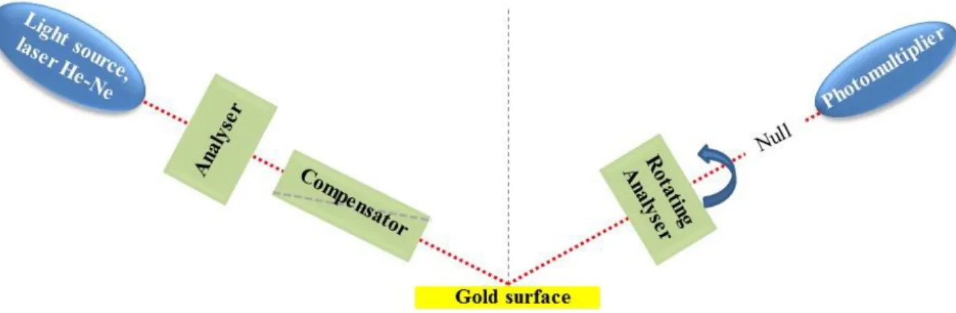
As shall be detailed in chapters V and VI, ellipsometry is an optical technique that can be
used for estimating the thickness of a thin film, by determining the change in the optical properties
of the surface before and after film formation. Its combination with AFM allows to effectively
evaluate the quality of the lipid bilayers prepared. Ellipsometric measurements rely on the
incidence of a polarized light beam on a surface and on its re-polarization after reflection.
When light is traveling in one environment (air or solution) and encounters a new one
(substrate) part will be reflected and part will be refracted
31-33(Figure 5a). The interaction of light
with this new medium can be described by the complex index of refraction Ñ, as follows:
𝑁
̃ = 𝑛 − 𝑖𝑘
eq.1
where n is the index of refraction and k is the extinction coefficient and i denotes the imaginary
number. The extinction coefficient is related to the absorption coefficient (
) through the following
expression:
𝑘 =
𝜆4𝜋