Essential Roles of
Drosophila
Inner Centromere Protein (INCENP) and
Aurora B in Histone H3 Phosphorylation, Metaphase Chromosome
Alignment, Kinetochore Disjunction, and Chromosome Segregation
Richard R. Adams, Helder Maiato, William C. Earnshaw, and Mar Carmena
Wellcome Center for Cell Biology, Institute for Cell and Molecular Biology, University of Edinburgh, Edinburgh EH9 3JR, Scotland, United Kingdom
Abstract.
We have performed a biochemical and dou-ble-stranded RNA-mediated interference (RNAi) anal-ysis of the role of two chromosomal passenger proteins, inner centromere protein (INCENP) and aurora B ki-nase, in cultured cells of Drosophila melanogaster. IN-CENP and aurora B function is tightly interlinked. The two proteins bind to each other in vitro, and DmIN-CENP is required for DmAurora B to localize properly in mitosis and function as a histone H3 kinase. DmAu-rora B is required for DmINCENP accumulation at centromeres and transfer to the spindle at anaphase. RNAi for either protein dramatically inhibited the abil-ity of cells to achieve a normal metaphase chromosome alignment. Cells were not blocked in mitosis, however,and entered an aberrant anaphase characterized by de-fects in sister kinetochore disjunction and the presence of large amounts of amorphous lagging chromatin. Anaphase A chromosome movement appeared to be normal, however cytokinesis often failed. DmINCENP and DmAurora B are not required for the correct local-ization of the kinesin-like protein Pavarotti (ZEN-4/ CHO1/MKLP1) to the midbody at telophase. These ex-periments reveal that INCENP is required for aurora B kinase function and confirm that the chromosomal pas-sengers have essential roles in mitosis.
Key words: mitosis • cytokinesis • chromosomal pas-sengers • chromosomes • RNAi
Introduction
Successful mitosis depends on the coordination of chro-mosomal and cytoskeletal behavior. A group of proteins termed chromosomal passengers are prime candidates for a role in this coordination. Chromosomal passengers were originally defined by their dynamic distribution in mitosis (Earnshaw and Bernat, 1990). They are concentrated at centromeres during metaphase, transfer to the central spindle during early anaphase and to the cell cortex at the presumptive cleavage furrow shortly thereafter, and then concentrate at the midbody during cytokinesis.
To date, four chromosomal passenger proteins have been described in detail: inner centromere protein (INCENP)1 (Mackay et al., 1993), TD-60 (Andreassen et al., 1991), au-rora B kinase (Schumacher et al., 1998), and survivin (Uren et al., 2000; Skoufias et al., 2000). These proteins colocalize
throughout mitosis, and INCENP is stockpiled in a com-plex with aurora B in Xenopus eggs (Adams et al., 2000). An INCENP–aurora B complex has also been precipitated from cultured human cells (Kaitna et al., 2000). This com-plex appears to be functionally significant, as INCENP is required for the proper localization of aurora B on the chromosomes, central spindle, and midbody during mito-sis. Thus, the hypothesis has emerged that INCENP may be a targeting subunit for aurora B kinases. It is not pres-ently known whether this targeting is essential for aurora B function, whether aurora B has a role in targeting IN-CENP, or whether survivin and TD-60 are also part of this chromosomal passenger complex.
Two dominant-negative forms of vertebrate INCENP have been described. One of these, INCENP1–405, inter-feres with prometaphase chromosome congression, sister chromatid disjunction at anaphase, and the completion of cytokinesis (Mackay et al., 1998). The other, CENP-B1–158: INCENP45–839, does not appear to interfere with pro-metaphase congression but inhibits the closing stages of cytokinesis, causing persistence of an intercellular bridge with a prominent midbody (Eckley et al., 1997). INCENP is an essential gene in the mouse (Cutts et al., 1999) and
Address correspondence to William C. Earnshaw,Center for Cell Biology, Institute for Cell and Molecular Biology, King’s Building, University of Edinburgh, Mayfield Road, Edinburgh EH9 3JR, Scotland, UK. Tel.: 44-0-131-650-7101. Fax: 44-0-131-650-7100. E-mail: bill.earnshaw@ed.ac.uk
1Abbreviations used in this paper: CB, cytoskeleton buffer; dsRNA,
double-stranded RNA; GST, glutathione S-trasferase; INCENP, inner centromere protein; KLP, kinesin-like protein; RNAi, dsRNA-mediated interference.
on January 10, 2011
jcb.rupress.org
Downloaded from
chicken (Vagnarelli, P., D. Hudson, and W.C. Earnshaw, unpublished material). Mouse embryos homozygous for a partial deletion of the INCENP gene die at the 32–64 cell stage, with multinucleate cells and abnormal microtubule bundling.
Aurora kinases were discovered in a screen of mitotic mutants in Drosophila (Glover et al., 1995). Budding yeast has a single aurora kinase, called Ipl1p (Chan and Bot-stein, 1993). Ipl1p is required for efficient chromosome segregation and appears to work, at least in part, by phos-phorylating the kinetochore protein Ndc10p (Biggins et al., 1999). In addition, Ipl1p is required for the phosphory-lation of histone H3 on serine10 during mitosis, a modifica-tion that is thought to be correlated with chromosome condensation (Hsu et al., 2000). Ipl1p interacts both genet-ically and physgenet-ically with the budding yeast INCENP, Sli15p (Kim et al., 1999).
Metazoans, including Drosophila, have two (three in vertebrates) aurora-related kinases (Giet and Prigent, 1999; Adams et al., 2001). Aurora A kinases are centroso-mal during mitosis, and mutants exhibit monopolar spin-dles with duplicated but not separated centrosomes (Glover et al., 1995). The role of Drosophila aurora B (ial) in mitosis has not previously been studied, however the aurora B kinases in mammals and Caenorhabditis elegans are essential for several aspects of mitotic progression, particularly cytokinesis (Schumacher et al., 1998; Terada et al., 1998). More recently, inactivation of C. elegans au-rora B/AIR-2 by double-stranded RNA (dsRNA)–medi-ated interference (RNAi) has revealed that this kinase is required for histone H3 phosphorylation on serine10 and sister chromatid separation (Hsu et al., 2000). C. elegans aurora B is also required for normal localization and func-tion of the ZEN-4/MKLP-1/PAV kinesin–like protein (KLP) during mitosis (Severson et al., 2000). It is aurora B that is in a complex with INCENP in Xenopus eggs.
Here, we report studies of chromosomal passenger func-tion in Drosophila embryos and cultured cells. Drosophila INCENP and aurora B both behave as classical chromo-somal passenger proteins, however they exhibit subtle dif-ferences in their targeting to chromosomes. Inactivation of INCENP and aurora B by RNAi in Drosophila cultured cells dramatically disrupted mitotic events with several sig-nificant differences from the results of recent studies in C. elegans (Schumacher et al., 1998; Kaitna et al., 2000). Our results demonstrate that chromosomal passenger function is interlinked and is essential for mitotic chromosome as-sembly, chromosome congression to the metaphase plate and segregation at anaphase, and cytokinesis.
Materials and Methods
Molecular Biology Methods and DNA Constructs Standard molecular biology methods were followed throughout this study. INCENP and aurora B/ial Drosophila cDNAs were purchased from Re-search Genetics. INCENP1–755, INCENP1–348, and INCENP654–755 were
amplified by PCR and cloned into pGEX 4T3 (Amersham Pharmacia Biotech). To produce pGEX-INCENP1–755–His6, an oligonucleotide
en-coding an His6 tag flanked by NotI adapters was inserted into the NotI site
of pGEX-INCENP. DmAurora B was subcloned into pET 22b (Novagen) into the NdeI site at the 5⬘ end and the XhoI site at the 3⬘ end. All con-structs were fully sequenced. After expression in E. coliBL21 (DE3) pLysS,DmAurora B was purified by Ni 2⫹-agarose chromatography.
Antibodies
Polyclonal anti-DmINCENP and anti–DmAurora B antibodies were pro-duced in rabbits. Rabbits were immunized with the following gel-purified proteins: glutathione S-transferase (GST)-INCENP 1–348 (R801),
GST-INCENP654–755 (R803), or GST–DmAurora B1–58 (R963). Anti-INCENP
sera were diluted 1:500 for use in immunoblotting and immunofluores-cence. Anti–aurora B antibodies were affinity purified by incubating serum with Affigel 10 beads (Bio-Rad Laboratories) to which aurora B1–58 had
been bound. After washes in 0.5 M NaCl, 10 mM phosphate (pH 7.4) anti-bodies were eluted in 0.2 M glycine, pH 2.0, and the eluate was neutralized with one-tenth volume 1 M Tris, pH 8.0, and dialyzed overnight into PBS. Antibodies were concentrated by ultrafiltration and stored in 50% glycerol at 20⬚C. For immunofluorescence, antibodies were used at 2 g/ml.
Other antibodies used were anti–␣-tubulin (YL1/2 rat monoclonal, used at 1:50; Harlan Sera Labs; or mouse mAb B512, used at 1:2,000; Sigma-Aldrich), anti–PAV-KLP (R3301, used at 1:500; Adams et al., 1998), anti–phosphorylated H3 (rabbit polyclonal, used 1:200; Upstate Biotechnology; or mouse monoclonal, used 1:100; NEB), anti-Cid (used at 1:2,000; gift of Steve Henikoff, Fred Hutchinson Cancer Research Center, Seattle, WA), anti–cyclin A and B (Rb270 and Rb271, used at 1:500; Whitfield et al., 1990; gift of W. Whitfield, University of Dundee, Dundee, Scotland), and mouse monoclonal antilamin (used at 1:50; gift of Paul Fisher, State University of New York, Stony Brook, NY). All fluores-cently conjugated secondary antibodies (Jackson ImmunoResearch Labo-ratories) were used according to the manufacturer’s instructions. FITC– phalloidin was used at 200 nM.
For the blocking experiment, the affinity-purified anti–aurora B anti-body was diluted 1:50 in PBS ⫹ 0.1% Triton X-100 (Bio-Rad Laborato-ries) with 10% FBS in the presence of GST–DmAurora B1–58 (0.3 mg/ml)
and then incubated for 1 h at room temperature before being added di-rectly to the cells and processed as usual.
Drosophila Embryos and Cells
Drosophila embryos derived from w1118 adults were fixed and processed for
immunostaining exactly as described previously (Adams et al., 1998). Immu-nostaining of Dmel-2 cells for the RNAi experiments was performed as fol-lows. Cells were grown in an incubator at 27⬚C in LAB-TEK Permanox chamber slides (177429; GIBCO BRL) or transferred onto poly-Lys–treated slides and left to attach for 20 min at each time point. In both cases, slides were centrifuged for 10 min at 4,000 rpm before fixation. Cells were fixed in 4% paraformaldehyde in cytoskeleton buffer (1.1 mM Na2HPO4, 0.4 mM
KH2PO4, 137 mM NaCl, 5 mM KCl, 2 mM MgCl2, 2 mM EGTA, 5 mM
Pipes, 5.5 mM glucose, pH 6.1) for 10 min at room temperature, permeabi-lized in 0.2% Triton X-100 in cytoskeleton buffer, and rinsed in PBS. Block-ing was performed for ⱕ30 min at room temperature in PBS ⫹ 10% FBS. Antibody incubation was done in PBS ⫹ 0.1% Triton X-100 for 1 h at 37⬚C, followed by 4–6-min washes in PBS five times. DNA was counterstained with 0.1 g/ml DAPI for 5 min at room temperature and rinsed in PBS. Slides were mounted in Vectashield and sealed using nail varnish. Three-dimensional data sets of selected cells were collected using a DeltaVision mi-croscope (Applied Precision), based on an Olympus IX-70 inverted micro-scope with a cooled couple-charged device camera (CH350L, Photometrics). Data sets were deconvolved if required, projected onto a single plane and, exported as TIF files to be processed using Adobe® Photoshop™.
Microtubule Binding Assay
30 g pure bovine tubulin (Cytoskeleton) was polymerized at 37⬚C in 20 l BRB80 (80 mM Pipes, pH 6.8, 1 mM MgCl2, 1 mM EGTA, 1 mM GTP)
by stepwise addition of taxol to a final concentration of 20 M. Either BRB80, 5 g GST-DmINCENP, or 5 g GST, was added to preformed microtubules to a final volume of 30 l. Reactions were incubated at 20⬚C for 30 min and then centrifuged at 50,000 g for 20 min through a 30% (wt/ wt) sucrose cushion containing 1 mM GTP and 5 M taxol. The superna-tant was aspirated and retained, and the cushion washed three times with BRB80. The pellets were resuspended in protein sample buffer before analysis on 10% SDS–polyacrylamide gels.
In Vitro Assay for DmINCENP/DmAurora B Binding Full-length GST-DmINCENP–His6 was expressed in E. coli and purified by
standard methods onto Ni2⫹-agarose beads (QIAGEN). After elution in
0.25 M imidazole, the protein was dialyzed into TEN buffer (10 mM Tris: HCl, pH 8.0, 100 mM NaCl, 2 mM EGTA) and bound to glutathione–
on January 10, 2011
jcb.rupress.org
Downloaded from
sepharose beads (Amersham Pharmacia Biotech). This procedure increased the yield of full-length INCENP over truncated INCENP. Beads were washed and incubated with TEN alone or 5 g DmAurora B for 30 min at 4⬚C. To remove nonspecifically bound protein, beads were washed five times in 20 volumes of TEN200 (TEN ⫹ 100 mM NaCl) containing 0.05% Triton X-100. As a control, DmAurora B was incubated with GST-coated sepharose beads. After washing, proteins were eluted by boiling in protein sample buffer and electrophoresed in 10% SDS polyacrylamide gels. Measurement of Relative Levels of Phospho-H3 and Mitotic Chromosome Condensation
Three-dimensional data sets were collected with the DeltaVision micro-scope and were in the linear range of the CH350L camera, deconvolved, and the image intensities of four image planes bisecting the midplane of the mitotic chromosomes were averaged using the Quick Projection algo-rithm. We then measured the integrated intensity of a box, 5 pixels on a side, at the appropriate wavelengths for detection of phospho-H3 and DAPI, respectively, using the Data Inspector tool. This assumes that the amount of DNA within the 25-pixel box is proportional to the overall level of condensation of the chromatin within that box. Three measure-ments were taken per metaphase, as were three measuremeasure-ments of the local background. These values were entered in a Microsoft® Excel™
spread-sheet, and the intensities at 617 and 457 nm were corrected by subtracting the local background. For control cells at prometaphase, metaphase, ana-phase, teloana-phase, and interana-phase, this method yields the following relative degrees of condensation: 2.4 ⫾ 0.55, 2.8 ⫾ 1.26, 2.7 ⫾ 0.49, 1.7 ⫾ 0.49, and 1.0 ⫾ 0.27, respectively. To produce the plot shown in Fig. 7 G, we aver-aged the three corrected values for each cell, sorted the DAPI measuments based on increasing DAPI fluorescence, and performed a linear re-gression on each data set. For this experiment, we obtained deconvolved three-dimensional data sets from 44 prometaphase cells, of which 34 were normal and 10 were “dumpy.” Average corrected phospho-H3 staining in-tensities were 2194 ⫾ 1779 and 74 ⫾ 123 for normal and dumpy chromo-somes, respectively. These corresponded to DNA condensation values of 317 ⫾ 109 and 474 ⫾ 249, indicating that dumpy chromosomes were not less condensed than normal-looking chromosomes.
RNA Interference
RNAi in Drosophila S2 and Dmel2 tissue culture cells was performed ac-cording to the published protocol (Clemens et al., 2000). Similar pheno-types were observed in both cell lines, only the Dmel2 data are shown here. 700-bp PCR fragments from the 5⬘ end of the DmINCENP and
DmAurora B genes were used as templates for RNA synthesis using the Megascript kit (Ambion). Experiments were performed in six-well plates or chamber slides. At each time point, cells from experiments and controls were collected and processed for immunoblotting or immunofluorescence. For immunoblotting, 106 cells were collected by centrifugation,
resus-pended in 50 l sample buffer, sonicated, and boiled for 3 min before loading on 10% SDS–polyacrylamide gels. Cell samples for immunostain-ing were processed as above.
Scoring of INCENP/Aurora-B Phenotype
Trypan blue (Sigma-Aldrich) was used to score nonviable cells. Growth curves were plotted considering only viable cells (levels of cell death were equivalent in all cultures). Microsoft® Excel™ was used to plot all
quanti-fications. Error bars represent the standard deviation, and all the values were plotted as numerical average. To determine the doubling time of the cells in the four experiments, we calculated the best fit for the growth curves (semilog scale), and the doubling time was calculated from the cor-responding equations. Mitotic Index was determined as a percentage of mitotic cells in the total population. Mitotic stages were quantified in terms of percentage of mitotic cells. To determine the mitotic index in the presence of microtubule poisons, Dmel2 cells were incubated for 12 h in the presence of 1 g/ml of colcemid.
Results
Identification of DmINCENP
We identified a candidate DmINCENP cDNA (LD24414) by querying the Berkeley Drosophila Genome Project
da-tabase with the conserved COOH-terminal 90 amino acids of vertebrate INCENP. LD24414 was completely se-quenced, and the cDNA sequence was mapped onto ge-nomic P1 clone AC005425. The candidate DmINCENP gene maps to cytological region 43B1, contains six exons, and encodes a 2,441-bp cDNA with a continuous ORF of 2,265 bp (Fig. 1). Our sequence and exon/intron analysis agrees exactly with that described by Celera Genomics for the hypothetical gene CG12165. (Sequence data are avail-able from GenBank/EMBL/DDBJ under indicated acces-sion nos.)
DmINCENP has a predicted molecular mass of 83.5 kD and a calculated isoelectric point of 9.63. Between residues 540 and 660, a coiled coil–forming region is predicted. The major region of homology with vertebrate INCENPs is in the COOH-terminal IN-BOX (Adams et al., 2000), a 40– 50-amino acid domain that defines the INCENP family from yeasts to humans. The NH2-terminal 540 amino acids are poorly conserved relative to vertebrate INCENPs, however it is this region that contains all the previously known functional domains for INCENP, such as the het-erochromatin protein 1 (HP-1) and -tubulin binding do-mains, the centromere targeting region and the spindle targeting domain (Mackay et al., 1993; Ainsztein et al., 1998; Wheatley et al., 2001).
DmINCENP and DmAurora B Kinase Are Chromosomal Passenger Proteins
To demonstrate whether the characteristic INCENP be-havior was conserved in Drosophila mitosis, we raised an-tibodies to two nonoverlapping regions of the protein (Fig. 1). Both sera recognized a single protein of 110 kD on im-munoblots of embryo extract (Fig. 2 A). This is larger than the predicted molecular mass of 83.5 kD but consistent with the behavior of INCENPs from other species, which also migrate anomalously on protein gels.
In syncytial embryos, DmINCENP associates with con-densing chromatin during prophase (Fig. 2, B–E), before becoming focussed to the centromeric regions of meta-phase chromosomes (Fig. 2, F–I, arrows). Upon entry into anaphase, the protein leaves the chromosomes to form a ring of spots between the segregating chromatids (Fig. 2, J–M). Each spot seems to be at the converging focus of bundles of microtubules (Fig. 2 K, arrow). As telophase progresses, the DmINCENP ring decreases in diameter un-til it becomes a single midbody-like structure between the central spindle microtubule bundles. A similar distribution of INCENP was also observed in cellularized embryos (Fig. 2 N) and Dmel2 cultured cells (Fig. 2, O–Q).
To localize DmAurora B in embryos and tissue culture cells, we raised antibodies against the NH2-terminal 58 amino acids of the protein fused to GST. Although neither of the two sera we raised detected a protein on immuno-blots of embryo extract, both recognized the recombinant protein expressed in bacteria (data not shown). The affin-ity-purified antibodies worked well for indirect immuno-fluorescence in both embryos and cells. This immunostain-ing is likely to be specific for DmAurora B for three reasons. First, affinity-purified antibodies gave a staining pattern essentially identical to the highly characteristic pattern seen with antibodies to aurora B kinases in higher
on January 10, 2011
jcb.rupress.org
Downloaded from
eukaryotes (Fig. 3, A–K) (Schumacher et al., 1998; Terada et al., 1998). Second, preincubation of the antibody with purified recombinant GST-aurora B1–58 abolished all spe-cific staining in cultured cells (Fig. 3 L). Third, treatment of Dmel2 cells with DmAurora B dsRNA obliterated all staining in many mitotic cells after 24 h, confirming that the epitope visualized in fixed cells is the product of the DmAurora B/ial gene (see Fig. 7 I).
The distribution of DmAurora B resembled that of DmINCENP throughout mitosis. However, interphase nu-clei showed no detectable staining for DmAurora B (they did for INCENP), and as nuclei entered prophase, DmAu-rora B appeared first at the centromere: no staining was observed along the chromosome arms. Fig. 3 A shows a re-gion of the embryo being traversed by a wave of mitosis. Nuclei just entering mitosis (i.e., adjacent to interphase nuclei) accumulated DmAurora B only at the centromere (Fig. 3, compare enlarged nuclei in C and D). DmAurora B remained at the centromere until the metaphase to ana-phase transition, when it transferred to the central spindle and subsequently to the midbody (Fig. 3, E–H). This dis-tribution was also observed in cellularised embryos (Fig. 3 B) and in cultured cells (Fig. 3, I–K). We conclude that DmINCENP and DmAurora B are chromosomal passen-ger proteins whose distribution in mitosis resembles their vertebrate counterparts.
DmINCENP Binds Directly to Microtubules and to DmAurora B
During anaphase, INCENP colocalizes with microtubules of the central spindle. Furthermore, INCENP overexpres-sion in cultured vertebrate cells or disruption of murine INCENP leads to a dramatic remodeling of the microtu-bule network (Mackay et al., 1998; Cutts et al., 1999). To test whether DmINCENP binds microtubules directly, we expressed soluble full-length GST-DmINCENP in bacte-ria, purified it, and incubated it with taxol-stabilized mi-crotubules prepared from purified tubulin. As a control, GST alone was incubated with polymerized tubulin. The reaction mixture was then sedimented through a sucrose cushion. GST-DmINCENP, but not GST, cosedimented
with the microtubules (Fig. 4 A). This suggests that the binding of DmINCENP to microtubules in mitosis is likely to be direct.
INCENP is stockpiled in Xenopus eggs in a complex with aurora B kinase (Adams et al., 2000). Drosophila contains two recognizable aurora-like protein kinases: the founder member of the family, Aurora (DmAurora A), which is required for centrosome separation, and the re-cently described DmAurora B/ial (Reich et al., 1999), whose function and localization are unknown. To deter-mine whether DmAurora B can associate with INCENP, we incubated DmAurora B expressed in bacteria with beads laden with GST-DmINCENP. As a control, the ki-nase was incubated with beads carrying GST alone. Under these conditions, DmAurora B bound specifically to GST-DmINCENP but not to GST alone (Fig. 4 B).
The RNAi Method
We have used RNAi to eliminate DmINCENP and DmAurora B/ial from cultured cells. dsRNA was added to exponentially growing cultures of Dmel-2 tissue culture cells (see Materials and Methods), and at different time points samples were taken for analysis by immunoblotting and indirect immunofluorescence. Two negative controls were analyzed in parallel: cells to which no dsRNA had been added and cells to which we added dsRNA synthe-sized from a human intronic sequence, chosen at random to rule out unspecific effects of dsRNA on the cell cycle. The latter caused no defects and was indistinguishable from the untreated control.
Analysis of RNAi experiments is complicated by the fact that this technique causes a gradual depletion of the proteins under study, and that proteins are not necessar-ily lost from all cells in the population at the same rate. Furthermore, inhibition of INCENP or aurora B function causes cultures to become polyploid, and it is difficult to exclude that some of the aspects of aberrant mitosis seen in these experiments are caused by complications arising during polyploid mitosis. To minimize these complica-tions, we examined the phenotypes described here at
var-Figure 1. Map of the
DmIN-CENP locus and constructs
used in this study.
DmIN-CENP (CG12165) is located
on P1 clone, AA005425 and contains six exons. A gene encoding a bromodomain protein (CG1845) is tran-scribed in the opposite direc-tion to the DmINCENP gene; the ORFs are separated
by ⵑ700 nucleotides.
IN-CENP NH2- and
COOH-ter-minal constructs were expressed to make antigens for immunization, and full-length INCENP was expressed for microtubule binding and DmAurora B binding experiments. (Sequence data are available from GenBank/EMBL/DDBJ under indicated accession nos.)
on January 10, 2011
jcb.rupress.org
Downloaded from
ious times after the onset of RNAi treatment and could show that certain phenotypic aspects, such as defects in histone H3 phosphorylation and mitotic chromosome as-sembly, are observed in cells in some cases within the first cell cycle, before cultures become highly polyploid. In addition, where possible, we limited our phenotypic conclusions to cells that were demonstrably lacking IN-CENP or aurora B, detectable by indirect immunofluo-rescence.
Elimination of DmINCENP and DmAurora B/ial Function by RNAi
Immunoblotting analysis of cells treated with DmIN-CENP dsRNA showed that the levels of DmINDmIN-CENP in the culture became greatly decreased 36–48 h after the ad-dition of dsRNA to the culture (Fig. 5 A⬘, top). In the best experiments, ⱕ95% of the protein was lost. The RNAi treatment for aurora B took effect more rapidly, the pro-tein becoming undetectable by indirect
immunofluores-Figure 2. (A) Immunoblot of total fly embryo protein extract probed with antibody R801, raised against
GST-INCENP1–348. A single band
of 110 kD is visible. (B–Q) Localization of DmINCENP in embryos and cultured cells. All panels are stained for INCENP with R801
(red), ␣-tubulin (green), and
DNA (blue). (B) Syncytial prophase (note that DmIN-CENP is distributed along the arms of the condensing chromosomes as well as at centromeres); (F) syncytial metaphase and early ana-phase; (J) syncytial anaphase. (C, G, and K) Enlargements of selected mitotic figures from B, F, and J, respec-tively. (D, H, and L) Corre-sponding INCENP staining; (E, I, and M) the correspond-ing DNA staincorrespond-ing. Arrows show centromere staining in H and I, and midzone stain-ing in K. (N) Mitotic domain of cellularized embryo show-ing cells at different stages of mitosis. m, metaphase; a, ana-phase; t, telophase. (O–Q) Dmel2 cells at metaphase, anaphase, and telophase, re-spectively. Bars, 5 m.
on January 10, 2011
jcb.rupress.org
Downloaded from
cence in most mitotic cells by 24 h (Fig. 5 A⬙, and see Fig. 7 H). In both cases, the loss of protein was transient, with levels beginning to recover at later times.
The phenotypes observed after INCENP and aurora B RNAi were complex, revealing defects at multiple stages of the mitotic cycle. To follow the appearance of the vari-ous phenotypes after the onset of RNAi, cultures were harvested at 24, 36, 48, and 72 h after exposure to dsRNA and assessed for the following parameters: cell number, frequency of dead (Trypan blue-positive) cells, frequency of overtly polyploid cells, mitotic index, percentage of mi-totic cells negative for INCENP or DmAurora B by indi-rect immunofluorescence, and for the cells in mitosis, the distribution of the various mitotic phases.
RNAi treatment caused an increase in the cell doubling time from 21 h in Dmel2 cells (21.6 h in cells after expo-sure to control dsRNA) to 36.1 and 27.5 h in cultures after exposure to dsRNA to DmAurora B and DmINCENP, re-spectively (Fig. 5 B). This was accompanied by an increase in polyploid cells starting at 24 h in the DmAurora B ex-periment (Fig. 5 C, 36 h for DmINCENP). These corre-spond to the first times when significant numbers of mi-totic cells were observed to be lacking DmAurora B and DmINCENP, respectively.
Strikingly, RNAi of INCENP abolished the ability of cells to achieve a metaphase chromosome alignment (Fig. 5, compare D with E). A similar phenotype was observed in the aurora B RNAi (Fig. 5 F). Instead, the population of
Figure 3. (A–L) Immunolocalization of DmAurora B in embryos and cultured cells. DNA (blue), DmAurora B (red), and tubu-lin (green) are shown. (A) Syncytial nuclei entering mitosis. Black arrow indicates a nucleus (enlarged in C) just ahead of the mitotic wave, lacking detectable aurora B staining. White arrow points to nucleus (en-larged in panel D) entering prophase, with DmAurora B accumulating at the cen-tromere. (E–H) Localization of DmAurora B in syncytial nuclei at various mitotic stages. Note that DmAurora B is located solely at the centromeres from prophase to metaphase. DmAurora B redistributes to the spindle midzone at anaphase. (B) Mi-totic domain from a cellularized cycle 14 embryo showing DmAurora B at prometaphase (pm), metaphase (m), late anaphase (a), and telophase (t). (I–K) Dmel2 staining at metaphase, anaphase, and telophase, respectively. White arrows point to centromeric (I) and midbody (K) staining. (L) Immunostaining of Aurora B is prevented by coincubation with recom-binant Aurora B protein (Black arrows
show absence of staining). Bars, 5 m.
on January 10, 2011
jcb.rupress.org
Downloaded from
mitotic cells came to be dominated by cells with a pro-metaphase-like chromosome arrangement. Importantly, this increase in the percentage of prometaphase cells did not reflect an arrest in mitosis, as the mitotic index of the culture remained constant at the control level of ⵑ5% throughout the entire experiment (Fig. 5, D–F). As dis-cussed below, we believe that many cells in these cultures must exit mitosis directly from prometaphase without achieving a metaphase chromosome alignment.
DmINCENP and DmAurora B Are Mutually Dependent for Their Correct Localization in the Cell Cycle
INCENP is required for the correct localization of aurora B kinases in human cells and C. elegans early embryos (Adams et al., 2000; Kaitna et al., 2000). However, it was not known whether aurora B kinases have a role in IN-CENP localization.
Elimination of DmINCENP by RNAi completely abol-ished DmAurora B localization throughout mitosis: the protein was not detected on chromosomes, central spindle microtubules, or midbodies (Fig. 6, E and F). In contrast, DmAurora B RNAi did not block DmINCENP association with the chromosome arms during prometaphase but im-paired its ability to concentrate at centromeres (Fig. 6 B) and eliminated the transfer to the midbody (Fig. 6 C).
Thus, aurora B function is not required for the initial stages of INCENP targeting to chromosomes during prophase, but it is necessary for INCENP behavior later in mitosis.
DmINCENP and DmAurora B Function Is Required for Histone H3 Phosphorylation, Mitotic Chromosome Structure, and Metaphase Chromosome Alignment
In untreated and control cultures, mitotic chromosomes invariably showed high levels of histone H3 phosphoryla-tion on serine10, detected with a specific antibody (Fig. 7, B–D). Inhibition of DmAurora B or DmINCENP func-tion led to both a decrease in the levels of detectable his-tone H3 phosphorylation (Fig. 7, A and E) and an increase in the incidence of malformed chromosomes starting as early as 24 h after exposure to dsRNA (Fig. 7 F). The phospho-H3 staining varied from cell to cell, but by 24 h after DmAurora B RNAi, the level of H3 phosphorylation was significantly reduced in 79% of the aurora-null prometaphase cells (74% of the INCENP-null cells at 36 h after DmINCENP RNAi; Fig. 7, A and E). This result sug-gests that, as in C. elegans (Hsu et al., 2000), DmAurora B is at least partially responsible for the histone H3 kinase activity in Drosophila cells.
Importantly, the level of histone H3-serine10 phosphory-lation showed only a weak correphosphory-lation with the overall de-gree of chromatin condensation. Fig. 7 G shows a plot in which quantitative measurements of phospho-H3 label-ing and DAPI stainlabel-ing were compared for a series of prometaphase cells, showing a wide range of phospho-H3 labeling (see Materials and Methods). Although levels of histone H3 phosphorylation on serine10 do tend to in-crease with increasing chromatin condensation, it is evi-dent that there is a huge scatter in the data from cell to cell. In other studies, we observed chromosomes com-pletely lacking detectable aurora B kinase, which showed an apparently normal level of condensation (Fig. 7, com-pare H with I).
An aberrant dumpy prometaphase chromosome mor-phology (Fig. 7 F) was seen in 46% of INCENP-negative and 60% of aurora B–negative cells after RNAi. These dumpy chromosomes had a 28-fold lower level of phos-pho-H3 staining, as detected with specific antibody, than did the chromosomes with a normal morphology (see Ma-terials and Methods). Dumpy chromosomes had an amor-phous shape, and defined sister chromatids were not seen. In many cases, the dumpy chromosomes appeared to cor-respond to an abnormal prometaphase arrangement, char-acterized by a disassembled nuclear lamina (data not shown) and persistent high levels of cyclin B protein (Fig. 7, compare J with K). Although they initially appeared less condensed than normal mitotic chromosomes (Figs. 2 O, 3, I and L, and 7, B–D), in fact, their level of condensation is normal (Fig. 7 G, includes measurements from both nor-mal and dumpy chromosomes; see also Materials and Methods). Instead, it appears that other aspects of chro-mosome higher order structure and behavior are aberrant. This may be due to defects in condensin binding (Giet and Glover, 2001).
Dumpy chromosomes have kinetochores, as defined by the presence of double dots of CENP-A/Cid staining. Cid is the Drosophila CENP-A orthologue (Henikoff et al.,
Figure 4. (A) DmINCENP binds directly to microtubules. (lane M) Molecular weight markers. (lanes 1–5) Coomassie blue– stained gel of microtubule pellet after sedimentation through a sucrose cushion. 64% of the added INCENP (lane 4) but no GST (lane 5) cosediments with microtubules. INCENP does not sedi-ment in the absence of microtubules but remains in the superna-tant (lanes 2 and 7). (lanes 6–10) Coomassie blue–stained gel of the supernatant fraction. 36% of the INCENP, but all the GST, remained unbound to microtubules (lanes 9 and 10). (B) DmIN-CENP binds DmAurora B in vitro. Coomassie blue–stained gel showing DmAurora B input (lane 1, 5% loaded). (lanes 2–3) GST-bound beads incubated with buffer (lane 2) or DmAurora B (lane 3, 50% loaded). No DmAurora B associates with GST beads. (lanes 4–5) GST-INCENP–bound beads incubated with buffer (lane 4) or DmAurora B (lane 5, 50% loaded). 19% of the
added DmAurora B associates with GST-INCENP. on January 10, 2011
jcb.rupress.org
Downloaded from
2000), and provides a marker for the kinetochore inner plate (Warburton et al., 1997).
Role of DmINCENP and DmAurora B in Anaphase and Telophase
As expected, given the lack of normal metaphase cells, we saw few if any normal anaphase cells that were negative for INCENP or aurora B. Instead, the anaphase/telophase cells had a range of abnormalities, including anaphase-like spindles with chromosomes distributed along their length (Fig. 8, A and B), cells in various states of attempted cy-tokinesis with large amounts of amorphous lagging chro-matin draped out behind the segregating chrochro-matin (Fig. 8, C and D), and bizarre cells in which banana-shaped nu-clei were surrounded by a mitotic-like bipolar microtubule array (Fig. 8, E–G). Fig. 8 E shows two adjacent mitotic cells in the INCENP RNAi, one of which is still expressing INCENP and is at normal metaphase, and the other of which is INCENP negative and is forming an elongate ba-nana-shaped nucleus.
In cells with chromosomes distributed along the spindle or with banana-shaped nuclei, centromeres were seen to cluster either near opposite poles (Fig. 8, A–D) or at the opposing pointed ends of the elongate nuclei (Fig. 8, F and G). This strongly suggests that kinetochores had attached to microtubules and that anaphase A movement of chro-mosomes had occurred.
In cells that appeared to be in telophase, we often no-ticed one or more pairs of centromeres that appeared to be
stalled midway between the spindle poles (Fig. 8, C and D; and data not shown). This organization is what would be predicted if these centromeres had successfully become bioriented but were then unable to disjoin at the onset of anaphase chromosome movement. This was never seen in normal anaphases where the centromeres were typically grouped in a tight cluster at the leading edge of the segre-gating chromatids (Fig. 7, C and D). Consistent with diffi-culties in disjunction of sister kinetochores, we also saw nu-merous paired kinetochore spots near the spindle poles, as though nondisjoined chromatid pairs had moved together to a single pole (Fig. 8, A⬙, B⬙, and F⬙, double arrows).
With increasing time after RNAi treatment, we ob-served a dramatic increase in the number of polyploid cells in both the INCENP and aurora B RNAi so that by 72 h most of the cell population had become highly polyploid (Fig. 5 C). The simplest explanation for the origin of the many binucleate cells that we observed (Fig. 9, C and F) is that chromosome segregation and nuclear reassembly oc-curred, but that cytokinesis was then defective. We also observed cells with one giant nucleus (Fig. 9 G). These are likely to have arisen as a consequence of repeated failures in chromatid segregation. In addition to the chromosomal defects, we also observed spindle abnormalities in IN-CENP and aurora B RNAi.
Together, these observations suggest that DmINCENP and DmAurora B might be essential for a variety of ana-phase/telophase events, including sister chromatid and ki-netochore disjunction, chromosome structure during ana-phase, and mitotic spindle architecture.
Figure 5. Summary of effects of RNAi on cell
growth and mitosis. (A⬘ and A⬙) Efficacy of
RNAi was assessed by immunostaining (INCENP and aurora B RNAi) and by immunoblotting (IN-CENP RNAi only). Graph shows percentage of cells lacking detectable INCENP or aurora B
staining (n ⫽ 75). Partially depleted cells were
not scored as nulls. INCENP immunoblots were quantitated in NIH Image and normalized with respect to a tubulin-loading control, and levels
were expressed as a percentage of t ⫽ 0 levels.
(B) Growth curve of control and dsRNA-treated cells. (C) Time course of percentage of polyploid (multinucleate and abnormally large) cells. (D–F) Histograms showing the percentage of control, INCENP null, or aurora B null mitotic cells at
different mitotic stages. For INCENP at t ⫽ 0,
24; and aurora B at t ⫽ 0, the whole mitotic
pop-ulation was scored due to the scarcity of null cells. For each time point, n ⫽ 50.
on January 10, 2011
jcb.rupress.org
Downloaded from
DmINCENP Is Not Essential for the Initiation of Cytokinesis
It was possible to observe cells lacking detectable DmIN-CENP in which constriction of the cleavage furrow had advanced considerably and a midbody had formed (Fig. 9 B). These cells showed an accumulation of actin at the cleavage furrow similar to that in untreated cells (Fig. 9, D and E), although more actin was dispersed throughout the remainder of the cell than normal. In binucleate cells, there was no longer a focus of actin staining between the nuclei, indicating that the contractile ring had disassem-bled (Fig. 9 F). In contrast, binucleate cells consistently showed an abnormally high density of tubulin between the two nuclei (Fig. 9 C). This is likely to be a remnant of the central spindle.
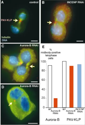
Pavarotti, an Essential Spindle Midzone KLP Localizes Normally to the Midbody Despite Disruption of the Chromosomal Passenger Complex
In aurora B/AIR-2 ts mutants of C. elegans, the kinesin-related protein ZEN-4 fails to localize properly, and a spindle midzone fails to form (Severson et al., 2000). As a result, cytokinesis begins, but the furrow regresses, and binucleate cells are produced. A similar phenotype is seen with the ZEN-4 ts mutant. In Drosophila, however, the ZEN-4 homologue PAV-KLP appears to act at an earlier stage, as pavarotti mutants do not form a stable contractile ring and fail to initiate cleavage (Adams et al., 1998).
In untreated cells, PAV-KLP was invariably associated with the central spindle throughout cytokinesis (Fig. 10 A). In DmINCENP depleted cells, PAV-KLP staining was present at the midbody of 94% of cells undergoing cytoki-nesis (n ⫽ 95), however the staining was occasionally weaker than in untreated cells. To monitor the effect of the DmAurora B RNAi on PAV-KLP localization, dsRNA-treated cells from the same well were split and stained for DmAurora B and PAV-KLP on the same slide. In 90% of telophases, PAV-KLP was detected at the mid-body, whereas DmAurora B staining was absent from 80% of telophases (Fig. 10, C and E). PAV-KLP was occa-sionally present in binucleate cells, where the cleavage fur-row had regressed (Fig. 10 D).
We conclude that PAV-KLP localization is relatively unchanged after the loss of DmINCENP or DmAurora B, at least in cells that form recognizable midbody structures.
Discussion
We have characterized the mitotic distribution and func-tion of Drosophila homologues of the chromosomal pas-sengers INCENP and aurora B in early embryos and cul-tured cells. These studies reveal that INCENP and aurora B have essential roles in mitotic chromosome assembly, chromosome alignment at the metaphase plate, chromatid segregation at anaphase, and the completion of cytokinesis.
DmINCENP and DmAurora B Are Chromosomal Passenger Proteins That Are Interdependent for Their Targeting during Mitosis
INCENP targeting in mitosis depends on motifs at the NH2 terminus of the protein (Mackay et al., 1993; Ainsz-tein et al., 1998). Thus, despite its highly divergent NH2 -terminal amino acid sequence, DmINCENP acts like a classical chromosomal passenger protein, appearing first along the chromosome arms during prophase and then subsequently accumulating at centromeres and transfer-ring to the central spindle and midbody. This is true both during the early syncytial mitoses in Drosophila and in cul-tured Dmel2 cells. This is the first localization of INCENP in normal cells during development: previous studies have all used a variety of transformed cell lines. DmAurora B is also a chromosomal passenger but shows a subtle differ-ence from DmINCENP in its behavior early in embryonic mitoses: DmAurora B is not chromosome associated dur-ing early prophase and, when it is first detected durdur-ing late prophase/early prometaphase, it is already concentrated at centromeres.
Figure 6. INCENP and DmAurora B are mutually dependent for their correct localization. (A) Control metaphase showing IN-CENP (red) in discrete centromeric spots. (B) Rare metaphase from DmAurora B dsRNA-treated cells. INCENP is more dif-fusely distributed on the chromosome arms than in controls. Boxes show INCENP staining alone. (C) After DmAurora B RNAi, INCENP does not transfer to the spindle midzone or mid-body (arrowhead) at telophase. (D) Control telophase with DmAurora B (red) at the midbody. After INCENP RNAi, DmAurora B is completely delocalized from the chromosomes (E) or from the midbody at telophase (F). Throughout,
microtu-bules are green; DNA, blue). Bars, 5 m.
on January 10, 2011
jcb.rupress.org
Downloaded from
Here, we have shown for the first time that DmAurora B is required for some, but not all, aspects of INCENP lo-calization in mitosis. In the absence of DmAurora B, INCENP localizes normally to chromosomes during pro-phase; however, it is subsequently unable to concentrate at centromeres and transfer to the central spindle or midbody. As predicted from previous studies, INCENP is essential for aurora B targeting: after INCENP RNAi, DmAurora B does not localize to chromosomes, midzone microtubules, or mid-bodies. Thus, the chromosomal passenger proteins are inter-dependent on one another for proper targeting during mitosis. This interdependence, plus the fact that the two proteins are stockpiled in an 11S complex in Xenopus eggs, sug-gests that they could function in vivo in a protein complex.
DmINCENP binds microtubules in vitro and may be re-sponsible for targeting aurora B to the central spindle, as the kinase appears to lack microtubule binding activity of its own (data not shown). However, the differences in cen-tromere targeting in Drosophila early embryos suggest that the two proteins may not function in an obligate com-plex, at least during prophase.
DmINCENP Is Required for DmAurora B to Function Efficiently as a Histone H3 Kinase and for Mitotic Chromosome Assembly
S. cerevisiae aurora/Ipl1p and C. elegans aurora B/AIR-2 are required for H3-serine10 phosphorylation in mitosis
Figure 7. INCENP and DmAurora B are required for histone H3 phosphoryla-tion, but not binding of ki-netochore protein CENP-A/ Cid. (A–F) Phospho-H3 (green) and CENP-A/Cid (red). (A) Condensed chro-mosomes lacking detectable phospho-H3 (two cells from the same image, INCENP RNAi, 36 h). (B–D) Normal mitotic chromosomes labeled for CENP-A/Cid and phos-pho-H3: metaphase (B), early anaphase (C), late ana-phase (D). (E) Condensed chromosomes lacking detect-able phospho-H3 (two cells from the same image, au-rora B RNAi, 36 h). (F) Dumpy chromosomes with decreased phospho-H3 (aurora B RNAi, 36 h). (G) There is only a weak correlation be-tween levels of detectable phospho-H3 (green boxes) and local chromatin conden-sation (blue circles). Note the extreme scatter in the levels of phospho-H3. Ordinate: av-erage pixel intensity. Ab-scissa: each paired blue circle and green box represent the average for a different cell of three background-corrected measurements of the pixel intensity at 457 and 617 nm, respectively. 457-nm mea-surements were sorted in as-cending order based on pixel intensity. (H and I) Similar levels of chromatin conden-sation in mitotic cells ex-pressing (H) or lacking (I) Dm Aurora-B (green, both from aurora B RNAi, 36 h). (J and K) High levels of cyclin B protein (red) in prometaphase cells: control RNAi (J) and aurora B RNAi
(K). ⬘, indicates merge; ⬙, indicates phospho-H3 or aurora B; , indicates DNA (DAPI). Bars, 5 m.
on January 10, 2011
jcb.rupress.org
Downloaded from
(Hsu et al., 2000). Here, we have shown that not only is INCENP essential for the proper targeting of aurora B in mitotic cells, but this targeting is required for normal lev-els of histone H3 phosphorylation on serine10. This is the first evidence that INCENP is an essential cofactor re-quired for aurora B kinase function in vivo.
The availability of mitotic cells containing chromosomes with a range of levels of H3 phosphorylated on serine10 en-abled us to assess the widely held hypothesis that H3 phos-phorylation is correlated with the degree of chromatin condensation. When phospho-H3 levels and the degree of chromatin compaction were compared by quantitative fluorescence microscopy, only a weak correlation between the two values was observed. Instead, interference with INCENP and aurora B function appears to correlate much more strongly with difficulties in assembling mitotic chro-mosomes of normal morphology. Mitotic chrochro-mosomes deficient in phospho-H3 had a characteristic dumpy mor-phology, with no evidence of resolved sister chromatids. This resembles the defects seen in Drosophila mutants in the SMC4 subunit of condensin (Steffensen et al., 2001) and when a ts mutant in C. elegans aurora B/AIR-2 en-tered mitosis at nonpermissive temperature (Severson et al., 2000). Phosphorylation of histone H3 or another chro-mosomal substrate by aurora B might be required for the
binding of condensins (Giet and Glover, 2001) or other chromosomal proteins that give mitotic chromosomes their characteristic morphology.
INCENP and Aurora B Are Required for Mitotic Chromosomes to Achieve a Stable Metaphase Alignment
At later times, after addition of dsRNA, we observed a dramatic increase in the percentage of mitotic cells in prometaphase coupled with a corresponding decrease in the number of metaphase cells. This was particularly dra-matic in the INCENP RNAi, where we failed to observe any INCENP-negative cells in metaphase. Surprisingly, this did not lead to an increase in the mitotic index of the cultures. Therefore, in the absence of INCENP and/or au-rora B function, Drosophila Dmel2 cells must exit mitosis from prometaphase. Elimination of INCENP and aurora B function does not trigger a mitotic checkpoint in Dmel2 cells. However, as these cells do not arrest in mitosis in colcemid (Fig. 5 D), they appear to lack a robust meta-phase checkpoint anyway.
What is the ultimate fate of these prometaphase cells? We have excluded the possibility that they are removed by cell death. Although the number of dying cells in the cul-ture showed a transient increase at 12–24 h in all culcul-tures,
Figure 8. Aberrant centromere disjunction and lagging chromatin in the INCENP and aurora B RNAi. (A and B) Aberrant anaphases with paired CENP-A/Cid spots (red) at poles and lagging chromatin (INCENP RNAi, 36 h). (C and D) Aberrant telophases with Cid spots at the midbody/midzone (INCENP RNAi, 36 h). (E) Metaphase and telophase/G1 cell, the latter showing an elongate nucleus with a bipolar microtubule array (INCENP RNAi, 36 h). The metaphase cell is still expressing INCENP, which is not detected in the ab-errant telophase/G1 cell. (F and G) Banana-shaped nuclei similar to those shown in E, showing well-separated clusters of Cid spots
joined by a continuum of chromatin (aurora B RNAi, 36 h). ⬘, indicates merge (green, ␣-tubulin); ⬙, indicates CENP-A/Cid with double
arrows showing paired Cid spots; , indicates DNA (DAPI). Bars, 5 m.
on January 10, 2011
jcb.rupress.org
Downloaded from
including controls (presumably due to some aspect of the serum-shock protocol used to induce dsRNA uptake), it then declined and remained constant in all cultures at ⵑ5% throughout the remainder of the experiment (data not shown).
An alternative explanation for the lack of an increase in mitotic index would be if the cells transit directly from prometaphase into anaphase or telophase, as is the case for budding yeast cells mutant in the essential kinetochore protein Ndc10p (Goh and Kilmartin, 1993). Consistent
with this, we saw a variety of striking abnormalities in cells either undergoing anaphase, or early in the next cell cycle (see below). Although we could observe anaphase/telo-phase cells with kinetochores at opposite poles of the chromatin mass, the kinetochores were often not clustered as tightly as normal (compare Fig. 7 D with Fig. 8 D). This may reflect the initiation of anaphase movement without prior alignment of the chromosomes at a metaphase plate. Why does abrogation of INCENP and/or aurora B func-tion prevent cells from attaining a stable metaphase chro-mosome alignment? One obvious possibility is that kineto-chore function is impaired, much as when anticentromere
Figure 9. Removal of DmINCENP causes cytokinesis defects.
(A, D, and D⬘) Telophase figures from untreated cells. All other
panels show cells after INCENP RNAi treatment. (A–C) Merged images with INCENP (red), tubulin (green), and DNA (blue). After INCENP RNAi, INCENP (red) is absent from the mid-body at telophase in B (arrowhead) compared with untreated cell in A. (C) Example of microtubule structure remaining between two nuclei after a failed cleavage. (D–F) Merged images with ac-tin (red) and DNA (blue). Acac-tin accumulation at the cleavage furrow in late telophase is unaffected by removal of INCENP (compare D with E). However, actin is not present between
nu-clei in binucleate cells (F). (D⬘–F⬘) The corresponding INCENP
images. INCENP is efficiently removed by the RNAi procedure. (G) Grossly polyploid cell 72 h after INCENP RNAi. Radial ar-rays of microtubules extend to the cell cortex in these greatly en-larged cells. Bars, 5 m.
Figure 10. Pavarotti-KLP distribution is normal after DmIN-CENP or DmAurora B RNAi. (A) Wild-type cell showing PAV-KLP (red) at the midbody, tubulin (green), and DNA (blue). (B) PAV-KLP (arrow) is present at the midbody after INCENP RNAi. (C and D) Aberrant telophases after DmAurora B RNAi. (C) A highly abnormal polyploid cell with two midbodies, both positive for PAV-KLP (arrowhead). (D) A binucleate cell, after a failed cytokinesis, in which a spot PAV-KLP is visible at an inter-nalized midbody remnant (arrowhead). (E) Histogram showing the percentage of telophase cells with positive midbody staining for DmAurora B or PAV-KLP in control or dsRNA-treated cells. Although DmAurora B RNAi removes DmAurora B stain-ing from 80% of cells (left), PAV-KLP stainstain-ing is removed from only 10% of telophase cells compared with control cells (right,
red, n ⫽ 30). In the INCENP RNAi, PAV-KLP is detected at the
midbody in 94% of telophase cells (blue bar, n ⫽ 95). Bars, 5 m.
on January 10, 2011
jcb.rupress.org
Downloaded from
antibodies from patients with scleroderma spectrum dis-ease are microinjected into human cultured cells (Bernat et al., 1990) or when cells express the Herpes virus protein ICP0, which causes the destruction of kinetochore pro-teins CENP-A and CENP-C (Everett et al., 1999). In bud-ding yeast, the aurora kinase Ipl1p phosphorylates the es-sential kinetochore component Ndc10p (Biggins et al., 1999). It is therefore possible that, in metazoans, one or more kinetochore components must be phosphorylated by aurora B in order for kinetochores to function in mitosis. An obvious candidate for this is CENP-A/Cid. CENP-A retains a site homologous to serine10, which is serine5 in Cid (Henikoff et al., 2000). It will be important to deter-mine whether CENP-A/Cid is phosphorylated in an au-rora B kinase–dependent manner.
Arguing against this model is our observation that kinet-ochores assemble correctly, at least as far as CENP-A/Cid binding is concerned, and move to the spindle poles at anaphase/telophase. This implies that the ability of kineto-chores to bind microtubules and to undergo anaphase A movement are preserved after abrogation of INCENP and aurora B function. However, other aspects of kinetochore function, namely the ability to form bipolar spindle attach-ments and disjoin at anaphase (see below), appear to be defective. How RNAi of INCENP or aurora B leads to de-fects in chromosome biorientation is unknown, but this is unlikely to be a result of interference with binding of the condensin subunit barren, as barren mutants successfully complete metaphase chromosome alignment (Bhat et al., 1996). Furthermore, we cannot exclude the possibility that some of the abnormal aspects of chromosome behavior re-flect an impairment of microtubule and/or spindle func-tion. The detailed behavior of the mitotic spindle after RNAi of INCENP and aurora B requires further analysis.
Normal INCENP and/or Aurora B Function May Be Required for Sister Kinetochore Disjunction and Chromosome Stability in Anaphase
Anaphase/telophase cells after RNAi for INCENP or au-rora B exhibited three highly unusual chromosomal phe-notypes. First, they often had one or more pairs of sister kinetochores located in the central spindle or flanking the midbody. Second, the foci of CENP-A/Cid staining at or near the spindle poles were often present as pairs, suggest-ing that sister kinetochores remained paired despite hav-ing undergone anaphase A–like poleward movement. Third, separated masses of chromatin were often con-nected by a mass of lagging chromatin. We refer to this as chromatin and not chromosomes because the material was amorphous, and little or no evidence of a condensed mi-totic chromosome morphology could be observed.
The first two phenotypes can be explained if cen-tromeres fail to disjoin at anaphase onset. Under these cir-cumstances, centromeres of bioriented chromosomes would tend to accumulate near the spindle equator—later, near the midbody—and be stretched apart by the spindle forces. Monooriented kinetochores would move as pairs to one or the other spindle pole. If this occurred in cells that had attained metaphase, then the bulk of kinetochores would remain as pairs in the spindle midzone. However, as described above, abrogation of INCENP and/or aurora B
function prevents cells from reaching metaphase and would therefore be expected to lead to the observed phe-notype, with most centromeres at poles and only a few re-maining in the midzone. Defects in sister kinetochore dis-junction could arise if INCENP and/or aurora B is involved in regulation of the cohesin complex at centro-meres; experiments are under way to determine whether cohesin components are substrates for aurora B.
The presence of massive amounts of lagging chromatin is highly characteristic of anaphase/telophase after loss of IN-CENP and/or aurora B function. This lagging chromatin is distinct from that seen when massive problems with sister chromatid disjunction are caused by loss of topoisomerase II function. Loss of topo II function in S. pombe resulted in anaphase cells with a small subset of the chromatin, includ-ing centromeres at the poles and the bulk of the tangled chromatin trapped at the spindle midzone (Funabiki et al., 1993). In vertebrates, inhibition of topo II function with drugs causes a similar phenotype (Gorbsky, 1994). In con-trast, RNAi for INCENP or aurora B did not appear to prevent the bulk of the chromatin from moving towards the spindle poles. However, this material trailed behind it a smear of decondensed chromatin that in extreme cases ap-peared to form a continuum across the dividing cell.
This lagging chromatin might arise from difficulties in sister chromatid disjunction, but we believe it more likely that it represents a failure of the chromosomes to move as integral units under the physical stress of anaphase movement. If the dumpy chromosomes observed in prometaphase cells lack an organized infrastructure then when centromeres begin to move polewards, the chroma-tin of the arms might simply unravel and be left behind as a smear of amorphous chromatin. This would be consis-tent with the observation that interference with aurora B function in Drosophila cells interferes with the binding of the condensin subunit barren to mitotic chromosomes (Giet and Glover, 2001). Indeed, barren mutants exhibit dramatic chromatin bridges during syncytial mitosis (Bhat et al., 1996), however such a dramatic defect was not seen in mutants affecting the condensin subunit SMC4 in Drosophila (Steffensen et al., 2001). It is possible that ac-tion of INCENP/aurora B on other chromosomal compo-nents in addition to condensin subunits contributes to a loss of chromosomal integrity during anaphase.
Cells with Reduced Levels of DmINCENP Can Assemble a Contractile Ring with Targeted pavarotti KLP but Are Defective in the Completion of Cytokinesis
After RNAi for DmINCENP or DmAurora B, the dra-matic increase of binucleate and polyploid cells in the cul-tures suggests strongly that a large percentage of attempts at cytokinesis ultimately fail. However, we did observe cells lacking detectable INCENP and aurora B in the final stages of cytokinesis with a contractile ring and well-devel-oped midbody structure (Fig. 9, B and E). This suggests that normal levels of INCENP cannot be essential for for-mation of a central spindle and contractile ring. Because of the nature of the RNAi method, however, we cannot ex-clude that at least a portion of these cells initiated mitosis with low levels of INCENP, and that this enabled them to assemble the central spindle and contractile ring.
on January 10, 2011
jcb.rupress.org
Downloaded from
These observations appear to exclude an essential role for the chromosomal passenger proteins in the earliest stages of cytokinesis. This is consistent with the pheno-types induced by two dominant-negative INCENP mu-tants in cultured cells, both of which caused a failure in the completion of cytokinesis (Eckley et al., 1997; Mackay et al., 1998). Similar late cytokinesis phenotypes were ob-served when aurora B (Schumacher et al., 1998; Severson et al., 2000), survivin/BIR-1 (Speliotes et al., 2000), and INCENP (Kaitna et al., 2000) were inactivated in C. ele-gans. In C. elegans, interference with INCENP or aurora B function blocks stable localization of the ZEN-4 kinesin– related protein in the central spindle, and it was proposed that regulation of the function of kinesin family members may be a major role of aurora B kinases (Severson et al., 2000). Because ZEN-4 ts mutants cause a failure in the completion of cytokinesis (Severson et al., 2000), the cy-tokinesis defect in aurora B depleted cells was proposed to result from central spindle instability caused by this lack of ZEN-4 targeting (Kaitna et al., 2000).
The situation in Drosophila cells is apparently different. The Drosophila ZEN-4 orthologue, Pavarotti KLP (PAV-KLP) is essential for cytokinesis but functions earlier in the process. PAV-KLP mutants do not form central spin-dles, do not assemble a stable contractile ring, and do not initiate furrowing (Adams et al., 1998). Furthermore, in contrast to another report (Giet and Glover, 2001), we consistently detected the Pavarotti kinesin (PAV-KLP/ ZEN-4/CHO1) in midbody structures of Dmel2 cells lack-ing detectable INCENP or aurora B protein. This suggests that the cytokinesis defects seen after inhibition of IN-CENP or aurora B function cannot be entirely due to in-terference with the targeting of this highly conserved KLP, and that the chromosomal passengers act on other essen-tial targets in order to promote the completion of cytoki-nesis. The identification of these targets, as well as those required for events earlier in mitosis, is an important goal of future research.
We thank P. Vagnarelli for communicating her results before publication; W. Whitfield for cyclin A and B antibodies; S. Henikoff for Cid anti-bodies; P. Fisher for antilamin antianti-bodies; and M. Heck, A. Merdes, C. Morrison, H. Ohkura, K. Sawin, and S. Vass for scientific discussions and for criticism of the manuscript.
R.R. Adams is a postdoctoral fellow of the Medical Research Council. H. Maiato was supported by a studentship from the Programa Gulbenian de Doutoramento em Biologia e Medicina and the Portuguese govern-ment. Work in the W.C. Earnshaw laboratory was supported by the Wellcome Trust, of which W.C. Earnshaw is a Principal Research Fellow. Submitted: 6 December 2000
Revised: 29 March 2001 Accepted: 29 March 2001
References
Adams, R.R., A.A. Tavares, A. Salzberg, H.J. Bellen, and D.M. Glover. 1998. pavarotti encodes a kinesin-like protein required to organize the central spindle and contractile ring for cytokinesis. Genes Dev. 12:1483–1494. Adams, R.R., S.P. Wheatley, A.M. Gouldsworthy, S.E. Kandels-Lewis, M.
Car-mena, C. Smythe, D.L. Gerloff, and W.C. Earnshaw. 2000. INCENP binds the aurora-related kinase AIRK2 and is required to target it to chromo-somes, the central spindle and cleavage furrow. Curr. Biol. 10:1075–1078. Adams, R.R., M. Carmena, and W.C. Earnshaw. 2001. Chromosomal
passen-gers and the (Aurora) ABCs of mitosis. Trends Cell Biol. 11:49–54. Ainsztein, A.M., S.E. Kandels-Lewis, A.M. Mackay, and W.C. Earnshaw. 1998.
INCENP centromere and spindle targeting: identification of essential
con-served motifs and involvement of heterochromatin protein HP1. J. Cell Biol. 143:1763–1774.
Andreassen, P.R., D.K. Palmer, M.H. Wener, and R.L. Margolis. 1991. Telo-phase disk: a new mammalian mitotic organelle that bisects teloTelo-phase cells with a possible function in cytokinesis. J. Cell Sci. 99:523–534.
Bernat, R.L., G.G. Borisy, N.F. Rothfield, and W.C. Earnshaw. 1990. Injection of anticentromere antibodies in interphase disrupts events required for chro-mosome movement in mitosis. J. Cell Biol. 111:1519–1533.
Bhat, M.A., A.V. Philp, D.M. Glover, and H.J. Bellen. 1996. Chromatid segre-gation at anaphase requires the barren product, a novel chromosome-associ-ated protein that interacts with Topoisomerase II. Cell. 87:1103–1114. Biggins, S., F.F. Severin, N. Bhalla, I. Sassoon, A.A. Hyman, and A.W. Murray.
1999. The conserved protein kinase Ipl1 regulates microtubule binding to ki-netochores in budding yeast. Genes Dev. 13:532–544.
Chan, C.S., and D. Botstein. 1993. Isolation and characterization of chromo-some-gain and increase-in-ploidy mutants in yeast. Genetics. 135:677–691. Clemens, J.C., C.A. Worby, N. Simonson-Leff, M. Muda, T. Maehama, B.A.
Hemmings, and J.E. Dixon. 2000. Use of double-stranded RNA interference in Drosophila cell lines to dissect signal transduction pathways. Proc. Natl. Acad. Sci. USA. 97:6499–6503.
Cutts, S.M., K.J. Fowler, B.T. Kile, L.L. Hii, R.A. O’Dowd, D.F. Hudson, R. Saffery, P. Kalitsis, E. Earle, and K.H. Choo. 1999. Defective chromosome segregation, microtubule bundling and nuclear bridging in inner centromere protein gene (Incenp)-disrupted mice. Hum. Mol. Genet. 8:1145–1155. Earnshaw, W.C., and R.L. Bernat. 1990. Chromosomal passengers: towards an
integrated view of mitosis. Chromosoma. 100:139–146.
Eckley, D.M., A.M. Ainsztein, A.M. Mackay, I.G. Goldberg, and W.C. Earn-shaw. 1997. Chromosomal proteins and cytokinesis: patterns of cleavage fur-row formation and inner centromere protein positioning in mitotic het-erokaryons and mid-anaphase cells. J. Cell Biol. 136:1169–1183.
Everett, R.D., W.C. Earnshaw, J. Findlay, and P. Lomonte. 1999. Specific de-struction of kinetochore protein CENP-C and disruption of cell division by herpes simplex virus immediate-early protein Vmw110. EMBO (Eur. Mol. Biol. Organ.) J. 18:1526–1538.
Funabiki, H., I. Hagan, S. Uzawa, and M. Yanagida. 1993. Cell cycle–depen-dent specific positioning and clustering of centromeres and telomeres in fis-sion yeast. J. Cell Biol. 121:961–976.
Giet, R., and C. Prigent. 1999. Aurora/Ipl1p-related kinases, a new oncogenic family of mitotic serine-threonine kinases. J. Cell Sci. 112:3591–3601. Giet, R., and D.M. Glover. 2001. Drosophila aurora B kinase is required for
histone H3 phosphorylation and condensin recruitment during chromosome condensation and to organize the central spindle during cytokinesis. J. Cell Biol. 152:669–681.
Glover, D.M., M.H. Leibowitz, D.A. McLean, and H. Parry. 1995. Mutations in aurora prevent centrosome separation leading to the formation of monopo-lar spindles. Cell. 81:95–105.
Goh, P.-Y., and J.V. Kilmartin. 1993. NDC10: a gene involved in chromosome segregation in Saccharomyces cerevisiae. J. Cell Biol. 121:503–512. Gorbsky, G.J. 1994. Cell cycle progression and chromosome segregation in
mammalian cells cultured in the presence of the topoisomerase II inhibitors ICRF-187 [(⫹)-1,2-bis(3,5-dioxopiperazinyl-1-yl)propane; ADR-529] and ICRF-159 (Razoxane). Cancer Res. 54:1042–1048.
Henikoff, S., K. Ahmad, J.S. Platero, and B. van Steensel. 2000. Heterochro-matic deposition of centromeric histone H3-like proteins. Proc. Natl. Acad. Sci. USA. 97:716–721.
Hsu, J.Y., Z.W. Sun, X. Li, M. Reuben, K. Tatchell, D.K. Bishop, J.M. Grush-cow, C.J. Brame, J.A. Caldwell, D.F. Hunt, et al. 2000. Mitotic phosphoryla-tion of histone H3 is governed by Ipl1/aurora kinase and Glc7/PP1 phos-phatase in budding yeast and nematodes. Cell. 102:279–291.
Kaitna, S., M. Mendoza, V. Jantsch-Plunger, and M. Glotzer. 2000. Incenp and an aurora-like kinase form a complex essential for chromosome segregation and efficient completion of cytokinesis. Curr. Biol. 10:1172–1181.
Kim, J.H., J.S. Kang, and C.S. Chan. 1999. Sli15 associates with the ipl1 protein kinase to promote proper chromosome segregation in Saccharomyces cerevi-siae. J. Cell Biol. 145:1381–1394.
Mackay, A.M., D.M. Eckley, C. Chue, and W.C. Earnshaw. 1993. Molecular analysis of the INCENPs (inner centromere proteins): separate domains are required for association with microtubules during interphase and with the central spindle during anaphase. J. Cell Biol. 123:373–385.
Mackay, A.M., A.M. Ainsztein, D.M. Eckley, and W.C. Earnshaw. 1998. A dominant mutant of inner centromere protein (INCENP), a chromosomal protein, disrupts prometaphase congression and cytokinesis. J. Cell Biol. 140:991–1002.
Reich, A., A. Yanai, S. Mesilaty-Gross, A. Chen-Moses, R. Wides, and B. Motro. 1999. Cloning, mapping, and expression of ial, a novel Drosophila member of the Ipl1/aurora mitotic control kinase family. DNA Cell Biol. 18: 593–603.
Schumacher, J.M., A. Golden, and P.J. Donovan. 1998. AIR-2: an Aurora/Ipl1-related protein kinase associated with chromosomes and midbody microtu-bules is required for polar body extrusion and cytokinesis in Caenorhabditis elegans embryos. J. Cell Biol. 143:1635–1646.
Severson, A.F., D.R. Hamill, J.C. Carter, J. Schumacher, and B. Bowerman. 2000. The aurora-related kinase AIR-2 recruits ZEN-4/CeMKLP1 to the mi-totic spindle at metaphase and is required for cytokinesis. Curr. Biol. 10: 1162–1171.
on January 10, 2011
jcb.rupress.org
Downloaded from
Skoufias, D.A., C. Mollinari, F.B. Lacroix, and R.L. Margolis. 2000. Human survivin is a kinetochore-associated passenger protein. J. Cell Biol. 151: 1575–1582.
Speliotes, E.K., A. Uren, D. Vaux, and H.R. Horvitz. 2000. The survivin-like C. elegans BIR-1 protein acts with the aurora-like kinase AIR-2 to affect chro-mosomes and the spindle midzone. Mol. Cell. 6:211–223.
Steffensen, S., P.A. Coelho, N. Cobbe, S. Vass, M. Costa, B. Hassan, S.N. Prokopenko, H. Hugo Bellen, M.M.S. Heck, and C.E. Sunkel. 2001. A role for Drosophila SMC4 in the resolution of sister chromatids in mitosis. Curr. Biol. 11:295–307.
Terada, Y., M. Tatsuka, F. Suzuki, Y. Yasuda, S. Fujita, and M. Otsu. 1998. AIM-1: a mammalian midbody-associated protein required for cytokinesis. EMBO (Eur. Mol. Biol. Organ.) J. 17:667–676.
Uren, A.G., L. Wong, M. Pakusch, K.J. Fowler, F.J. Burrows, D.L. Vaux, and
K.H.A. Choo. 2000. Survivin and the inner centromere protein INCENP show similar cell-cycle localization and gene knockout phenotype. Curr. Biol. 10:1319–1328.
Warburton, P.E., C. Cooke, S. Bourassa, O. Vafa, B.A. Sullivan, G. Stetten, G. Gimelli, D. Warburton, C. Tyler-Smith, K.F. Sullivan, et al. 1997. Immunolo-calization of CENP-A suggests a distinct nucleosome structure at the inner kinetochore plate of active centromeres. Curr. Biol. 7:901–904.
Wheatley, S.P., S.E. Kandels-Lewis, R.R. Adams, A.M. Ainsztein, and W.C. Earnshaw. 2001. INCENP binds directly to tubulin and requires dynamic mi-crotubules to target to the cleavage furrow. Exp. Cell Res. 262:122–127. Whitfield, W.G., C. Gonzalez, G. Maldonado-Codina, and D.M. Glover. 1990.
The A- and B-type cyclins of Drosophila are accumulated and destroyed in temporally distinct events that define separable phases of the G2-M transi-tion. EMBO (Eur. Mol. Biol. Organ.) J. 9:2563–2572.
on January 10, 2011
jcb.rupress.org
Downloaded from