This thesis is dedicated to my parents for their love, patience, endless support and encouragement.
“O êxito começa no momento exato em que o homem decide o que quer e começa a trabalhar para o conseguir.”
O sucesso de qualquer projeto depende do esforço e trabalho realizado, mas também do apoio, incentivo e diretrizes de muitas outras pessoas. A realização desta tese de Mestrado foi apenas possível devido ao contributo, direto e indireto, de uma série de pessoas a quem gostaria de expressar a minha gratidão.
Em primeiro lugar, expresso o meu profundo agradecimento ao meu orientador, Doutor Nuno Cerca, pela excepcional orientação, conhecimentos transmitidos, exigência e apoio constante que sem dúvida, muito estimularam a minha vontade de querer saber mais e fazer melhor. Agradeço também a oportunidade que me deu de integrar no seu grupo de investigação e reconheço, com gratidão, não só a confiança que em mim depositou, desde o início, mas também, o sentido de responsabilidade que me incutiu em todas as fases do projecto.
Um agradecimento especial a todos os meus colegas do Grupo NC, pela forma como me acolheram e me integraram na equipa, pela ajuda e cooperação, pela discussão e troca de conhecimentos, mas também pelos memoravéis momentos de convívio. Agradeço particularmente à Ângela França, quer pelos conhecimentos transmitidos, quer pelo auxílio prestado, disponibilidade e acompanhamento que fez de todo meu trabalho.
Agradeço a todos os meus colegas de laboratório pela ajuda, companheirismo, boa disposição, e ótimo ambiente de trabalho.
A todos os meus colegas de Mestrado pelos momentos passados, pela entreajuda, boa disposição e ambiente agradável gerado ao longo destes dois anos.
A todos os meus amigos, particularmente ao Nuno Quintão e Jerina Nogueira que apesar da distância, sempre me incentivaram e acreditaram no meu trabalho. Obrigada pela vossa amizade e apoio em momentos mais difíceis.
Ao André Afonso, um agradecimento especial pelo apoio e carinho diários, pelas palavras doces e pela transmissão de confiança e de força, em todos os momentos. Por tudo, o meu enorme obrigado.
que agora termino, possa, de alguma forma, retribuir e compensar todo o carinho, apoio e dedicação que, constantemente, me oferecem. A eles, dedico todo este trabalho.
Financial support
This thesis was partially supported by the Project “BioHealth - Biotechnology and Bioengineering approaches to improve health quality" Ref. NORTE-07-0124-FEDER-000027, co-funded by the Programa Operacional Regional do Norte (ON.2 – O Novo Norte), QREN, FEDER and the Project RECI/BBB-EBI/0179/2012 (FCOMP-01-0124-FEDER-027462)
| ABSTRACT
Coagulase-negative staphylococci (CoNS) are frequently found in healthy human skin and mucosae, and hence, have long been considered non-pathogenic bacteria. However, in recent years, they have been recognized as important etiological agents of healthcare-associated infections, being particularly healthcare-associated with patients with indwelling medical devices. Despite their importance as nosocomial pathogens, very little is known regarding their virulence determinants. Nevertheless, it is recognized that the ability of CoNS to form biofilms play the most important role in their pathogenesis. Biofilms are communities of microorganisms attached to a surface and encased in an extracellular polymeric matrix, which among other functions, protect bacteria from antimicrobial agents allowing, consequently, the progression of infection. In addition, the overuse of antibiotics led to a dramatic rise of resistant bacteria, resulting in an increase in patients’ morbidity and in the costs associated with the treatment of these infections. Hence, major efforts need to be made toward the development of new prophylactic and therapeutic strategies for the management of CoNS biofilm-associated infections. The objective of this work is, therefore, to study the susceptibility of CoNS biofilms to carvacrol, a monoterpenic phenol, which presents a wide antimicrobial activity spectrum and is considered as one of the latest novelties in biofilm disruption studies. Carvacrol displayed a significant effect in the viability of planktonic and biofilm cells after a short period of time. Small concentrations (2 µM) were sufficient to exhibit antibacterial effect on these cells. Interestingly, the effect of carvacrol at 2 µM was greater to the effect of vancomycin at peak serum concentration in planktonic cells.
Overall, the results indicate a potential antibacterial effect of carvacrol against CoNS, and therefore the possible action of this molecule on the prevention and treatment of S.
epidermidis and other CoNS related infections.
Key words: Healthcare-associated infections, Coagulase negative staphylococci, Biofilms, Antibiotic resistance, Carvacrol.
| RESUMO
Os stafilococus coagulase-negativos (CoNS) são frequentemente encontrados na pele e mucosas de pessoas saudáveis e portanto, são normalmente considerados bactérias não patogénicas. No entanto, nos últimos anos foram reconhecidos como agentes etiológicos importantes de infecções nosocomiais, estando particularmente associados a pacientes com dispositivos médicos invasivos. Apesar da sua importancia como patógenos nosocomiais, muito pouco se sabe acerca da sua virulência. Contudo, reconhece-se que a capacidade dos CoNS de formar biofilmes, desempenha o papel mais importante na sua patogénese. Biofilmes são comunidades de microrganismos aderidos a uma superfície e cobertos por uma matriz extracelular polimérica, que, entre outras funções, protege as bactérias de agentes antimicrobianos permitindo, consequentemente, a progressão da infecção. Além disso, a utilização excessiva de antibióticos conduziu a um aumento dramático de bactérias resistentes, resultando num aumento da morbidade e dos custos associados com o tratamento destas infecções. Assim, esforços devem ser feitos para o desenvolvimento de novas estratégias profiláticas e terapêuticas para o controlo de infecções causadas por biofilmes de CoNS. O objetivo deste trabalho foi, portanto, estudar a suscetibilidade dos biofilmes dos CoNS ao carvacrol, um fenol monoterpénico, que apresenta um largo espectro de atividade antimicrobiana e é considerado como uma das mais recentes novidades em estudos de rutura de biofilmes. O carvacrol exibiu um efeito significativo na viabilidade das células planctónicas e de biofilme após um curto período de exposição. Pequenas concentrações (2 µM) foram suficientes para exibir efeito antibacteriano sobre as células. Curiosamente, o efeito de carvacrol a 2 µM em células planctónicas foi superior ao efeito da vancomicina na concentração máxima no soro humano. No geral, os resultados indicam um potencial efeito antibacteriano do carvacrol contra os CoNS, e, portanto, o possível uso desta molécula na prevenção e tratamento de infecções relacionadas com S. epidermidis e outros CoNS.
Palavras chave: Infecções associadas aos cuidados de saúde, Stafilococus coagulase-negativos, Biofilmes, Resistência a antibioticos, Carvacrol.
Page Index
| AGRADECIMENTOS ... i
| ABSTRACT ... iii
| RESUMO ... iv
| LIST OF IMAGES ... vii
| LIST OF TABLES ... ix
| ABBREVIATIONS AND ACRONYMS... x
Chapter 1| General Introduction ... 1
1.1| Healthcare–associated Infections ... 3
1.2| Coagulase-Negative Staphylococci and its pathogenesis ... 4
1.3| Impact of CoNS Biofilms ... 7
1.3.1| Mechanism of Biofilm formation ... 8
1.4| Biofilms and antibiotics resistance ... 9
1.4.1| Mechanisms of antibiotic resistance ... 10
1.4.1.1| Antimicrobial penetration failure ... 11
1.4.1.2| Slow growth rate ... 12
1.4.1.3| Resistance genes ... 12
1.5| New approaches for biofilm infections prevention and therapy ... 13
1.5.1| Essential oils ... 13
1.5.2| Carvacrol ... 16
Chapter 2| Materials and Methods ... 19
2.1| Bacteria and growth conditions ... 21
2.2| Optical density versus colony forming units ... 21
2.3| Determination of carvacrol minimum inhibitory concentration (MIC) ... 22
2.4| Planktonic cells and biofilms susceptibility to carvacrol and vancomycin assessed by CFU counting... 22
2.5| Assessment of bacteria death by flow cytometry ... 23
2.6| Confocal laser scanning microscopy of biofilms exposed to carvacrol ... 24
Chapter 3| Results and Discussion ... 25
3.1| Carvacrol MIC determination ... 27
3.2| Evaluation of carvacrol activity on planktonic cultures ... 28
3.3| Evaluation of carvacrol activity on biofilm cultures ... 36
3.4| Antibiotic susceptibility assay in planktonic cultures ... 42
Chapter 4| Conclusions and Future work ... 45
4.1| Conclusions ... 47
4.2| Future work ... 48
| Reference list ... 49
| Supplementary information ... 65
| LIST OF IMAGES
Figure 1.1. Staphylococcus under the microscope………3
Figure 1.2. Model of Staphylococcus biofilm development………8
Figure 1.3. Three hypotheses proposed for antibiotic tolerance in biofilms………..10
Figure 1.4. Mechanisms of action and target sites of EOs in microbial cells…………...14
Figure 3.1. Biofilm CFU quantification of all six CoNS………...25
Figure 3.2. Biofilm biomass quantification of the biofilms formed by all the six CoNS……….26
Figure 3.3. Planktonic cells susceptibility to carvacrol……….………..27
Figure 3.4. S. epidermidis 20A.1 CFU counting after 5, 50 and 95 minutes of exposure to carvacrol……….………...28
Figure 3.5. CFU counting after S. epidermidis 20A.1 planktonic cells treatment with different concentrations of carvacrol……….……..39
Figure 3.6. CFU counting after treatment with 2 µM of carvacrol with and without cell wash………...30
Figure 3.7. Susceptibility of planktonic cells of all CoNS strains to carvacrol………31
Figure 3.8. Percentage of dead cells after treatment with carvacrol………32
Figure 3.9. CoNS biofilms CFU reduction after exposure to carvacrol………..…33
Figure 3.10. S. epidermidis 20A.1 confocal laser scanning microscopy structure images……….…………35
Figure 3.11. S. equorum 060A confocal laser scanning microscopy images structure………35
Figure 3.12. S. haemolyticus 065A confocal laser scanning microscopy structure images……….…………36
Figure 3.13. S. capitis 52A confocal laser scanning microscopy structure images………..………..36
Figure 3.14. S. hominis M11 confocal laser scanning microscopy structure images……….………...37
Figure 3.15. S. warneri F16 confocal laser scanning microscopy structure images…..38 Figure 3.16. The susceptibility of CoNS isolates to vancomycin and carvacrol…...40 Figure 3.17. Susceptibility of S. epidermidis 20A.1 and S. epidermidis 9142 to vancomycin peak serum concentration……….………...41
| LIST OF TABLES
Table 1.1. Estimated number of HAI occurring in acute care hospitals in the U.S.A. during 2011………..……….……..……..2
Table 1.2. List of implanted medical devices more often colonized by CoNS………...4
Table 1.3. Rank of nosocomial bloodstream pathogens and the associated crude mortality among 49 hospitals throughout the U.S.A……….…….……….5
Table 1.4. Major chemical components of EOs that exhibit antibacterial properties………13
Table 1.5. Carvacrol structure and its physico-chemical properties………...16
Table 3.1. Evaluation of the minimum inhibitory concentration of carvacrol on the six CoNS isolates under study……….24
Table 3.2. Evaluation of the minimum inhibitory concentration of vancomycin on the six CoNS isolates under study………..39
| ABBREVIATIONS AND ACRONYMS
ATP Adenosine triphosphateCDC Centre for Disease Control and Prevention CFU
CSLM
Colony forming units
Confocal scanning laser microcopy
CoNS Coagulase-negative staphylococci DNA
EOs EPS
Deoxyribonucleic acid
Essential oils
Extracellular polymeric substances
EUCAST European Commission for Antimicrobial Susceptibility Testing HAI
IUPAC
Healthcare-associated infection
International Union of Pure and Applied Chemistry
MIC Minimum inhibitory concentration NaCl
LD
Sodium chloride
Lethal dose
OD Optical density
PBS Phosphate buffered saline
PNAG Poly-N-acetylglucosamine TSA Tryptic soy agar
TSB Tryptic soy broth
1.1| Healthcare–associated Infections
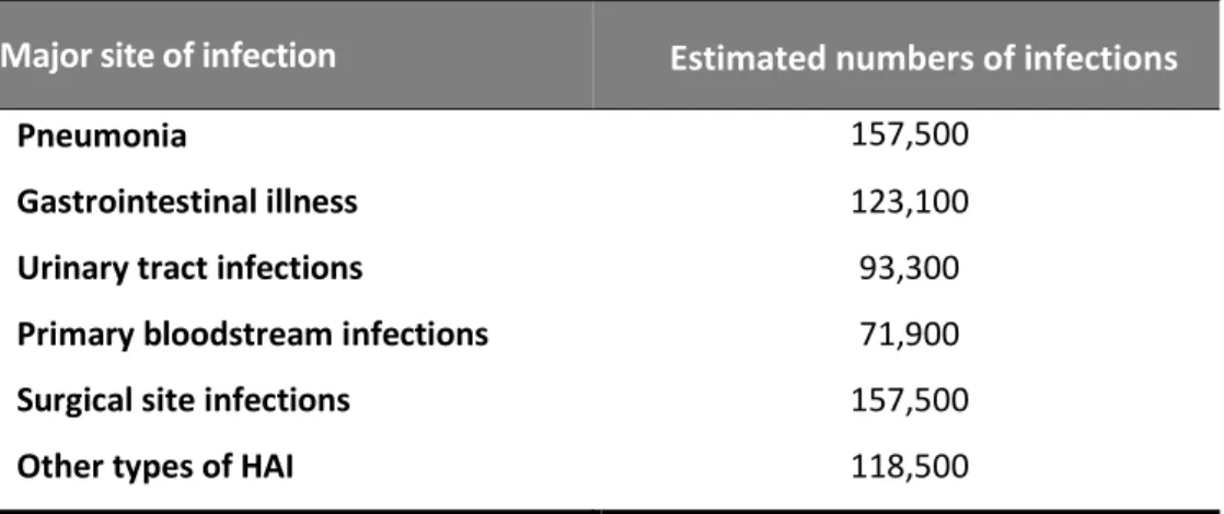
Healthcare–associated infections (HAI) are defined as a condition that results from an adverse reaction to the presence of an infectious agent(s), or its toxin(s), that occurs during a hospital or clinic stay, for which there is no evidence of the infection, or incubation, at admission (Horan et al., 2008). In general, these infections become manifest 48 hours after initial patient care (Wenzel, 2007). The last HAI prevalence survey provided by the Centre for Disease Control and Prevention (CDC) estimated, that in 2011, 1 in 25 patients hospitalized in U.S.A. acute care hospitals contracts at least one HAI and about 75,000 die during their hospitalization.
Table 1.1. Estimated number of HAI occurring in acute care hospitals in the U.S.A. during 2011.
Adapted from CDC HAI Prevalence Survey (Magill et al., 2014).
Major site of infection Estimated numbers of infections
Pneumonia 157,500
Gastrointestinal illness 123,100
Urinary tract infections 93,300
Primary bloodstream infections 71,900
Surgical site infections 157,500
Other types of HAI 118,500
Despite the progress observed in healthcare in the last years, infections continue to rise in hospitalized patients affecting, as well, hospital staff (Stone et al., 2008). Several factors such as decreased immunity and the increasing use of invasive techniques or implantation of medical devices (Von Eiff et al., 2005) can promote infection among hospitalized patients. The incidence of medical devices-associated infections is higher in the case of skin penetrating devices as these may bring together members of the normal skin flora. Coagulase-negative staphylococci (CoNS), are considered one of the main causes of medical devices-related nosocomial infections due to their ubiquity in the normal skin flora and mucosae (Rogers et al., 2009). The contamination of medical devices can also occur from the hands of the
a) b)
surgical or clinical staff, contaminated disinfectants, other patients or distant local infections (McCann et al., 2008; Pittet et al., 1999; von Eiff et al., 2002).
1.2| Coagulase-Negative Staphylococci and its pathogenesis
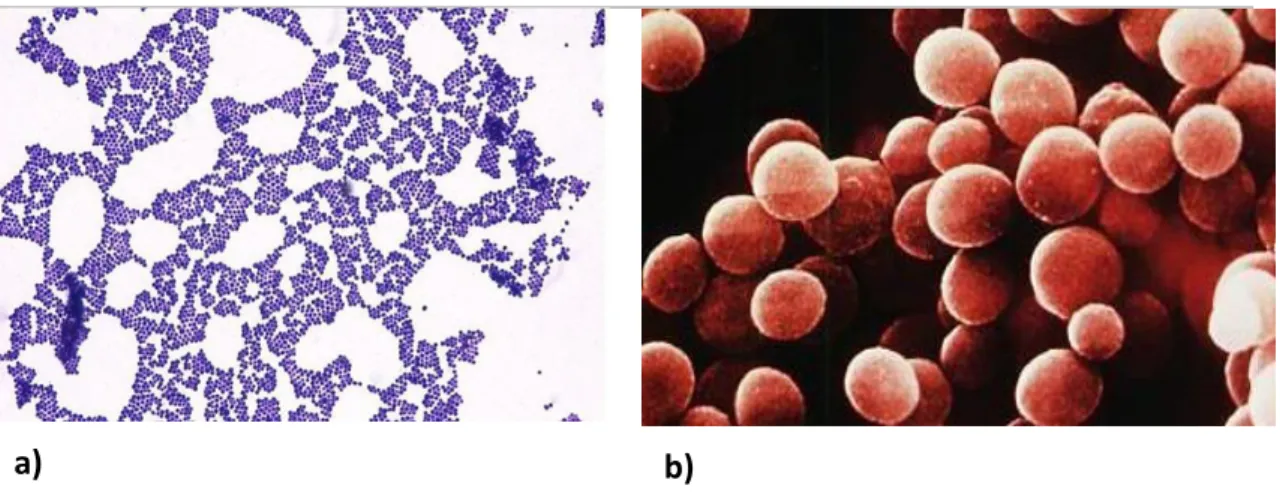
Staphylococci are members of the family Micrococcaceae and belong to genusStaphylococcus. They are non-motile, non-spore-forming, Gram-positive, facultative
anaerobic and clustering cocci that produce catalase (Pfaller and Herwaldt, 1988). On a solid culture medium, staphylococcal species exhibit different colony morphologies. While S. aureus produces large yellow colonies, CoNS species usually form relatively small white-greyish colonies. Until 1975, CoNS were grouped together as S. albus or
S. epidermidis, and were distinguished from S. aureus by their inability to clot blood
(Huebner and Goldmann, 1999; Mansour et al., 2009). Nowadays, there are 49 recognized CoNS species (Euzéby, 1997).
Figure 1.1. Staphylococcus under the microscope (a) Gram-stain of a staphylococcus culture (b)
Scanning electron microscopy image of staphylococci showing their clustered organization.
Adapted, respectively, from http://www.microbiologyinpictures.com/staphylococcus%20aureus.html
and http://textbookofbacteriology.net/staph.html [Accessed at 25 March 2015].
As referred before, CoNS are an important integrant and integrant part of the normal flora of human skin and mucosae. Certain CoNS species prefer particular niches, while others occur almost everywhere on our body (Grice and Segre, 2011). S.
epidermidis, the most frequently isolated CoNS, colonizes several areas such as the
1999). In addition, the colonization by S. epidermidis plays an important role in the maintenance of a healthy flora, by competing with potentially harmful microorganisms such S. aureus (Fey and Olson, 2010).
The insertion or implantation of foreign bodies, such as prosthetic heart valves, cardiac pacemakers, total artificial hearts and joint replacements, intravascular catheters, cerebrospinal fluid and renal dialysis shunts or continuous ambulatory peritoneal dialysis catheters, have become indispensable in modern medicine (Von Eiff et al., 2005). Nowadays, for instance, a considerable number of patients have one or even more than one vascular catheters in place during their hospitalization (Service and Services, 2003). However, unfortunately, the use of foreign material has been associated with the onset of severe bacterial and fungal infections (Bonadio, 2006; Halebeedu et al., 2014).
While a variety of Gram-positive and negative bacteria, as well as fungi, have been recognized as important causative agents of foreign body-related infections, staphylococci, particularly S. epidermidis and other CoNS, account for the majority of infections, including both temporarily inserted and permanently implanted material (Donlan, 2001; Hugonnet et al., 2004; Kleeman et al., 1993).
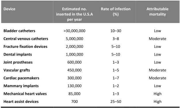
Table 1.2. List of implanted medical devices more often colonized by CoNS. Adapted from (Darouiche,
2001; Lynch and Robertson, 2008).
Device Estimated no.
inserted in the U.S.A per year
Rate of infection (%)
Attributable mortality
Bladder catheters >30,000,000 10–30 Low
Central venous catheters 5,000,000 3–8 Moderate
Fracture fixation devices 2,000,000 5–10 Low
Dental implants 1,000,000 5–10 Low
Joint prostheses 600,000 1–3 Low
Vascular grafts 450,000 1–5 Moderate
Cardiac pacemakers 300,000 1–7 Moderate
Mammary implants 130,000 1–2 Low
Mechanical heart valves 85,000 1–3 High
However, in recent years, S. epidermidis and other CoNS have become important opportunistic pathogens being especially problematic in patients with indwelling medical devices (Otto, 2011) (Table 2). S. epidermidis, for instance, is one of the most frequent causative agents of indwelling medical devices-associated infections, being particularly associated with peripheral and central intravenous catheters (Otto, 2009; Rogers et al., 2009). These infections usually begin with the introduction of this bacterium from the skin of the patient, or healthcare personnel, during catheter insertion (Rogers et al., 2009) being particularly increased in the last years, due to the increased use of such devices in the clinical setting (Nnis, 2004; Toole et al., 2000). Bloodstream infections occur in at least 4–5 out of every 1,000 catheters insertions performed on patients in intensive care in the U.S.A, and at least 22% of these infections are caused by S. epidermidis (Hall-Stoodley and Stoodley, 2009; Nnis, 2004; Otto, 2009).
Table 1.3. Rank of nosocomial bloodstream pathogens and the associated crude mortality among 49
hospitals throughout the U.S.A. Adapted from (Edmond et al., 1999).
S. epidermidis is also frequently associated with prosthetic joint, vascular graft,
surgical site, central nervous system shunt and cardiac device infections (Otto, 2009). Nevertheless, other CoNS are often implicated in HAI onset. S. haemolyticus, for instance, it is second only to S. epidermidis in its frequency of isolation and has been associated with the development of important infections such as peritonitis, otitis media, urinary tract infections and septicemia (Akinkunmi and Lamikanra, 2010).
Rank Pathogen Number of
isolates % Crude mortality (%) 1 Coagulase-negative staphylococci 3,908 31.9 21 2 Staphylococcus aureus 1,928 15.7 25 3 Enterococci 1,354 11.1 32 4 Candida spp 934 7.6 40 5 Escherichia coli 700 5.7 24 6 Klebsiella spp 622 5.4 27 7 Pseudomonas spp 557 4.5 28
Other CoNS species with clinical significance include S. hominis, S. warneri, S. capitis and S. equorum (Delmas et al., 2008), being the 3 first found less frequently (John and Harvin, 2007). To what concerns CoNS virulence determinants, the most important characteristic is their capability to form biofilms on the surface of implanted foreign bodies (Goldmann and Pier, 1993; Longauerova, 2006).
1.3| Impact of CoNS Biofilms
Biofilms are defined as a sophisticated community of matrix-encased, surface-attached bacteria that exhibit a particular phenotype. They are highly hydrated structures composed of 73 to 98% of extracellular material (Freitas, 2003), and contain channels that allow the inward diffusion of nutrients and oxygen to maintain the viability and capacity to proliferate. Biofilms may also contain non-microbial host derived components (Donlan, 2002). Among others functions, it was shown that biofilm formation in S. epidermidis and CoNS protects bacteria from antibiotics (Cerca et al., 2005a) and also from the host immune system (Cerca et al., 2005b; Costerton et al., 2005; Longauerova, 2006) resulting in infections that can linger for months, years, or even a lifetime (Costerton et al., 1999). Evidently this has serious clinical implications as it results in the emergence of chronic and persistent infections (Costerton et al., 1999). Indeed, it was shown that biofilms are implicated in more than 80% of the diagnosed chronic infections caused by bacteria including S.
epidermidis and others CoNS (Otto, 2009; Sauer et al., 2007). Although S. epidermidis
infections are rarely fatal, they significantly compromise patients’ quality of life often implicating the removal of the infected device in order to treat the infection. In addition to the clear human consequences, these infections increase healthcare system outgoings, due to the costs associated with the diagnosis and treatment of such infections (Stewart and William Costerton, 2001).
Henceforth, in order to better understand the origin of biofilm-related infections, the study of the mechanisms underlying biofilm formation has been extensively explored in the last years.
1.3.1| Mechanism of Biofilm formation
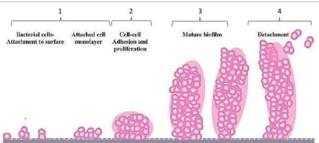
Biofilm formation is classically defined as a four-step process which includes: (1) adherence, (2) accumulation of cells in multiple layers, (3) maturation and (4) detachment (Fey and Olson, 2010; Stoodley et al., 2002). The initial step in biofilm formation, the attachment, can occur through direct binding to the native surface of a medical device or, via interaction with host extracellular matrix proteins present in the conditioning film that rapidly forms on the surface of medical devices following implantation (Vacheethasanee et al., 1998). The bacterial proteins that mediate binding to medical devices surfaces have been identified. In S. epidermidis, these include the bifunctional adhesins/autolysins AtlE and Aae (Heilmann et al., 2003, 1997; Williams et al., 2002) and multiple adherence factors, known as microbial surface components recognizing adhesive matrix molecules, which specifically interact with human serum proteins coating the implanted medical device (Clarke and Foster, 2006).
After adherence, bacterial cells rapidly grow leading to the accumulation of cellular aggregates, which results in small adherent micro colonies coalescing to form large coherent bacterial biofilm (Choong and Whitfield, 2000). These aggregates are then embedded within a matrix of extracellular polymeric substances, which is composed, among other components, by extracellular DNA, proteins, lipids and polysaccharides (Hall-Stoodley and Stoodley, 2009).
The maturation stage is facilitated by the increased transcription of genes that codify specific adhesion molecules, such as poly-N-acetylglucosamine (PNAG) (Mack et al., 1996), which cement the bacterial cells to the surface and to each other, giving rise to the characteristic biofilm structure (Choong and Whitfield, 2000). Finally, bacteria slowly proliferate and detach from the biofilm into the surrounding environment, enabling bacteria to colonize new surfaces and renew biofilm formation cycle elsewhere.
Figure 1.2. Model of Staphylococcus biofilm development. At stage (1), the bacterial cells attach to
the surface. At the next stage (2), bacterial cells accumulate in multiple layers resulting in the formation of micro colonies. Then, at stage (3), the maturation phase is reached, as indicated by the complex biofilm architecture obtained. At the detachment stage (4), single or clusters of cells leave the biofilm in to the involving environment. Image adapted from (Vuong and Otto, 2002).
1.4| Biofilms and antibiotics resistance
Since the discovery of penicillin in 1928s, antibiotics have been considered one of the wonder discoveries of the 20th century. Antibiotics have been critical in fighting infectious diseases caused by bacteria and other microbes, consequently increasing the average of life expectancy. However, concomitantly, antibiotic resistance has risen (Davies and Davies, 2010). Over the past several decades, the frequency of antimicrobial resistance and its association with the onset of serious infectious diseases have increased at alarming rates, resulting in an upsurge of morbidity, mortality, although less frequent in CoNS, and the costs associated with diagnosis and treatment of these infections (Davies and Davies, 2010). The way antibiotics are available to the population played an important role in the current massive use of antibiotics and, subsequently, in the emergence of an antibiotic resistance Era (Turnidge and Christiansen, 2005).
Antimicrobial resistance and/or tolerance in CoNS became an issue in the 1970s when CoNS species were isolated, for the first time, from prosthetic valve endocarditis and cerebrospinal fluid shunt infections (Odonkor and Addo, 2011). Since then, the treatment of CoNS infections has become increasingly difficult due to the high
prevalence of antibiotic-resistant strains and the increased antibiotic tolerance inherent to biofilm formation.
1.4.1| Mechanisms of antibiotic resistance
By interacting with specific bacterial targets, antibiotics are able to inhibit, among others, bacterial cell-wall synthesis, protein synthesis or nucleic acid replication and transcription (Kohanski et al., 2010). Evidently, toaccomplish its function, antibiotics must have access to and bind to its bacterial target. Over the years, bacteria have developed several mechanisms to circumvent antibiotics action. Most of the mechanisms employed by bacteria do not destroy or inactivate antibiotics but rather changes the antibiotic target or export it out of the cell (e.g. efflux pumps). In the last case, antibiotics are released into the surrounding environment in their active form, continuing to exert their selective pressure (Levy and Marshall, 2004).
In general, antibiotic resistance mechanisms fall into three broad categories: 1) direct inactivation of the active molecule, 2) alteration of the organism sensitivity to the antibiotic by modification of the target site, and 3) reduction of the concentration of drug that reaches the target (Hogan and Kolter, 2002). The mechanisms within the first category include the production of enzymes that degrade or modify the drug itself (Dzidic et al., 2008). Regarding the second category, the decreased sensitivity to antibiotics due to target modification, comprehend diverse microbial strategies such as the production of structure-altering or inactivating enzymes (e.g. β-lactamases or amino glycoside-modifying enzymes) (Paterson, 2001), alteration of penicillin-binding proteins or other cell-wall target sites (Brakstad and Maeland, 1997), altered DNA gyrase targets (Bearden and Danziger, 2001), mutations and ribosomal modification (Dzidic and Bedeković, 2003). However, most of the recent excitement in the field of antibiotic resistance is focused in the third category of antibiotic resistance mechanisms, the reduction of the antibiotic concentration at the target site.
In biofilms, despite the above referred mechanisms, there are other particular factors contributing to the inconsistent success of antimicrobial therapy (Alekshun and Levy, 2007). Although is still unclear which are all the biofilm-specific mechanisms that result in antibiotics tolerance, the barrier diffusion created by the biofilm matrix
(Donlan and Costerton, 2002), slow growth rate of the cells within biofilms (Costerton et al., 1999; Gilbert et al., 2002; Percival et al., 2011), the induction of antibiotic tolerant phenotypes (Percival et al., 2011), as well as the acquisition of genetic material are some of the factors known so far (Kokare et al., 2009).
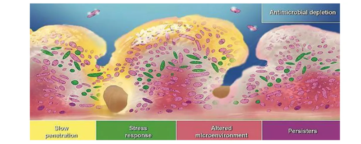
Figure 1.3. Three hypotheses proposed for antibiotic tolerance in biofilms. 1) Antibiotic (yellow) may
fail to penetrate beyond the surface layers of the biofilm; 2) some of the biofilm cells may differentiate into a protected phenotypic state (green); 3) in zones of nutrient depletion or waste accumulation (pink), antibiotic action may be antagonized and 4) persisters (purple) are cells that neither grow nor die in the presence of bactericidal agents, exhibiting multidrug tolerance. Image adapted from (Stewart and William Costerton, 2001).
1.4.1.1| Antimicrobial penetration failure
When microorganisms grow in planktonic culture, diffusion is not an issue. Planktonic cultures are generally agitated, and the fluid flow rapidly transports solutes to all cells. Diffusion limitation arises in biofilms. Fluid flow is reduced and the diffusion distance is increased (Stewart, 2003) in biofilms due to the high cell density and the presence of extracellular polymeric substances that arrest the flow of water. Since diffusion is the principal transport process in cells aggregates (De Beer et al., 1997), antimicrobial agents have difficulty to penetrate the biofilm structure. Some studies showed, however, that the extracellular polymeric matrix does not form an impenetrable barrier to the diffusion of antimicrobial agents (Cho, 2007; Donlan and Costerton, 2002; Percival and Bowler, 2004), and hence, other mechanisms must be occurring in order to protect biofilm cells from antibiotics action (Dunne et al., 1993; Percival and Bowler, 2004).
1.4.1.2| Slow growth rate
The transition from exponential to slower or no growth is generally accompanied by an increase in tolerance to antibiotics (Butler et al., 2010; Stewart, 2003). Antibiotics can kill bacteria by interfering with their cell wall, causing the cell to break open or lyse (Kohanski et al., 2010; Neu and Gootz, 1996). Hence, the antimicrobials that target bacterial cell wall are more effective in rapidly than slow growing cells. The efficacy of penicillin and ampicillin, for instance, is directly proportional to the rate of growth, being unable to kill non-growing cells (Lewis et al., 2001).Some of the more advanced β-lactams, cephalosporins, aminoglycosides and fluoroquinolones can kill non-growing cells, but they are clearly more effective in rapidly dividing cells (Costerton et al., 1999). However, other studies suggested that some determinants other than growth rate may be responsible for tolerance, and slow growth just enhances the effect (Duguid et al., 1992).
1.4.1.3| Resistance genes
Antibiotic resistance may be 1) intrinsic or 2) acquired (Neu, 1992). Intrinsic resistance is normally associated with the occurrence of genetic mutations in the bacterial chromosomal DNA, whereas acquired resistance is often related with horizontal gene transfer, which involves the acquisition of new genetic material. Mutations are generally uncommon events but may result in the development of antibiotic resistance in organisms that were initially susceptible (Mulvey and Simor, 2009). However, the development of resistance through the acquisition of new genetic material is of greater concern in the biofilm mode of growth since biofilms are an ideal niche for the exchange of extrachromosomal DNA (Donlan, 2002). Hence, this mechanism plays an important role in the dissemination of antibiotic resistance among strains or among different bacterial species or genera constituting a biofilm (Dzidic and Bedeković, 2003). Mechanisms associated with genetic transfer include conjugation, transformation and transduction (Tenover, 2006). Conjugation, the mechanism by which the bacterial plasmid may be transferred to other bacteria, occurs at a greater rate within biofilms than among planktonic cells (Hausner and Wuertz, 1999; Roberts et al., 1999) as the biofilm environment provides minimal
shear and closer cell-to-cell contact (Donlan, 2002; McCarty et al., 2014; Prakash et al., 2003). Since plasmids may encode several resistance genes, conjugation provide an extraordinary mechanism to the spread of antimicrobial resistance genes (Donlan, 2002). Because antibiotic resistance occurs as part of a natural process in which bacteria evolve, it cannot be stopped (Frieden, 2013). There is, therefore, an urgent need to find alternative strategies in order to control resistant bacteria.
1.5| New approaches for biofilm infections prevention and
therapy
The rapid development of antimicrobial drug-resistant pathogens is amongst the most serious threats to public health nowadays (Lai et al., 2011; Lambert et al., 2011). As a result, during the last few years the scientific community has shown considerable interest in the study of plant elements as antimicrobial agents in order to be used for new prophylactic and therapeutic strategies (Nostro and Papalia, 2012).
1.5.1| Essential oils
Essential oils (EOs) are aromatic oily liquids, most commonly obtained by steam distillation, found in plant constituents such as flowers, buds, seeds, leaves, twigs, bark, herbs, wood, fruits and roots (van de Braak et al., 1994). In the past, several EOs were identified and commercialized by their flavors and fragrances (van de Braak et al., 1994), as dental canal sealers (Manabe et al., 1987), antiseptics (Cox et al., 2000) and also used as food supplements (Ilsley et al., 2002). In addition, EOs are known, for a long time, by their antimicrobial and antioxidant properties (Guenther, 1948) and several studies have been conducted in order to evaluate their action in several bacteria, viruses and fungi (Kalemba and Kunicka, 2003; Reichling et al., 2009; Solórzano-Santos and Miranda-Novales, 2012).
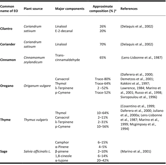
Table 1.4. Major chemical components of EOs that exhibit antibacterial properties. Common
name of EO Plant source Major components
Approximate composition (% )a References Cilantro Coriandrum sativum Linalool E-2-decanal 26% 20% (Delaquis et al., 2002) Coriander Coriandrum
sativum Linalool 70% (Delaquis et al., 2002)
Cinnamon Cinnamomum
zeylandicum
Trans-cinnamaldehyde 65% (Lens-Lisbonne et al., 1987)
Oregano Origanum vulgare
Carvacrol Thymol λ-Terpinene p-Cymene Trace-80% Trace-64% 2 –52% Trace-52% (Daferera et al., 2000; Demetzos et al., 2001; Kokkini et al., 1997; Lawrence, 1984; Marino et al., 2001; Russo et al., 1998; Sivropoulou et al., 1996)
Thyme Thymus vulgaris
Thymol Carvacrol λ-Terpinene p-Cymene 10–64% 2–11% 2–31% 10–56% (Cosentino et al., 1999; Daferera et al., 2000; Juliano et al., 2000a; Lens-Lisbonne et al., 1987; Marino et al., 1999; Mcgimpsey et al., 1994)
Sage Salvia officinalis L.
Camphor α-Pinene β-pinene 1,8-cineole α-tujone 6–15% 4–5% 2–10% 6–14% 20–42% (Marino et al., 2001)
aPercentages of total volatiles rounded up to the nearest whole number. EO-essential oil.
The composition of EOs from different plants is distinct and, in particular species, can also differ among harvesting seasons and geographical sources (Cosentino et al., 1999; Faleiro et al., 2003; Juliano et al., 2000b; Marotti et al., 1994; Mcgimpsey et al., 1994). Generally, EOs produced from herbs harvested during, or immediately after flowering processes, present the strongest antimicrobial activity (Marino et al., 1999). However, the composition, and consequently, the action of the EOs obtained from different parts of the same plant can vary widely (Delaquis et al., 2002). Although the antimicrobial properties of EOs and their components have been reviewed in the past (Nychas, 1995; Shelef, 1984), their exact mechanism of action is still unknown
(Lambert et al., 2001). Considering the large number of different groups of chemical compounds that compose EOs, it is most likely that their antibacterial activity is not determined by one specific mechanism or target, but by several mechanisms or targets in the cell (Carson et al., 2002; Skandamis et al., 2001).
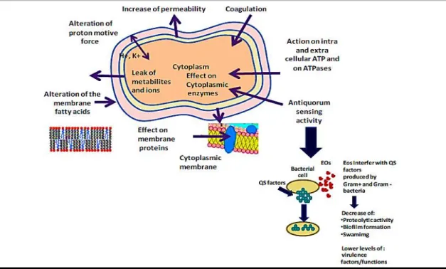
The mechanisms of action of the EOs known so far include: 1) the degradation of the cell wall (Gill and Holley, 2006; Helander et al., 1998), 2) cytoplasmic membrane damage (Oosterhaven et al., 1995; Ultee et al., 2002, 2000), 3) cytoplasm coagulation (Gustafson et al., 1998; Ultee et al., 2002, 2000), 4) membrane proteins damage (Juven et al., 1994; Ultee et al., 1999), 5) increased permeability due to increased hydrolysis and reduced membrane potential resulting in leakage of the cells contents (Burt, 2004; Cox et al., 2000; Gustafson et al., 1998; Helander et al., 1998; Juven et al., 1994; Lambert et al., 2001; Oosterhaven et al., 1995; Ultee et al., 2002), 6)reduced proton
motive force (Ultee and Smid, 2001; Ultee et al., 1999), and 7) reduced intracellular ATP pool via decreased ATP synthesis (Burt, 2004). It is important to emphasize that not all of the enumerated mechanisms are separate targets, some are affected indirectly, as a consequence of another mechanism being targeted (Burt, 2004).
Figure 1.4. Mechanisms of action and target sites of EOs in microbial cells: 1) degradation of the cell
wall; 2) cytoplasmic membrane damage; 3) coagulation of cytoplasm; 4) membrane proteins damage; 5) increased permeability resulting in leakage of cell contents; 6) depletion of the proton motive force;
It has been shown that EOs with higher percentage of phenolic compounds, such as carvacrol, eugenol and thymol, retain the strongest antibacterial properties (Cosentino et al., 1999; Dorman and Deans, 2000; Juliano et al., 2000b; Lambert et al., 2001). Carvacrol has proven to have better antimicrobial properties when compared with parent compounds (Mastelić et al., 2008). Because of its promising antimicrobial effect, carvacrol became a compound of interest in the search for new approaches for biofilms infections treatment (Borges et al., 2013; Iannitelli et al., 2011).
1.5.2| Carvacrol
Since biofilm resistance depends, at some extent, on their multicellular structure, one possible strategy for the management of biofilm-associated infections is to develop therapies that disrupt its multicellular structure (Percival et al., 2011). If its multicellularity is defeated the efficacy of antibiotics might be restored and the host defenses might also be able to resolve the infection (Stewart and William Costerton, 2001).
Potential therapies under evaluation comprise, among others, the use of enzymes that degrade the polymers of the biofilm matrix (Nemoto et al., 2000; Parsek and Greenberg, 2000; Yasuda et al., 1993) and the use of molecules capable of interfering with cell-cell interaction required for normal biofilm formation.
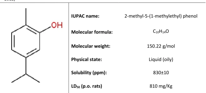
Carvacrol, (2-methyl-5-(1-methylethyl)-phenol), is one of the most common components of the EOs of the family Labiatae (Başer and Demirci, 2007; Kirimer et al., 1995; Nostro et al., 2012; Solórzano-Santos and Miranda-Novales, 2012). As a monoterpenic phenol, carvacrol is mainly found in several aromatic plants such as thyme, oregano, wild bergamot and pepperwort (Singh and Chittenden, 2010). Due to carvacrol wide range of biological properties, which includes: 1) antibacterial (Aligiannis et al., 2001; Pérez-Conesa et al., 2011; Rivas et al., 2010; Svoboda and Hampson, 1999), 2) antifungal (Ahmad et al., 2011; Dalleau et al., 2008), 3) antitumor (Arunasree, 2010; Karkabounas et al., 2006), 4) antioxidant (Aristatile et al., 2011; Jayakumar et al., 2012; Prieto et al., 2007), 5) antiinflammatory (Hajhashemi et al.,
2002; Hotta et al., 2010; Landa et al., 2009), 6) anti-obesity (Cho et al., 2012), 7) antidepressant/anxiolytic (Melo et al., 2010), 8) insecticidal, (Karpouhtsis et al., 1998; Park et al., 2005), 9) antispasmolytic (Boskabady et al., 2011) and 10) vasorelaxant (Earley et al., 2010; Peixoto-Neves et al., 2010) activities, the interest in its use as therapeutic agent has been increasing.
The antibacterial activity of carvacrol has been attributed to its hydrophobic nature, the presence of a free hydroxyl group and a delocalized electron system (de Sousa et al., 2012; Nostro et al., 2009). Its free hydroxyl group interacts with the lipid bilayer of the cytoplasmatic membrane causing the expansion and destabilization of the membrane structure increasing membrane fluidity and permeability to protons and ions, resulting in loss of integrity (Ben Arfa et al., 2006; Cristani et al., 2007; Helander et al., 1998; Lambert et al., 2001; Ultee et al., 1999). Moreover, studies performed in
Bacillus cereus demonstrated that carvacrol damages bacterial cell membrane by
pushing apart the fatty acid chains of the phospholipids layer, creating channels that allow ions leakage from the cytoplasm (Ultee et al., 2000). In addition, numerous reports have shown that carvacrol has a potent action against bacteria, yeasts and fungi (Altintas et al., 2013; Camele et al., 2014; Kalemba and Kunicka, 2003; Nostro et al., 2009; Reichling et al., 2009; Solórzano-Santos and Miranda-Novales, 2012), even in drug-resistant species (Chami et al., 2005; Dorman and Deans, 2000; Ultee et Table 1.5. Carvacrol structure and its physico-chemical properties. Adapted from (Nostro and Papalia,
2012)
IUPAC name: Molecular formula:
2-methyl-5-(1-methylethyl) phenol
C10H14O
Molecular weight: 150.22 g/mol
Physical state: Liquid (oily)
Solubility (ppm): 830±10
al., 1998). It has also been shown that both S. aureus and S. epidermidis, including methicillin-resistant strains, are susceptible to carvacrol (Nostro et al., 2004). Furthermore, it has been reported that carvacrol is able to inhibit the growth of preformed biofilms and to interfere with biofilm formation of numerous species, including S. epidermidis (Knowles and Roller, 2001; Knowles et al., 2005; Nostro et al., 2009, 2007, 2004).
Taking into consideration the increasing incidence and prevalence of CoNS biofilm-related infections and the emergence of multidrug resistance, there is an urgent need to find new and effective therapies for the management of CoNS biofilm-associated infections. Herein, we investigated the susceptibility of different CoNS biofilms to the action of carvacrol.
2.1| Bacteria and growth conditions
For this study the biofilm-forming species S. epidermidis strain 20A.1, S. equorum
strain 060A, S. haemolyticus strain 065A.1, S. hominis strain M11, S. capitis strain 52A and S. wameri strain F16 were used. These strains were previously isolated and
characterized by (Oliveira and Cerca, 2013). All strains were prepared by inoculating a single colony into 2 mL of Tryptic Soy Broth (TSB) (Liofilchem, Roseto Degli Abruzzi, Italy) and incubating at 37°C with shaking at 200 rpm for 24 (±2) hours.
Planktonic cultures were attained by diluting (1:200) the 24 hours-old culture in 2 mL of fresh TSB and by incubating at 37°C and 200 rpm for additional 6 or 24 (± 2) hours in order to obtain, respectively, exponential or stationary phase planktonic cells. Biofilms were formed as described previously (Cerca et al., 2012b) with some modifications. In brief, in a 96-well microtitre plate (Orange Scientific, Braine- l’Alleud, Belgium), 2 μL of the 24 hours-old culture was inoculated into 200 μL of TSB supplemented with 0.25% of glucose to promote biofilm formation. The plates were then incubated for 24 hours at 37°C with shaking at 120 rpm to allow biofilm formation. Thereafter, prior to any experiment, spent medium, was carefully removed and biofilms washed once with 200 μL of 0.9% NaCl.
2.2| Optical density versus colony forming units
In order to be able to adjust the same concentration of bacteria in the planktonic or biofilms cultures, the optical density, at 640 nm, and the number of colony forming units per mL (CFU) were determined. Summarily, after 24 hours of growth, biofilms were suspended in 0.2 mL of 0.9% NaCl solution by scrapping the biomass attached to the bottom of the well. For each strain, a pool of three biofilms was performed. Then, both biofilm and planktonic cells were harvested by centrifugation at 13500 g during 10 minutes, suspended in 1 mL of 0.9% NaCl supplemented with 0.05% Tween20 (0.9% NaCl tween) (Applichem, Darmstadt, Germany) and sonicated, on ice, for 3 seconds at 40% amplitude (Cole Parmer Ultrasonic Processor, Illinois, USA). This procedure eliminated bacterial aggregates that would interfere with posterior CFU
counting (Cerca et al., 2011). Subsequently, 10-fold dilutions were performed in 0.9% NaCl tween and plated on Tryptic Soy Agar plates. Plates were then incubated at 37°C overnight to allow CFU counting. For optical density readings at 640 nm, both planktonic and biofilm cultures were diluted 1:10, respectively, in TSB and 0.9% Nacl. The values of optical density and the respective number of CFU per mL were plotted in order to allow the correlation between OD readings and concentration of cells. This experiment was performed three independent times.
2.3| Determination of carvacrol minimum inhibitory concentration (MIC)
The determination of carvacrol minimum inhibitory concentration in planktonic cultures of the different CoNS isolates under study was evaluated by the broth microdilution method, adapted from (Clinical and Laboratory Standards Institute, 2012). Briefly, in a 96-well microtiter plate (Orange Scientific, Braine-l’Alleud, Belgium), carvacrol (Sigma, MO, USA), initially dissolved in dimethyl sulfoxide was serially diluted (1:2) in TSB in order to obtain carvacrol concentrations ranging from 4 µM to 0 µM. Then, each dilution was inoculated with 2 μL of a planktonic cells suspension with approximately 1 × 10⁸ CFU/mL, equivalent to an OD
640nm of 0.16. The plate was incubated at 37°C during 24 (±2) hours without shaking. Bacterial growth, in the presence or absence of carvacrol, was evaluated by direct visualization of medium turbidity. Carvacrol MIC was defined as the lowest concentration of carvacrol that prevented bacterial growth. This experiment was repeated three independent times.
2.4| Planktonic cells and biofilms susceptibility to carvacrol and vancomycin assessed by CFU counting
For the analysis of the effect of both carvacrol and vancomycin in the different CoNS isolates under study, planktonic cells, grown as described above, were incubated, for 5, 50, 95 and 185 minutes, with carvacrol (2 µM, 4 µM, 8 µM, 16 µM) or vancomycin (40 µL/mL) which represent vancomycin peak serum concentration (Boselli et al.,
2005). Biofilms cultures were exposed for 5, 50 and 95 minutes, with carvacrol MIC (2 µM) or vancomycin (40 µL/mL). A control was achieved by plating both planktonic and biofilm cells before addition of carvacrol or vancomycin.
After exposure to carvacrol and vancomycin, the number of culturable cells in both planktonic cells and biofilms was assessed by CFU counting. Prior to cells collection, biofilms were washed once and suspended in 0.2 mL of 0.9% NaCl. For each species, a pool of three biofilms was performed. Thereafter, biofilm and planktonic cells were collected by centrifugation, suspended in 1 mL of 0.9% NaCl tween, sonicated and plated as described in “Optical density versus colony forming units calibration curve”. This experiment was performed five times for planktonic cells and three times for biofilms.
2.5| Assessment of bacteria death by flow cytometry
The viability of planktonic cells exposed to carvacrol was evaluated by flow cytometry, using Live/Dead staining, as previously optimized (Cerca et al., 2011) with minor modifications. In brief, a control with dead cells was obtained by incubating bacteria, 15 min on ice, with 1 mL of 99% methanol (Fisher, LE, UK). Subsequently, bacteria were collected by centrifugation and suspended in 1 mL of phosphate buffered saline (PBS). Then, 10 µL aliquots were collected, from both controls and treated planktonic cells, and mixed with 90 µL of PBS containing 1:80000 SYBR®Safe DNA gel stain (Invitrogen, CA, USA) and 20 µg/mL of propidium iodide (Sigma, MO, USA). Samples were then analyzed in an EC800TM flow cytometer (Sony Biotech Inc., CA, USA) using the 488 nm laser and at a 10 µL/min flow rate. SYBR green was detected in the FL1 (525/50 nm) and propidium iodide in the FL4 (615/30) filter. Data were acquired and analyzed using EC800TM software version 1.3.6 (Sony Biotech Inc). This experiment was performed once for confirmation of CFU counting results.
2.6| Confocal laser scanning microscopy of biofilms exposed to carvacrol
Biofilms were prepared for confocal scanning laser microcopy (CSLM) as described above, with some modifications. Shortly, in a 24-well polystyrene plate (Thermo Fisher Scientific, New York, USA), 5 µL of a 24 hours growth was inoculated into 1 mL of TSB supplemented with 0.25% glucose and incubated, for 24 hours (±2), at 37 oC and shaking at 120 rpm. Carvacrol treatment was performed using 200 µL of carvacrol at 2 µM concentration for 95 minutes of incubation. Then, biofilms were washed with 0.9% NaCl solution to remove planktonic cells and carvacrol residues. Afterwards, the bottom of each well, was removed using a metal heat cutter with a similar diameter of the wells. Biofilms were then stained with SYTO9 (485/498 nm) (Molecular Probe, city, USA) following the manufacturer’s instructions and placed on the top of a glass slide.
Biofilms images were obtained using an OlympusTM FluoView FV1000 (Olympus, Lisboa, Portugal) confocal scanning laser microscope. Biofilms formed by all CoNS strains were observed using a 60 × water-immersion objective (60 ×/ 1.2 W). Images were acquired with 512 × 512 resolution and in at least two different regions of each surface analyzed. For biofilm maximum thickness determination, at least ten different regions per surface were analyzed, by determining the top and bottom layer of the biofilm. This experiment was performed twice.
2.7| Statistical analysis
Statistical significance between control and treated samples was determined by Student’s t-test using Microsoft®Excel® 2013 software (Santa Rosa, CA, USA). Differences were considered significant when P values were less than 0.05.
Conventional treatment of S. epidermidis and others CoNS infections, including those caused by biofilms on implanted medical devices, involves the use of common antibiotic therapy (McCann et al., 2008). Biofilm infections are very difficult to treat, especially due to the increasing resistance to antibacterial agents (Folkesson et al., 2008; McCann et al., 2008). Treatment with antibiotics commonly fails to eradicate cells embedded within the biofilm, which can then result in the onset of recurrent and relapsing infections (Donlan and Costerton, 2002).Therefore, new strategies to control
S. epidermidis and other CoNS biofilm infections are required.
3.1| Carvacrol MIC determination
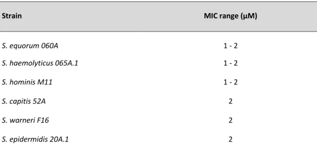
In the first stage of this research project it became essential to understand if strains were susceptible to the antimicrobial agent or not. Therefore, the first step was to assess the MIC of carvacrol. The results obtained for all six CoNS strains used are summarized in Table 1.
Table 3.1. Evaluation of the minimum inhibitory concentration of carvacrol on the six CoNS isolates
under study.
Strain MIC range (µM)
S. equorum 060A 1 - 2 S. haemolyticus 065A.1 1 - 2 S. hominis M11 1 - 2 S. capitis 52A 2 S. warneri F16 2 S. epidermidis 20A.1 2
The results shown in Table 3.1 indicate that all the CoNS strains used in this study were susceptible to low concentrations of carvacrol, which was consistent with the results found in the literature for S. epidermidis (Nostro et al., 2007).
3.2| Evaluation of carvacrol activity on planktonic cultures
Biofilm formation, as mentioned previously, is the major virulence factor of S.
epidermidis (Vuong and Otto, 2002), as well as other CoNS species being frequently
associated with biofilm-associated nosocomial infections (Otto, 2008). Therefore it is of particular interest to address the action of carvacrol in S. epidermidis biofilms and other CoNS species.
In order to correlate results from carvacrol effect on planktonic and biofilm cultures, we first characterized the biofilms formed by the selected CoNS species by quantifying the total number of cultivable cells (Figure 3.1) and the total biomass (Figure 3.2). Biofilms cultures were quantified after 24 h of growth.
Figure 3.1. Biofilm CFU quantification of all six CoNS. This experiment was performed in S. haemolyticus 065A.1, S. equorum 060A, S. capitis 52A, S. warneri F16, S. hominis M11 and S. epidermidis 20A.1. The values represent the mean and the error bars the standard deviation of six independent
experiments. 1,00E+00 1,00E+01 1,00E+02 1,00E+03 1,00E+04 1,00E+05 1,00E+06 1,00E+07 1,00E+08 1,00E+09 1,00E+10
S. haemolyticus S. equorum S. capitis S. warneri S. hominis S. epidermidis
Lo
g 10
C
FU/
Figure 3.2. Biofilm biomass quantification of the biofilms formed by all the six CoNS. This experiment
was performed in S. haemolyticus 065A.1, S. equorum 060A, S. capitis 52A, S. warneri F16, S. hominis
M11 and S. epidermidis 20A.1. The values represent the mean and the error bars the standard deviation
of two independent experiments.
According with biofilm CFU quantification assay, all strains formed biofilms with approximately 1 x 10⁸ CFU /mL, with exception of S. warneri strain F16 that showed, comparatively with the other CoNS isolates tested, lower number of cultivable cells after 24 hours of growth. As observed in Figure 3.2, the biofilms formed by this strain also showed inferior optical density, predicting a biofilm composed by fewer cells, polysaccharides, proteins and others matrix constituents (Sousa et al., 2009). Results from biofilm quantification assays allowed the preparation of planktonic cultures with the same number of cultivable cells as biofilms. This way, the results obtained in both planktonic and biofilms cultures can be compared. In order to proceed, a calibration curve was constructed to correlate the values of optical density (OD), at 640 nm, and the number of colony forming units (CFU) per mL of planktonic cells (Appendix A).
0 0,2 0,4 0,6 0,8 1 1,2 1,4 1,6 1,8
S. haemolyticus S. equorum S. capitis S. warneri S. hominis S. epidermidis
OD
6
4
0
To evaluate the effect of carvacrol on biofilms, the cultivability of the bacterial cells within the biofilms as well as its tridimensional structure were assessed after different exposure times to carvacrol. Because very little is known about carvacrol effect on CoNS, many tests were performed with the intention of optimizing carvacrol antimicrobial activity.
Firstly, for comparative purposes, planktonic cultures were tested. This approach assures that if carvacrol is not efficient killing bacteria in planktonic state, on biofilms it is most likely that it will not have any significant effects either (Cargill and Upton, 2009; Ceri et al., 1999; Farber et al., 1990; Mah and O’Toole, 2001). A preliminary experiment was performed to understand how carvacrol would affect cells cultivability. Hence, it was decide to use a concentration of carvacrol of 4 × the MIC determined previously.
Figure 3.3. Planktonic cells susceptibility to carvacrol (4 × MIC). This assay was performed using both
stationary and logarithmic planktonic cells of S. equorum 060A, S. capitis 52A and S. warneri F16 after 3 hours of incubation with carvacrol. Error bars and P values were not calculated since this was a single experiment. 1,00E+00 1,00E+01 1,00E+02 1,00E+03 1,00E+04 1,00E+05 1,00E+06 1,00E+07 1,00E+08 1,00E+09
Stationary Logarithmic Stationary Logarithmic
T0 min T185 min Lo g 10 C FU/ ml S. equorum S. capitis S. warneri
As can be seen in Figure 3.3, after 3 hours of incubation with carvacrol, no CFU were detected neither in stationary nor in logarithmic phases of growth. In order to better understand the results obtained, we have tested reduced exposure times (Figure 3.4). However, because this was a preliminary experiment, this was only tested on S.
epidermidis strain 20A.1.
Figure 3.4. S. epidermidis 20A.1 CFU counting after 5, 50 and 95 minutes of exposure to carvacrol (4 × MIC). Error bars and P values are not calculated since this was a single experiment.
The results obtained showed that even in lower exposure times, no CFU were observed. It was clear that at this concentration and for the tested exposure times, carvacrol was very efficient.
The following experiment was designed to determine how difference concentrations of carvacol would influence cell cultivability. On this new experiment, four different carvacrol concentrations, starting from MIC, and rising up to 2, 4 and 8 × MIC were tested, first, in S. epidermidis.
1,00E+00 1,00E+01 1,00E+02 1,00E+03 1,00E+04 1,00E+05 1,00E+06 1,00E+07 1,00E+08 1,00E+09
T0 min T5 min T50 min T95 min
Lo g 10 C FU/ mL S. epidermidis
Figure 3.5. CFU counting after S. epidermidis 20A.1 planktonic cells treatment with different concentrations of carvacrol. Concentrations are respectively MIC – 2 µM, 2 × MIC – 4 µM, 4 × MIC – 8
µM and 8 × MIC – 16 µM. Error bars and P values are not calculated since this was a single experiment.
CFU counting showed a decrease of cultivable cells with the increase of carvacrol concentration as well as exposure time. It appeared that after 50 minutes of exposure, cells lose their cultivability. In addition, it seems that for the concentration eight times higher than MIC, cells were immediately damaged, since no CFU were detected after 5 minutes of exposure.
Since this very quick effect was somewhat surprising, an extra experiment was performed. It’s known that carvacrol interacts with cells’ membrane by binding to the lipid bilayer (Nowotarska et al., 2014). So, it was essential to ensure that carvacrol was not carried out through the serial dilutions performed during CFU plating. To address that, before platting the cells exposed to carvacrol a washing step was performed. Figure 3.6 represents results from this experiment.
8 MIC 4 MIC 2 MIC MIC T0 min 1,00E+00 1,00E+02 1,00E+04 1,00E+06 1,00E+08 5 min 50 min 95 min Lo g 10 C FU/ ml
Figure 3.6. CFU counting after treatment with 2 µM of carvacrol with and without cell wash. The assay
was performed once using S. epidermidis 20A.1. Error bars and P values were not calculated since this experiment was performed once.
By analyzing these results is was clear that no significant changes were observed with or without a washing step. We did not assess the short time period of exposition (5 min), since it took 10 min to centrifuge the cells and it was not possible to guarantee that during this time carvacrol was not inducing any reduction in cells cultivability. Given this outcome, it is possible to conclude that carvacrol at 2 µM, is a powerful antimicrobial agent that is capable of quickly bind to cells resulting in an absence of growth.
Since small concentrations of carvacrol had a strong effect on S. epidermidis, even under a short exposure time, it would be of great interest to evaluate carvacrol MIC in all the six CoNS under study. Although, this group of six species belongs to a group with similar characteristics, they can respond differently to the same stress. To pursue our objective, the susceptibility of all CoNS strains to 2 µM of carvacrol was tested.
1,00E+00 1,00E+01 1,00E+02 1,00E+03 1,00E+04 1,00E+05 1,00E+06 1,00E+07 1,00E+08 1,00E+09
T0 min T50 min T95 min
Lo g 10 C FU /mL With wash Without wash T0
Activity of carvacrol against Coagulase-negative Staphylococci biofilm related infections
Figure 3.7. Susceptibility of planktonic cells of all CoNS strains to carvacrol. This test was performed in S. warneri F16, S. capitis 52A, S. equorum 060A, S. epidermidis 20A.1, S. hominis M11 and S. haemolyticus 065A.1 in stationary phase of growth. All cells were exposed for 90 min to 2 µM of
carvacrol. The values represent the mean and the error bars the standard deviation of five independent experiments. Significant differences are represented with: a = P < 0.05 and b = P < 0.001.
Although each strain responded slightly different to carvacrol MIC, all were affected by 2 µM of carvacrol. This effect can be due to the hydrophobic nature of carvacrol that favours its accumulation in the cellular membrane, possibly causing membrane disruption and resulting in the leakage of intracellular contents (Ultee et al., 1999). There was a significant decrease, in all strains, in the CFU counting after 5 min of exposure (P < 0.05 or P < 0.001). Analyzing each strain separately, S. warneri F16, S.
capitis 52A and S. hominis M11 had a reduction in CFU of proximally 1 Log (90% of
cells), S. haemolyticus 065A.1 2 Log (99% of cells) and S. equorum 060A and S.
epidermidis 20A.1 had a reduction superior to 3 Log (> 99.9% of cells). After 50 or 95
minutes of treatment with carvacrol, an abrupt decrease in CFU was observed in all six strains, in fact, no growth was detected. In conclusion, a carvacrol concentration of 2 µM was sufficient to promote a significant reduction in planktonic cells cultivability in all six CoNS under study. Furthermore, Figure 3.5 suggested that cell damaging is directly proportional to both exposure time and carvacrol concentration.
1,00E+00 1,00E+01 1,00E+02 1,00E+03 1,00E+04 1,00E+05 1,00E+06 1,00E+07 1,00E+08 1,00E+09
T0 min T5 min T50 min T95 min
Lo g 10 C FU/ ml S. warneri S. capitis S. equorum S. epidermidis S. hominis S. haemolyticus a a b a a a a a
Nevertheless, as previously shown, the decrease in cells cultivability is not always associated with cells dead but rather with the development of a dormant state (Cerca et al., 2014). Hence, in order to determine if carvacrol was inducing a viable but non-culturable state or killing bacterial cells, a LIVE/DEAD staining was performed and cells were analyzed by flow cytometry.
Figure 3.8. Percentage of dead cells after treatment with carvacrol. This assay was performed in all six
CoNS under study after 95 min of incubation with 2 µM of carvacrol. Positive control (dead cells), was achieved by incubating cells with methanol for 15 min; T0 min represents cells without any treatment and T95 min represent cells after 95 min of exposure to carvacrol. Strain S. equorum 060A was unable to stain, even after 1 hour of incubation with a mix of SYBR and propidium iodide. This assay was
performed once.
The results obtained showed that after 95 min incubation with 2 µM of carvacrol >95% of cells were dead. Since carvacrol acts on cell membrane, it is possible to conclude, that the membrane fluidity is an important factor affecting the viability of CoNS, but probably not the only factor (Ultee et al., 1998). The changes in the cellular membrane (lipid membrane composition, leakage of essential components) are definitely associated with loss of integrity and subsequent death, but more studies have to be done to obtain more insights into the mechanisms underlying carvacrol bactericidal effect (Ben Arfa et al., 2006; Cristani et al., 2007; Di Pasqua et al., 2007).
0 10 20 30 40 50 60 70 80 90 100 110 T0 min T95 min % o f d e ad c e lls S. warneri S. capitis S. hominis S. epidermidis S. haemolyticus
3.3| Evaluation of carvacrol activity on biofilm cultures
According to the main goal of this work, carvacrol was added to 24h biofilms of all six CoNS species under study and biofilm structure and cells cultivability was evaluated. Figure 3.9 demonstrate results from CFU counting after contact with carvacrol.
Figure 3.9. CoNS biofilms CFU reduction after exposure to carvacrol. Biofilms of S. capitis 52A, S. equorum 060A, S. warneri F16, S. hominis M11, S. epidermidis 20A.1andS. haemolyticus 065A.1, were
exposed to a concentration of 2 µM of carvacrol for 95 min. The values represent the average and the error bars the standard deviation of 3 independent experiments. Significant differences are represented with: a = P < 0.05 and b = P < 0.001, when compared with the control (T0 min).
As could be expected, biofilm cells were less sensitive to carvacrol than their planktonic counterparts. It should be noted that biofilm formation depends on many intrinsic and extrinsic factors, and vary from species to species and even among strains of the same species (McLandsborough et al., 2006). Furthermore, bacteria within a biofilm is organized in aggregates, physically joined together, and protected by an extracellular matrix that contains many different types of extracellular polymeric substances (EPS) including proteins, DNA and polysaccharides that block antimicrobial treatment allowing cells to proliferate (Abdel-aziz and Aeron, 2014; Anderson and O’Toole, 2008; del Pozo and Patel, 2007). These aggregates can withstand very high
1,00E+03 1,00E+04 1,00E+05 1,00E+06 1,00E+07 1,00E+08 1,00E+09 1,00E+10
T0 min T5 min T95 min
Lo g 10 C FU/ ml S. capitis S. equorum S. warneri S. hominis M11 S. epidermidis S. haemolyticus a a a a a a a a b a a