1. Rheumatology Unit - Evangelic University Hospital, Curitiba, PR-BRAZIL
Systemic Lupus Erythematosus activity
and serum bilirubins
ACTA REUMATOL PORT. 2013;38:242-246
AbstrAct
Background: Serum bilirubins play an important role
in controlling oxidative stress; there is increased oxi-dative stress during activity of rheumatic diseases such as systemic lupus erythematosus (SLE).
Objective: To study bilirubin levels in SLE (Systemic
Lupus Erythematosus) patients and relate them to di-sease activity.
Methods: We analyzed levels of total bilirubins (TB),
direct bilirubins (DB) and indirect bilirubins (IB), se-dimentation rate (ESR) and C reactive protein (CRP) in 143 SLE patients. Data were collected on the clinical and autoantibody profiles and patients underwent mea-surement of SLEDAI and SLICC .
Results: Correlation of indirect bilirubin values with
SLEDAI was negative (p=0.02; Spearman rho=-0.18). Comparing the levels of IB according to the clinical ac-tivity profile we observed associations with increase of anti DNA titer (p=0.027) and with decrease in com-plement levels (p=0.017). ESR correlated negatively with IB levels (p=0.01) but CRP did not (p=0.15). In a multiple linear regression analysis only the increase in ESR titer remained significant. SLICC values were not correlated with TB (p=0.30), DB (p=0.12) or IB (p=0.31).
Conclusions: We conclude that IB levels in SLE
corre-late negatively with disease activity. IB levels are lower in patients with higher ESR.
Keywords: Systemic lupus erythematosus; Oxidative
stress; bilirubins.
Brenda Hernandes dos Santos1, Camila Monteiro de R Almeida1, Thelma L Skare1
IntroductIon
Oxidative stress is a state in which there is an increase in reactive oxygen species (ROS) as result of an increa-se in its production or becauincrea-se of reduced antioxidati-ve defenses of the body1. This imbalance is associated
with a variety of inflammatory conditions or associated with the aging process and results in oxidation of cel-lular components such as DNA, proteins, lipids and carbohydrates1. In a normal physiological state there is
a balance between oxidizing and reducing agents, in-cluding the generation of active oxygen radicals and antioxidant defenses1,2. Substances such as
alpha-to-copherol, beta carotene, retinol and bilirubins act as antioxidants and avoid the deleterious consequences of oxidative stress such as accelerated atherogenesis2-4.
Interpretation of the role of serum bilirubins has changed recently. Initially considered just the final pro-duct of heme metabolism, they have emerged as subs-tances with potent cytoprotective action due to their antioxidant, anti-inflammatory and immunosuppres-sive roles when at low concentrations5,6. When indirect
bilirubin (IB) reacts with an oxidizing agent, this for-med oxidized IB is excreted in the urine7. Lower
bili-rubin serum levels have been demonstrated in patients with peripheral artery disease, increased intima-media thickness of carotid artery and with coronary calcifica-tion8. Their protective role has also been studied in the
context of metabolic syndrome X, myasthenia gravis, and systemic lupus erythematosus (SLE)3,4,9,10.
SLE is an autoimmune multisystemic inflammatory disease characterized by cyclic periods of activation and remission. During periods of activity, there is vascular inflammation and increased oxidative stress4. Vitek et
al4studying the levels of TB in SLE patients noticed that
they were lower in SLE than in controls and that there was an inverse correlation between serum levels and disease activity. Yang et al3confirmed that serum
were negatively associated with C-reactive protein and renal involvement.
In the present study we analyzed serum bilirubins of patients with SLE looking for its association with disease activity measured by SLEDAI and cumulative damage measured by SLICC.
Methods
This study was approved by the Ethics Committee in Research of the local institution and written consent was obtained from all participants.
We evaluated 143 patients with SLE, chosen in or-der of arrival to the Clinic of Rheumatology and willing to participate in the study, between September 2010 and September 2011. All participants had been diag-nosed with SLE according to the classification criteria of the American College of Rheumatology (ACR)11. We
excluded patients who had evidence of liver injury, use of potentially cholestasis inducing medication, patients younger than 18 years, smokers, with diabetes mellitus, renal failure, infections, hemolytic anemia at the time of data collection. Patients with tumors, pregnancy and alcohol users were also excluded.
To assess the functional integrity of patients’ liver, serum levels of aminotransferases (AST and ALT) and alkaline phosphatase were measured as well as serum bilirubins. Liver aminotransferases measurements were made by the method of Dry Chemical (QS)-Sys-tem VITROS (Johnson & Johnson®) and their refe-rence values were 17-59 U/L for AST in men, 14 to 36 U/L in women; 21-72 U/L for ALT in men and 9-52 U/L in women. Alkaline phosphatase was determined by the enzymatic method and its reference value is 38126 U/L. With regards to bilirubin, we used the me -thod of Dry Chemical (QS)-VITROS System (Johnson & Johnson ®). Both TB and DB were measured. The value of IB was obtained by subtracting the value of DB from TB. We considered as normal values: 0.2--1.3 mg/dL for the TB; 0.0 to 1.1 mg/dL for IB and 0 to 0.4 mg/dL for DB. At the same time sedimentation rate (ESR – by the Westergreen method) and C reacti-ve protein (CRP by immunoturbidimetry; normal va-lue <5 mg/L) were determined.
Patients underwent a demographic questionnaire and SLEDAI12and SLICC13measurements as tools for
assessing respectively disease activity and cumulative damage of the disease. We also collected data on cu-mulative clinical and autoantibody profile. Data
ob-tained were on joint involvement, skin (butterfly rash, discoid lesions, aphtae and photosensitivity), serositis, glomerulonephritis, leucopenia, lymphopenia, seizu-res and psychosis. The definition of each of these organ involvements followed those of the ACR classification criteria for SLE11. We also obtained an autoantibody
profile, composed of anti-DNA, anti Ro, anti La, anti RNP, anti Sm, anti aCl IgG, IgM anti aCl, LAC and rheu-matoid factor. In our institution dsDNA antibody is determined by indirect immunofluorescence method using Crithidia luciliae (IMMUNOCONCEPT, ALKA®), with a cutoff of 1/10. The values of anti Sm, anti Ro, La and anti Anti RNP are determined by ELI-SA kit (ORGENTEC, ALKA ®) and we considered po-sitive values equal to or above 25 U/mL.
SLEDAI12is a composite index for the evaluation of
24 clinical and laboratory items with different weights, present until 10 days before the visit the value of which ranges from 0 (no clinical activity) to 105 (maximum activity). Values up to 4 are considered as disease with low activity, between 4 and 10 with moderate activity and above 10 with high activity. The SLICC13is a
com-posite index of 12 items ranging from 0 to 46 and as-sesses the cumulative damage of SLE, zero meaning no damage and 46 the maximum damage.
Data on SLEDAI, SLICC, ESR, CRP, liver function and bilirubin levels were all obtained simultaneously, cross-sectionally. Data on population characteristics: clinical and auto antibody profile and treatment were obtained through chart review, retrospectively.
All data were grouped into frequency and contin-gency tables. Bilirubin levels of patients according to clinical and autoantibody profile were studied by t test or Mann Whitney test according to variable distribu-tion. Correlation studies were made for testing the the Pearson s coefficient or Spearman s rank order coeffi-cient according to variable distribution. Values that in univariate analysis showed p <0.1 underwent multiva-riate analysis by linear multiple regression (stepwise). Calculations were made with the aid of the software Medcalc version 12.1.3.0. The level of significance adopted was 5%.
results
A) AnAlysIs of the studIed populAtIon:
The study sample consisted of 143 patients aged 18-69 years (average 37.29 ± 11.88 years) and duration of di-sease 0.3 to 39 years (median 6.5 years). Seven of 143
patients (4.89%) were male and 136 (95.31%) fema-les. In relation to ethnicity, 4/143 (2.80%) were Asian descendents, 55/143 (16.08%) Afro descendents and 84/143 (58.74%) Caucasians.
Regarding the cumulative clinical profile 86/143 (60.13%) had joint involvement; 57/143 (39.86%) butterfly rash; 10/143 (6.99%) had discoid lesions; 62/143 (43.35%) oral ulcers; 96/143 (67.13%) pho-tosensitivity; 8/143 (5.59%) pleuritis; 13/143 (9.09%) pericarditis; 12/143 (8.39%) hemolytic anemia; 46/143 (32.16%) had leucopenia; 9/143 (6.29%) lymphopenia; 12/143 (8.39%) seizures and 5/143 (3.49%) psychosis. With regards to glomerulonephri-tis 52/143 (36.36%) had had this manifestation: 8/52 (15.38%) were glomerulonephritis class II; 10/52 (19.23%) Class III; 21/52 (40.38%) Class IV; 9/52 (17.3%) Class V and 4/52 (7.6%) Class VI.
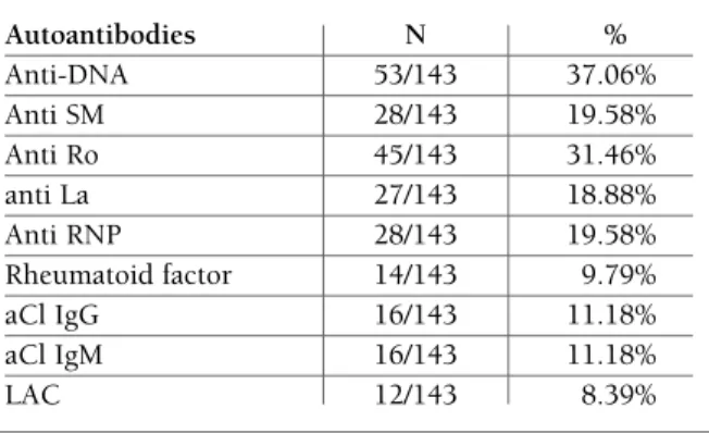
The profile of autoantibodies can be seen in Table I. Regarding treatment, 77/143 (53.8%) patients were on glucocorticoids (doses from 10 to 60 mg predniso-ne/day; median of 10 mg/day); 25/143 (17.8 %) on azathioprine; 29/143 (20.2%) on methotrexate; 13/143 (9.09%) on mofetil mycophenolate; 117/143 (81.8%) on antimalarials and 1/143 (0.69%) on cy-clophosphamide at time of data collection.
Erythrocyte sedimentation rate (ESR) varied from 1 to 115 mm with a median value of 18 mm; CRP values varied from 0.9 to 49.70 mg/L (median value 5 mg/L). The value obtained for SLEDAI varied from 0 to 18, with a median value of 2 and SLICC value varied from 0 to 14, with a median of 2.
Tests performed to verify liver integrity showed ALT values from 5 to 32 U/L (median 23 U/L); AST values
from 6 to 59 U/L (median 22 U/L) and alkaline phos-phatase from 38 to 123 (median 74 U/L). With respect to bilirubin results, they were: TB from 0.2 to 1.7 (mean 0.5), DB from 0 to 0.7 (mean 0.2) and IB from 0.1 to 1.3 (median 0.3).
b) studIes of the correlAtIon of bIlIrubIn VAlues WIth slIcc, sledAI And
InflAMMAtory ActIVIty tests:
Analyzing the correlation of bilirubin values with SLE-DAI, we observed that there was no correlation be tween this index with TB (p=0.17) and DB (p=0.47) (al -though) there was a negative correlation with IB (p=0.02, Spearman rho = -0.18, 95% CI -0.34 to -0.01). Examining the SLEDAI scores, we found that at time of data collection there were: 3/143 (2.09%) patients with convulsions (2.09%), 1/148 (0.69%) with peri -pheral vasculitis, 1/143 (0.69%) with cranial nerve in-jury; 15/143 (10.48%) with active arthritis; 54/143 (37.7%) with muco-cutaneous manifestations (skin rash, alopecia and ulcers); 9/143 (6.29%) with hema-tological manifestations (leucopenia and thrombocy-topenia); 17/143 (11.8%) with renal disease activity (increase in proteinuria, pyuria, hematuria and casts); 7/143 (4.8 %) with complement decrease and 21/143 (14.6%) with a 25% increase in the title of anti dsDNA. Comparing the levels of IB according to the clinical activity profile observed we obtained data shown in Table II.
tAble I. AutoAntIbody profIle In 143 pAtIents WIth lupus erytheMAtosus
Autoantibodies N % Anti-DNA 53/143 37.06% Anti SM 28/143 19.58% Anti Ro 45/143 31.46% anti La 27/143 18.88% Anti RNP 28/143 19.58% Rheumatoid factor 14/143 9.79% aCl IgG 16/143 11.18% aCl IgM 16/143 11.18% LAC 12/143 8.39%
tAble II. coMpArIson of IndIrect bIlIrubIn leVels AccordInG to the profIle of clInIcAl ActIVIty MeAsured by sledAI.
Clinical With Without
profile (*) activity (**) activity(**) P Arthritis 0.25 ± 0.20 0.33 ± 0.21 0.06 Muco-cutaneous 0.28 ± 0.17 0.35 ± 0.23 0.16 Hematological 0.23 ± 0.11 0.33 ± 0.21 0.23 activity Kidney 0.23 ± 0.11 0.33 ± 0.21 0.28 Decreased 0.24 ± 0.15 0.34 ± 0.22 0.01 complement Increased anti 0.24 ± 0.13 0.34 ± 0.221 0.02 ds DNA titer
(*)Other items were not studied due to the low prevalence in the sample.
(**)Activity was determinate according to criteria used by SLEDAI determination [12].
tivity and extent4. Others, like Yang et al3have found
a positive relationship of total and IB with CRP in pa-tients with SLE. We did not establish any association with CRP in our current work; only ESR maintained a negative correlation. Characteristically in SLE, unlike other rheumatic diseases, there is a relative failure of acute phase CRP response during active disease despite evident tissue inflammation20justifying the classical
observation that very high levels of CRP in a lupus pa-tient suggest associated infection21. According to
Ba-tuca et al22, CRP levels are elevated only in certain
ma-nifestations of disease activity such as pleuritis but not in other. None of our patients had serositis at the mo-ment of the study. In the same way, anti CRP antibo-dies have been found in lupus patients mainly in tho-se with renal activity19and may contribute to modify
the serum measurement of this inflammatory marker. Differences in the spectrum of clinical activity and le-vels of anti CRP auto antibodies may explain the dis-crepancy of results between our study and those of the previously mentioned work.
Moreover, in the present study it was not possible to relate any specific form of clinical lupus activity with IB levels.
Therapeutic use of IB infusions has been applied in animals17. In perfused rat heart, it reverses the effects
of ischemia in cardiac function23; intravenous
admi-nistration of bilirubin ameliorates pulmonary fibrosis induced by bleomycin24and injury to liver grafts in
rats is prevented by rinsing them with bilirubin25.
A limitation of our study is the fact that most of our patients had low values of SLEDAI (mean 2.0) not al-lowing us to observe changes in bilirubin levels that may happen with a very active disease. Furthermore, some forms of activity such as serositis are underre-presented in our sample. Taking into account that car-diovascular events are a leading cause of death in SLE patients26, it is very important to study the mechanisms
involved in the defense of oxidative stress generated by disease activity in order to better understand these complications.
In conclusion, we have shown that that the levels of IB in SLE patients negatively correlate with ESR. No as-sociation could be found with any specific manifesta-tion of disease activity.
correspondence to Thelma L Skare
Rua João Alencar Guimarães, 796 80310420 Curitiba PR – Brazil E-mail: [email protected] Studying the correlation of IB levels with ESR and
CRP we found a negative correlation for ESR (p=0.01; Spearman rho=-0.21; 95% CI=-0.37 to -0.046) but not for CRP (p=0.15).
In a multiple linear regression model where SLEDAI, joint activity data, reducing complement and increased anti dsDNA and ESR were placed, only the values of ESR remained significant (t=-2.81; B coeffi-cient =-0.019; p=0.005).
The SLICC values were not correlated with the TB (p=0.30), DB (p=0.12) or IB (p=0.31).
dIscussIon
The results of this study suggest that serum levels of IB correlate negatively with ESR. We speculate that this is probably due to a compensatory mechanism against oxidative stress resulting from inflammatory activity.
Bilirubins at physiological levels account for 10% of the total antioxidant capacity in adults14. Their
antioxidant capacity is higher than that of alpha tocophe -rol, ascorbic acid and catalase that have been recogni-zed for a long time as powerful antioxidants2,4. A study
in the Framingham cohort has examined the link be -tween serum bilirubin and coronary artery disease and found that higher bilirubin levels were associated with lower risk of myocardial infarction or other cardiovas-cular disease events15. It has also been demonstrated
that patients with Gilbert disease, a genetic disorder as-sociated with elevations of unconjugated bilirubins, have lower prevalence of ischemic heart disease than control population16. Smoking has also been
associa-ted with lower bilirubin levels and has attenuaassocia-ted its protective cardiovascular effect17. All these observations
points to the fact that bilirubins might may have a role in alleviating oxidative stress in the blood.
In the same way it is also believed that bilirubins play an immunoregulatory role18. It is also known that
they inhibit C1q complement activation by causing a pigment-protein interaction on C1q esterase18. As
mo-dification of DNA by ROS may turn it immunogenic, oxidative stress has been implicated in the appearan-ce of auto antibodies19. This hypothesis is supported by
the fact that anti-DNA antibodies have an increased reactivity against denatured DNA by ROS19. The
pro-tective action of IB might help decrease this immune stimulation.
Some authors have demonstrated that serum bili-rubin levels are negatively related to lupus disease
ac-references
1. Grimsrud PA, Xie H, Griffin TJ, Bernlor DA Oxidative Stress and Covalent Modification of Protein with Bioactive Aldehydes J Biol Chem 2008;283(:21837-21841.
2. Ozkan Y, Yardym Akaydyn S, Sepici A, Keskin E, Sepici V, Sim-sek B. Oxidative status in rheumatoid arthritis. Clin Rheuma-tol 2007; 26: 64-68
3. Yang Z, Liang Y, Li C, Xi W, Zhong R. Bilirubin levels in patients with systemic lupus erythematosus: increased or decreased. Rheumatol Int 2012; 32: 2423-2430.
4. Vitek L, Muchová L, Pesickova S, et al. Association of systemic lupus erythematosus with low a serum bilirubin levels. Scand J Rheumatol 2010; 39: 480-484.
5. Vitek L, Jhirsa M, Bodanova M, et al. Gilbert syndrome and is-chemic heart disease: a protective role of elevated bilirubin le-vels. Atherosclerosis 2002; 160:449-456.
6. Ilzecka J, Stelmasiak Z. Serum bilirubin concentration in pa-tients with amyotrophic lateral sclerosis. Clin Neurol Neurosurg 2003; 105:237-240.
7. Vitek L, Ostrow JD. Bilirubin Chemistry and Metabolism; Harmful and Protective Aspects Curr Pham Des 2009;15:2869--2883.
8. Reamer LH, Wannamethee G, Ebrahim S, Sharper AG. Serum bilirubin and risk of ischemic heart disease in middle aged Bri-tish men. Clin Chem 1995; 41:1504-1508.
9. Huang S-S, Huang P-H, Leu H-B, Wu T-C, Lin S-J, Chen J-W. Serum bilirubin predicts long term clinical outcomes in pa-tients with cardiac syndrome X. Heart 2010;96:1227-1232. 10. Fuhua P, Xuhui D, Zhiyang Z, et al. Antioxidant status of
bili-rubin and uric acid in patients with myasthenia gravis. Neu-roimmunomodulation 2012; 19:43-49.
11. Tutuncu ZN, Kalunian KC. The definition and Classification of systemic lupus erythematosus. In Wallace D, Hahn BH (eds) Dubois’ Lupus Erythematosus, 2007, Lippincot , Willians & Wilkins, Philadelphia, p.16-20.
12. Bombardier C, Gladman DD, Urowits MB, Caron D, Chan CH. Derivations of SLEDAI. A disease activity index for lupus pa-tients. Arthritis & Rheum 1992; 35: 630-640.
13. Stoll T, Seifert B, Isenberg DA. SLICC/ACR damage index is va-lid, and renal and pulmonary organ scores are predictors of se-vere outcome in patients with systemic lupus erythematosus. Br J Rheumatol 1996; 35:248-254.
14. Tell G, Gustincich S Redox State, Oxidative Stress, and Mole-cular Mechanisms of Protective and Toxic Effects of Bilirubin on Cells. Cur Pharm Des, 2009, 15, 2908-2914.
15. Djousse L, Rothman KJ, Cupples LA, Levy D, Ellison RC. Ef-fect of serum albumin and bilirubin on the risk of myocardial infarction (the Framingham Offspring Study). Am J Cardiol 2003; 91:485–488
16. Vitek L, Jirsa M, Brodanova M, et al. Gilbert syndrome and is-chemic heart disease: a protective effect of elevated bilirubin le-vels. Atherosclerosis 2002; 160: 449–456
17. Sedlak TW, Snyder S. Bilirubin benefits: Cellular protection by a biliverdin reductase antioxidant cycle. Pediatrics 2004; 113:1776-1782.
18. Basiglio CL, Arriaga SM, Pelusa F, Almar AM, Kapitulnik J, Mot-tino AD. Complement Activation and disease: protective effects of hyperbilirubianemia. Clin Sci 2010; 118:99-113. 19. Cooke MS, Mistry N, Wood, C, Herbert KE, Lunec J.
Immu-nogenicity of DNA damaged by reactive oxygen species- im-plication for anti- DNA antibodies in lupus. Free Radic Biol Med 1997; 22: 151-159.
20. Carvalho JF, Hanaoka B, Szyper-Kravitz M, Shoenfeld Y. C reac-tive Protein and its implication in systemic lupus erythemato-sus. Acta Rheum Port 2007;32:217-222.
21. Meyer O. Anti-CRP antibodies in systemic lupus erythemato-sus. Joint Bone Spine 2010; 77: 384–389
22. Batuca J, Delgado Alves J. C-reactive protein in systemic lupus erythematosus. Autoimmunity 2009; 42: 282-285.
23. Clark JE, Foresti R, Sarathchandra P, Kaur H, Green CJ, Mot-terlini R. Heme oxygenase-1-derived bilirubin ameliorates post ischemic myocardial dysfunction. Am J Physiol Heart Circ Phy-siol. 2000; 278: 643–651.
24. Wang HD, Yamaya M, Okinaga S, et al. Bilirubin ameliorates bleomycin-induced pulmonary fibrosis in rats. Am J Respir Crit Care Med. 2002; 165:406–411.
25. Kato Y, Shimazu M, Kondo M, et al. Bilirubin rinse: a simple protectant against the rat liver graft injury mimicking heme oxygenase-1 preconditioning. Hepatology 2003; 38: 364–73. 26. Pyrpasopoulous A, Chatzimichailidou S, Aslanidis S. Vascular disease in systemic lupus erythematosus. Autoimmune Dis 2012:876456. Epub 2012 Aug 22.