UNIVERSIDADE DE LISBOA
FACULDADE DE FARMÁCIA
ER Stress in Parkinson’s Disease
Miguel de Almeida Santos
Dissertação
Mestrado em Ciências Biofarmacêuticas
UNIVERSIDADE DE LISBOA
FACULDADE DE FARMÁCIA
ER Stress in Parkinson’s Disease
Miguel de Almeida Santos
Dissertação orientada por:
Professora Doutora Maria João Gama
Professora Doutora Elsa Rodrigues
Mestrado em Ciências Biofarmacêuticas
2015
i The studies presented in this thesis were performed within the Cellular Function and Therapeutic Targeting research group, at the Research Institute for Medicines (iMed.ULisboa), Faculty of Pharmacy, Universidade de Lisboa, under the supervision of Maria João Gama, Ph.D. and Elsa Rodrigues, Ph.D
ii
Publications
The studies included in this thesis were presented in the following publications:
Abstracts:
Oral presentations
Santos MA, Carvalho NA, Silva – Azevedo C, Castro – Caldas M, Rodrigues E, Gama
MJ. Characterization of the effects of MPTP insult on ER – stress response in C57/BL6 wild type and Gstp null mice brain. XXIX “Grupo de Estudo de Envelhecimento Cerebral e Demência” Meeting. 2015. Portugal.
Carvalho NA, Silva-Azevedo C, Santos MA, Rodrigues E, Castro-Caldas M, Gama MJ. “Unfolded Protein Response in Parkinson’s disease: a new neuroprotective role for Glutathione S-Transferase pi?” 40th FEBS Congress – The Biochemical Basis of Life. July 4-9, 2015 – Berlin, Germany
Selected as Speed-talk
Silva-Azevedo C, Carvalho AN, Santos MA, Nunes MJ, Rodrigues E, Castro-Caldas M, Gama MJ. “TUDCA modulates the ER stress response in the brain of C57BL/6 mice”. XLV SPF Meeting. Feb 4-6, 2015 - NOVA Medical School, Lisbon, Portugal
Poster communications
Santos MA, Carvalho NA, Nunes MJ, Castro – Caldas M, Rodrigues E, Gama MJ.
Endoplasmic Reticulum Stress Response in C57/BL6 wild type and Gstp null mice Brain under MPTP Oxidative Stress. 7th Post-Graduate iMed.ULisboa Students Meeting. 2015. Lisbon, Portugal
Neves Carvalho A, Silva-Azevedo C, A Santos M, Rodrigues E, Castro-Caldas M, Gama MJ. “Unfolded Protein Response in Parkinson’s disease: a new neuroprotective role for Glutathione S-Transferase pi?” 40th FEBS Congress – The Biochemical Basis of Life. July 4-9, 2015 – Berlin, Germany
iii M.J. NUNES, A.N. CARVALHO, C. SILVA-AZEVEDO, M.A. SANTOS, M. CASTRO-CALDAS, E. RODRIGUES, M.J. GAMA. “MPTP-Induced oxidative stress and NRF2-dependent regulation of GSTP gene promotor”. XIV Meeting of the Portuguese Society for Neurosciences 4-5-June 2015, Póvoa de Varzim, Portugal.
v
Table of Contents
Index of Figures ... vii
Resumo ... viii
Abstract ... x
List of abbreviations ... xii
I.INTRODUCTION AND OBJECTIVES ... 1
1. Parkinson’s Disease ... 1
1.1 Epidemiology and risk factors ... 3
1.2 Pathophysiology of PD ... 4
1.3 Diagnosis and treatment ... 5
1.4 Models of PD ... 7
1.4.1 Toxin-induced models of PD ... 7
1.4.2 Genetic models of PD ... 11
2. The Endoplasmic Reticulum and Oxidative Stress ... 13
2.1 Endoplasmic Reticulum Stress ... 14
2.2 The UPR ... 15 2.2.1 PERK signaling ... 17 2.2.2 IRE1α signaling ... 18 2.2.3 ATF6α signaling ... 19 2.3 ER Stress in PD ... 19 3. Gluthatione S-transferases ... 21 3.1 Glutathione S-transferase Pi ... 23 3.1.1GSTP in PD ... 25
4. TUDCA as a therapeutic approach ... 26
4.1 UDCA and TUDCA: endogenous function and therapeutic properties ... 26
4.2 TUDCA in neurodegenerative diseases ... 28
5. Objectives ... 31
II. MATHERIALS AND METHODS ... 32
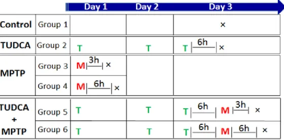
1. Animals and Treatment ... 33
2. Western Blot Analysis ... 35
3. Immunohistochemistry ... 36
vi
III RESULTS ... 38
1. The expressions levels of mediators of the UPR pathways are altered in GSTP KO mice ... 39
2. The expression levels of downstream effectors of the UPR pathways are altered in GSTP KO mice ... 44
3. Nrf2 expression levels are increased in wild type mice treated with MPTP... 47
IV DISCUSSION ... 50
Acknowledgements ... 58
vii
Index of Figures
Table 1 PARK-designated PD-related loci. 4
Table 2 List of primary antibodies used in the Western blot assays 36
Figure 1 Schematic representation of MPTP metabolism
and intracellular pathways affected by MPP+. 9
Figure 2 Schematic representation of the UPR 16
Figure 3 Detoxification scheme for glutathione conjugation 22
Figure 4 Schematic representation of C57BL/6 wild type and
GSTP null mice treatment course 34
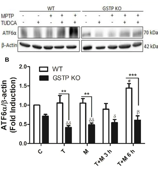
Figure 5 ATF6α expression levels in the brain cortex in response to
treatment with TUDCA, MPTP or TUDCA + MPTP 41
Figure 6 p-PERK expression levels in the brain cortex in response to
treatment with TUDCA, MPTP or TUDCA + MPTP 42
Figure 7 IRE1α expression levels in the brain cortex in response to
treatment with TUDCA, MPTP or TUDCA + MPTP 43
Figure 8 p-eIF2α expression levels in the brain cortex in response to
treatment with TUDCA, MPTP or TUDCA + MPTP 45
Figure 9 CHOP expression levels in the brain cortex in response to
treatment with TUDCA, MPTP or TUDCA + MPTP 46
Figure 10 Nrf2 expression levels in the brain cortex in response to
treatment with TUDCA, MPTP or TUDCA + MPTP 48
Figure 11 Nrf2 sub-cellular distribution in the brain cortex of C57/BL6 wild type mice in response to treatment with TUDCA, MPTP
viii Resumo
O desencadeamento da disfunção do metabolismo proteico nas formas esporádicas da doença de Parkinson (PD) poderá resultar do stress oxidativo mediado por espécies reactivas de oxigénio (ROS). O stress do reticulo endoplasmático (ER) pode levar à produção de ROS e ao desequilíbrio redox no ER mas a relação exacta entre o stress oxidativo e o stress do ER está pouco documentada. No entanto, sabe-se que a produção de ROS por neurotoxinas indutoras de parkinsonismo leva a uma rápida acumulação de proteínas oxidadas que por sua vez podem activar a unfolded protein response (UPR)
Um potencial mecanismo de defesa celular contra a toxicidade das ROS é a indução da expressão de enzimas de destoxificação de Fase II, nomeadamente a Glutationa S-Transferase Pi (GSTP), pelo factor de transcrição Nrf2. Já demonstrámos que a administração sub-aguda de 1-metil-4-fenil-1,2,3,6-tetrahidropiridina (MPTP) a ratinhos C57BL/6 induz a expressão da GSTP, e que a degeneração e morte de neurónios dopaminérgicos causadas pelo MPTP ocorre mais precocemente nos ratinhos knockout para a GSTP (GSTP KO) do que nos ratinhos wild type (wt). Para além disso, o nosso grupo também demonstrou que o ácido tauroursodeoxicólico (TUDCA), um ácido biliar endógeno com propriedades anti-apoptóticas e neuroprotectoras, diminui a morte celular em neurónios dopaminérgicos tratados com MPTP.
Neste estudo, avaliámos os níveis de expressão de marcadores de stress do ER no córtex de ratinhos C57/BL6 wt e ratinhos GSTP KO após o tratamento com MPTP. Em paralelo, investigámos também o papel do TUDCA na redução do stress do ER.
Os nossos resultados mostram que neste modelo de PD in vivo os ratinhos GSTP KO apresentam um decréscimo nos níveis de expressão de ATF6α e um aumento nos níveis de expressão de IRE1α quando comparados com os ratinho wt. Curiosamente, o efeito mais proeminente da deleção dos genes Gstp1/2 foi observado nas amostras de ratinhos tratados com MPTP, nas quais os níveis de expressão de Nrf2 se encontram reduzidos de forma significativa nos ratinhos GSTP KO quando comparadas com as amostras
ix
correspondentes de ratinhos wt. Verificámos também que o pré-tratamento com TUDCA modulou os níveis de expressão de diferentes mediadores da UPR após o insulto com MPTP.
Ainda que preliminares, os resultados aqui apresentados mostram que no encéfalo de ratinho, diferentes componentes da UPR apresentam diferentes susceptibilidades ao stress oxidativo induzido pelo MPTP. Essas diferenças estão relacionadas com o genótipo dos ratinhos (wt vs GSTP KO) o que indica que a GSTP possa desempenhar um papel na manutenção do equilíbrio redox no ER.
x Abstract
The trigger for dysfunctional protein metabolism, in sporadic Parkinson’s disease (PD), may be oxidative stress through damage caused by reactive oxygen species (ROS). The endoplasmic reticulum (ER) stress may trigger ROS production and redox imbalance in the ER but the precise interplay between oxidative stress and ER stress in neurons has been sparsely described. However, generation of ROS by PD triggering neurotoxins leads to a rapid accumulation of oxidized proteins that can activate the unfolded protein response (UPR).
One potential defence against the toxicity of ROS is the up-regulation of phase II detoxification enzymes, namely Glutathione S-Transferase Pi (GSTP), by the Nrf2 transcription factor. We have demonstrated that the sub-acute administration of 1-methyl-4-phenyl-1,2,3,6-tetrahydropyridine (MPTP) to C57BL/6 mice induced GSTP expression and that MPTP-induced dopaminergic neuronal degeneration is an earlier event when comparing GSTP null vs wild type (wt) mice. Furthermore, our group has also shown that tauroursodeoxycholic acid (TUDCA), an endogenous bile acid with anti-apoptotic and neuroprotective properties, rescued dopaminergic neurons from MPTP-induced damage.
In the present study, we evaluated the expression levels of ER stress markers in the cortex of C57/BL6 wt mice and GSTP null mice under MPTP-induced oxidative stress. In parallel, we investigated the role of TUDCA in reducing ER stress.
Our results show that in this in vivo model of PD GSTP null mice exhibit a decrease in ATF6α expression levels while exhibiting an increase in IRE1α expression levels when compared to the wild type. Interestingly, the most prominent effect of Gstp1/2 deletion was observed in the MPTP-treated samples, in which Nrf2 expression levels are significantly decreased in GSTP null mice when compared to their wt counterparts. We also observed that pre-treatment with TUDCA modulated the expression levels of the different UPR mediators following the MPTP insult.
xi
Although preliminary, the results present herein show that in the mice brain, different components of the UPR display different susceptibilities to MPTP-oxidative stress. These differences relay on the mice genotype (wt vs Gstp null) indicating that GSTP may have also a role in maintaining the ER redox balance.
xii
List of abbreviations
6-OHDA 6-hydroxydopamine
ARE Antioxidant-Responsive Element
ASK1 Apoptosis Signal-Regulating Kinase 1
ATP Adenosine Triphosphate
ATF4 Activating Transcription Factor 4
ATF6α Activating Transcription Factor 6α
BBB Blood Brain Barrier
bZIP Basic leucine zipper
CHOP C/EBP homologous protein
DA Dopamine
eIF2 Eukaryotic Translation Initiation Factor
ER Endoplasmic Reticulum
ERAD ER-Associated Protein Degradation
GRP78/BIP Glucose-Related Protein/Binding Immunoglobulin Protein
GSH Glutathione
GST Glutathione S-transferase
GSTP Glutathione S-transferase isoform Pi
GTP Guanosine Triphosphate
HD Huntington’s disease
IRE1α Inositol Requiring Enzyme 1α
JNK c-Jun N-terminal kinase
Keap1 Kelch-like ECH-associated protein 1
KO Knockout
MAPK Mitogen-Activated Protein Kinase
MAO-B Monoamine oxidase type B
MPDP+ 1-methyl-4-phenyl-2,3-dihydropyridinium
MPP+ 1-methyl-4phenylpiridinium
MPTP 1-methyl-4-phenyl-1,2,3,6-tetrahydropyridine
NADH Nicotinamide adenine dinucleotide
Nrf2 Nuclear factor (erythroid-derived 2)-like 2
PDP arkinson’s disease
PERK Double-stranded RNA-activated protein kinase-like endoplasmic reticulum kinase
xiii
RIDD Regulated IRE1-Dependent Decay
ROS Reactive Oxygen Species
RNase Ribonuclease
SN Substantia Nigra
TRAF2 Tumor necrosis factor Receptor-Associated Factor 2
TUDCA Tauroursodeoxycholic acid
UDCA Ursodeoxycholic acid
UPR Unfolded Protein Response
UPS Ubiquitin Proteasome System
XBP1 X-box binding protein 1
2 1. Parkinson’s Disease
PD is a chronic and progressive neurodegenerative disorder, characterized by a large number of motor and non-motor features (Jankovic, 2008). Firstly identified in 1817 by James Parkinson, it is the second most common neurodegenerative disease. Its pathophysiological hallmarks are loss or degeneration of dopaminergic neurons in the SN of the midbrain, resulting in a decrease of DA levels, and the development of neuronal LB, abnormal intracytoplasmic aggregates of protein that mostly contain α-synuclein, as well as ubiquitin and phosphorylated neurofilament proteins (Kim et al., 2014). DA is a neurotransmitter essential for normal movement, allowing information concerning motor control to be transmitted from the SN to the striatum, which then initiates and controls the ease and balance of movement (Segura-Aguilar et al., 2014).
Due to the decrease in DA levels, the symptomatology of PD is characterized by four major cardinal motor symptoms: tremor (e.g. hand tremor at rest), akinesia or bradykinesia (loss of spontaneous movements, facial expression), muscle rigidity and impaired balance and posture (stooping posture) (Hindle, 2010). Furthermore, LB can also occur in multiple areas of the central and peripheral autonomic nervous system, giving rise to a variety of symptoms in addition to the classical PD motor features (Sprenger and Poewe, 2013). There is a big spectrum of non-motor features that PD patients may suffer from, and that may reduce their quality of life, which are not only frequent but also often under-reported by patients and caregivers alike, remaining consequently under-treated (Maass and Reichmann, 2013). These include neurobehavioural disorders like depression, anxiety, apathy, hallucinations, cognitive impairment, impulse disorders (binge eating, pathological gambling) (Chaudhuri et al., 2011) and sleep disorders like difficulties with falling asleep, REM sleep behavior disorder and non-REM parasomnias (confusional wandering) (Maas and Reichmann, 2013). Also, PD patients report symptoms like constipation, genitourinary urgency, sensory pain and visual diplopia (Jellinger 2014).
3
Even though PD symptoms are well characterized, its ultimate causes are still unknown. Recent decades have witnessed a proliferation of medical pharmacological therapies and innovative surgical interventions like deep brain stimulation, but definitive disease modifying therapy is still lacking.
1.1. Epidemiology and risk factors
It is estimated that approximately 1-2% of the population over 65 years suffers from PD, with this figure increasing to 3% to 5% in people 85 years and older (Alves et al., 2008). Incidence rates of PD in population-based studies from Europe and the USA range from 8.6 to 19 per 100,000 inhabitants. PD can be sporadic, which comprises the majority of cases, or familial PD, in which patients with the disease report a positive family history. Indeed the highest risk factor for PD is considered to be family history, with monogenic forms, caused by single mutation in dominantly or recessively inherited gene, accounting for 30% of familial PD (Noyce et al., 2012). Other risk factors include pesticide exposure (Wilk et al., 2006) and aging.
There were identified 18 specific chromosomal regions, also called chromosomal locus, with a putative link to PD termed PARK (Table 1). However, the causative gene has not yet been identified for all of the loci, with some of them being identified by genetic linkage analysis in large families, and others discovered and established by genome wide association performed on a population level (Klein and Westenberger, 2012). Several genes have been found to be associated with PD (Table 1), namely SNCA, which encodes for α-synuclein, the main component of LB, LRRK2, a gene that encodes the cytoplasmic protein leucine-rich repeat kinase 2, PARK2, the second largest gene in the human genome that encodes for parkin, a protein that functions as an E3 ubiquitin ligase in the process of ubiquitination, PINK1, a phosphatase and tension homolog-induced putative kinase 1 that functions in a common pathway with parkin for sensing and selectively eliminating damaged mitochondria from the mitochondrial network, and DJ-1, a protein ubiquitously expressed that functions as a cellular sensor of oxidative stress (Klein and
4
Westenberger, 2012). Each gene has different effects on various aspects of the disease, such as the onset of the disease or the severity of the symptoms.
1.2 Pathophysiology of PD
As previously mentioned, PD is characterized by the degeneration of dopaminergic neurons in the SN of the midbrain, which results in a progressive nigrostriatal DA deficiency. It also features the presence of proteinaceous intracellular bodies containing aggregates of α-synuclein. This protein is considered to play a central role in the pathophysiology of PD, as its fibrillar form is known to be a major structural component of LBs in PD and other synucleinopathies (Kim et al., 2014). Accumulation of α-synuclein at the synapse leads to a loss of synaptic proteins and synaptic pruning with loss of
Table 1 – PARK-designated PD-related loci. Adapted from Klein and Westenberger,
5
connectivity. In vitro studies have shown that α-synuclein aggregates cause a series of secondary processes leading to neuroinflammation, neurodegeneration and cell death, providing compelling evidence that α-synuclein is involved in the pathogenesis of PD (Kim et al., 2014). However, it seems that the displacement of α-synuclein monomers from their physiological location in the cells may also contribute to neurodegeneration (Lashuel et al., 2013). Furthermore, it is also thought that oxidative damage may play a pertinent role in the aggregation of α-synuclein in PD due to a selective tyrosine nitration of this protein in lesions in PD and other synucleinopathies (Moore et al., 2005). The source of the increased oxidative stress is unclear but may include mitochondrial dysfunction, increased DA metabolism that can yield excess hydrogen peroxide and other ROS and impaired antioxidant defense pathways (Jenner 2003). Oxidative stress is considered to compromise the integrity of vulnerable neurons and thus to contribute to neuronal degeneration, and its role in PD will be latter discussed.
1.3 Diagnosis and treatment
Despite decades of research, the diagnosis and subsequent management of PD is hampered by suboptimal methods for detection and prognosis due to the lack of valid diagnostic biomarkers (tests or screening mechanisms) with high sensitivity and specificity that are critically needed for the correct identification of the disease. The definite and accurate diagnosis of PD can only be done through post-mortem neuropathological examination of brain tissue from PD patients. During lifetime, the diagnosis of PD relies on clinical criteria, based chiefly in the presence of parkinsonian symptoms. This proves to be a challenging task given the fact that the classic PD symptoms can be present in other neurodegenerative disorders. Careful history taking and astute physical assessment coupled with initial medical therapy are necessary to distinguish PD from other diseases or from secondary parkinsonism due to provoking drugs, toxins, infections and neurological damage (Beitz 2014).
6
Although there are no available disease-modifying therapies to alter the underlying neurodegenerative process, symptomatic therapies can improve the patients’ quality of life. These pharmacological approaches target chiefly the nigrostriatal dopaminergic pathway and attempt to replenish the DA content in the brain even though they do not prevent the progression of PD. L-DOPA remains as the gold standard because it can readily cross the BBB and is converted to DA through the
actions of the enzyme DOPA decarboxylase (Hornykiewicz, 2002). However, L-DOPA is also converted into DA in the peripheral nervous systems, causing undesirable adverse events such as nausea and vomiting. Given these effects, it is standard in clinical practice to co-administer L-DOPA with DOPA decarboxylase inhibitor such as carbidopa that does not cross the BBB, reducing the conversion of L-DOPA to dopamine in the peripheral tissue and therefore increasing L-DOPA delivery to the brain (Nagatsua and Sawadab, 2009). However, long-term L-DOPA treatment is associated with significant complications including involuntary movements, dyskinesias and response fluctuations (Tarazi et al., 2014).
Medication should be initiated when patients experience functional impairment from their symptoms. Initial therapy selection typically depends on patients’ specific symptoms and age. If motor symptoms are mild but require therapy, MAO-B inhibitors may be used before moving into more potent treatments such as L-DOPA. These inhibitors can increase synaptic dopamine by blocking its metabolization and are currently approved for the symptomatic treatment of early PD and to reduce off-time in patients with more advanced PD, having demonstrated a small symptomatic benefit and potential disease-modifying effects (Connolly and Lang, 2014). However, these agents have limited use in clinical management due to its low efficacy and possible drug interactions with medications that are contraindicated when taken with MAO inhibitors (Chen, 2011). Several promising new families of drugs are being evaluated for treatment of PD, including adenosine A2A receptor antagonists,
neurotrophic factors, glutamate antagonists and transdermal nicotine (Payami and Factor, 2014). However, none of these has been approved for general use
7
and often the reason is insufficient efficacy. In summary, current treatments for PD do not provide adequate neuroprotection and have limited efficacy, being incapable of slowing the progression of this disease and being associated with adverse motor and non-motor side effects. Accordingly, there is an urgent need to develop novel pharmacotherapies that are superior to the current ones, with improved efficacy, safety and long-term maintenance.
1.4 Models of PD
Currently, there is a plethora of animal models that mimic different aspects related to the pathology of PD. All of these experimental models can be categorized into two main groups: toxin-induced models and genetic models. None of the currently available models copy the phenotype of PD, mainly because they lack some specific neuropathological and/or behavioral feature of this disease, with each model having certain advantages and shortcomings. In addition to the classical motor abnormalities observed in PD, animal models are increasingly used to study non-motor symptoms (Campos et al., 2013). Both toxin-based and genetic models are suitable for studying these non-motor symptoms that are increasingly recognized as relevant in disease-state.
1.4.1. Toxin-induced models of PD
Toxin-based models of parkinsonism have been widely used and have yielded a wealth of insight into PD neuropathogenesis while also providing disease models in which to define putative pharmacological targets and to test potential therapies. These models have continued to evolve and have been used to study PD for more than half a century, starting with the 6-OHDA model of parkinsonism, introduced in the late 1950’s (Mendez and Finn, 1975). The structure of this selective catecholaminergic neurotoxin is very similar to that of DA, but an additional hydroxyl group on 6-OHDA renders it specifically toxic to dopaminergic neurons. Much of the biochemical, physiological and behavioral effects of nigral dopaminergic neuron loss and striatal DA depletion has been
8
collected from the 6-OHDA model of parkinsonism, with oxidative stress being widely regarded as the primary mechanism through which this molecule exerts its effects (Martinez and Greenamyre, 2012).
The molecule MPTP was first recognized as a neurotoxin when a group of intravenous heroin users developed an acute version of parkinsonism that was symptomatically indistinguishable from sporadic PD (Langston et al.,1983). MPTP was discovered to be a contaminant in the synthetic heroin that poisoned the drug users, and after it was recognized to function as a DA neurotoxicant, animal models were quickly developed, initially through repeated injections of MPTP in monkeys, providing the first effective non-human primate model of parkinsonism (Burns et al, 1983), and paving the way for the widespread use of MPTP as an in vivo neurotoxin. This molecule has been shown to be toxic in a large range of species and it is the tool of choice for investigations into the mechanisms involved in the death of dopaminergic neurons in PD, causing damage primarily to the nigrostriatal DA pathway with a profound loss of DA in the striatum and SN (Dauer and Przedborski, 2003). MPTP neurotoxicity arises from the formation of the toxic MPP+ metabolite, which decreases ATP production, inhibits mitochondrial enzymes and increases ROS production (Figure 1) (Nicklas et al, 1985). Several studies have demonstrated that JNK, a protein that belongs to a family of stress kinases subject to transient activation in response to ROS, heat or osmotic shock and growth factors or inflammatory cytokines (Davis, 2000), is a key mediator of MPTP/MPP+-induced neuronal apoptosis in animal models of PD (Nishi, 1997; Saporito et al., 2000). It has been shown that the SN of MPTP-treated mice presents increased levels of JNK and c-Jun (Silva et al., 2005; Castro-Caldas et al., 2012), supporting the idea that this kinase is somehow involved in the MPTP-induced degeneration of dopaminergic neurons. Also, it was shown that JNK null mice are resistant to MPTP neurotoxicity (Hunot et al, 2004) and selective inhibitors of JNK protect against the neurodegeneration in the nigrostriatal dopaminergic neurons caused by MPTP (Saporito et al., 1999; Wang et al., 2004). JNK activation is mediated through a sequential kinase cascade that results in the dual phosphorylation of the Thr-Pro-Tyr motif located in its activation loop (Davis, 2000), after which
9
JNK phosphorylates c-Jun, increasing its transcriptional activity and leading to the up-regulation of a number of genes involved in the control of cell survival and apoptosis (Kuan and Burke, 2005).
Among the toxic animal models of PD, rotenone represents one of the most recently used approaches (Betarbet et al., 2000). This substance is widely used around the world as an insecticide and pesticide, and it is a member of the rotenoids, a family of natural cytotoxic compounds extracted from various parts of Leguminosae plants (Hisata, 2002). Similarly to MPTP, rotenone is highly lipophilic and thus readily gains access to all organs including the brain. Furthermore, this neurotoxin also freely crosses all cellular membranes and can accumulate in subcellular organelles such as the mitochondria. After a single intravenous injection, rotenone reaches maximal concentration in the central nervous system within 15 minutes and decays to about half of this level in less
Figure 1 – Schematic representation of MPTP metabolism and intracellular pathways affected by MPP+. Following systemic injection, MPTP readily crosses the BBB and is metabolized through the action of MAO-B in glial cells, converting MPTP to MPDP+, which is then rapidly deprotonated (likely due to spontaneous oxidation) to the active neurotoxin MPP+ (Przedborski
et al, 2004). Afterwards, MPP+ is released into the extracellular space where it enters dopaminergic neurons through selective uptake by the DA transporter. Once inside the neuron, MPP+ enters the mitochondria through a mechanism dependent of mitochondrial transmembrane potential, where it binds to complex I, uncoupling the oxidation of NADH-linked substrates and consequently disrupting the flow of electrons along the electron transport chain. In Dauer and Przedborski (2003).
10
than 2 hours (Talpade et al., 2000). Once in the CNS, its brain distribution is heterogeneous, paralleling regional differences in oxidative metabolism.
Rotenone impairs oxidative phosphorylation by inhibiting NADH-ubiquinone reductase activity through its binding to the PSST subunit of the multipolypeptide enzyme complex I of the electron transport chain (Schuler and Casida, 2001). Beside its effects on mitochondrial respiration, rotenone also inhibits the formation of tubulin microtubules (Brinkley et al., 1974; Marshall and Himes, 1978), an effect relevant to the mechanism of dopaminergic neurodegeneration, as excess of tubulin monomers may be toxic to cells (Weinstein and Solomon, 1990). Rotenone has been used extensively as a prototypic mitochondrial poison in cell cultures, but less frequently in living animals. Behaviorally, rotenone-infused rats exhibit reduced mobility, flexed posture and in some cases rigidity and even catalepsy (Sherer et al., 2003).
The potent herbicide N,N’-dimethyl-4-4-4-bypiridinium, commonly known as paraquat, is another prototypic toxin known to exert deleterious effects through oxidative stress. Paraquat exhibits a striking structural similarity to MPP+ and its toxicity is mediated by redox cycling with cellular enzymes that present nicotinamide adenine dinucleotide phosphate-diaphorase activity, such as neuronal form of nitric oxide synthase, yielding ROS (Przedborski and Ischiropoulos, 2005). Although it poorly crosses the BBB spontaneously (Shimizu et al., 2001), significant damage to the brain is seen in individuals who died from paraquat intoxication and epidemiological studies have suggested an increased risk for PD due to paraquat exposure (Liou et al., 1997), but at this time, the data and clinical evidence are still inconclusive (Berry et al., 2010). Some investigators have reported reduced motor activity and dose-dependent loss of striatal dopaminergic nerve fibers and SN neuronal cell bodies in paraquat treated mice (Bové et al., 2005). However, this toxin model has shown some contradictory results, variable cell death and loss of striatal DA content (Miller, 2007) and the effect of this compound in the nigrostriatal dopaminergic system are somewhat ambiguous (Freira and Koifman, 2012).
11
1.4.2. Genetic models of PD
With the identification of PD-associated genes and given the fact that the chemical-induced animal models do not show all the classical phenotypes of PD, animal models based on PD patient gene mutations have been created as well. Currently there are many genetic animal models of PD due in different model organisms, including mice, Drosophila melanogaster and Caernorhabditis elegans. There are also a variety of cell models that have been used to glean insight into how mutations in the various PD-related genes lead to neuronal dysfunction. These genetic models may better simulate the mechanisms underlying the genetic forms of PD, even though their pathological and behavioral phenotypes are often quite different from the human condition. A number of cellular and molecular dysfunctions have been shown to result from these gene defects (Blesa and Przedborksi, 2014) and some studies have reported alterations in motor function and behavior in mice models (Hinkle et al., 2012; Hennis et al., 2013), as well as sensitivities to complex I toxins like MPTP that differ from those of wild type mice (Nieto et al., 2012; Dauer et al., 2002). However, studies evaluating the integrity of the nigrostriatal dopaminergic system in these genetic models failed to find significant loss of dopaminergic neurons (Goldberg et al., 2003; Hinkle et al., 2012), suggesting that the recapitulation of the genetic alterations in mice is insufficient to reproduce the final neuropathological feature of PD.
Mutations in the SNCA gene are known to cause a rare form of autosomal dominant PD, either through isolated point mutation or gene multiplication (Lee and Trojanowski, 2006). Numerous α-synuclein transgenic mice have been reported using a variety of promoters. It has been shown that these mice models may have motor dysfunction and filamentous inclusions that initiate neurodegeneration (Giasson et al., 2002), although none of them show robust and progressive nigrostriatal degeneration (Ted et al., 2010). The phenotypic outcome of the overexpression of α-synuclein in mice depends heavily on the promoter used. Of the many vertebrate models, only the mouse
12
prion promoter A53T synuclein transgenic mice exhibit the full range of α-synuclein pathology that is observed in humans, including progressive age-dependent neurodegeneration (Chesselet et al., 2008). Other transgenic models also exhibit gradations of α-synuclein aggregation, but they lack the characteristic α-synuclein fibrils that are present in humans with PD and related α-synucleinopathies (Chesselet, 2008; Dawson et al, 2010).
The most frequent genetic cause of autosomal dominant PD is mutations in LRKK2 (Zimprich et al., 2004). Several animal and cellular models of LRKK2 have been reported and they have provided important insight into how mutations in this gene may lead to neurodegeneration in PD. Mutations in LRKK2 that segregate with PD are concentrated in the GTPase and kinase domains but LRKK2 binding partners and regulators of kinase and GTPase activity have yet to be confirmed or clarified (Biskup and West, 2009). The majority of cases of LRKK2 related PD is characterized pathologically by the presence of α-synuclein inclusions, suggesting that these two proteins share common pathogenic mechanisms (Ross et al., 2006). In cellular models, overexpression of disease causing mutations of LRKK2 are toxic and toxicity is kinase and GTP-binding dependent (Smith et al., 2006; West et al., 2007). Knockout of LRRK2 in mice suggest that LRRK2 does not play a role in the development or maintenance of dopaminergic neurons (Andres-Mateos et al., 2009) and current transgenic mouse models are not very robust PD models. Most of these models have abnormalities in the nigrostriatal system such as stimulated DA neurotransmission or behavioral deficits, which are DA responsive (Dawson et al., 2010) but it is not yet clear why LRRK2 transgenic models do not exhibit more substantial pathology. The utility of the current models is likely to be focused on how mutations in LRRK2 lead to early dysfunction of the nigrostriatal dopaminergic system.
Regarding parkin, mutations in this gene are the most common genetic cause of early-onset PD (Marder et al., 2010). Knockout of this gene in Drosophila leads to mutant flies with reduced lifespan, male sterility and severe defects in both flight and climbing abilities, with decreased tyrosine hydroxylase levels observed in aged flies and specific degeneration of a subset of
13
dopaminergic neurons (Greene et al., 2003). However, none of the parkin KO mice have any substantial dopaminergic or behavioral abnormalities (Goldberg et al., 2005) and it has been shown that LB formation and neurodegeneration are parkin independent in a mouse model of α-synucleinopathy (von Coelln et al., 2006). Some of the parkin knockout mice have subtle abnormalities in the dopaminergic nigrostriatal circuit or the locus coeruleus noradrenergic system (von Coelln et al., 2004). Interestingly, overexpression of mutant human parkin in both Drosophila and mice lead to a progressive degeneration of dopaminergic neurons (Lu et al., 2009; Sang et al., 2007).
2. The Endoplasmic Reticulum and Oxidative Stress
It has been proposed that mitochondrial dysfunction and oxidative damage may play a critical role in both aging and neurodegenerative diseases (Beal, 2005). Mitochondrial oxidative phosphorylation is the primary source of high energy compounds in the cell and the dysfunction of mitochondrial metabolism leads to reduced ATP production, impaired calcium buffering and generation of ROS (Surmeier et al., 2011). In regards to PD, accumulating evidence indicates that oxidative stress contributes to the cascade of events leading to the degeneration of dopaminergic neurons in the SN. Postmortem brain analyses show increased levels of 4-hydroxyl-2-nonenal, a by-product of lipid peroxidation, carbonyl modifications of soluble proteins and DNA and RNA oxidation products (Dias et al., 2013). Also, the link between oxidative stress and dopaminergic neurons is further supported by modeling the motor aspects of PD in animals with toxins that cause oxidative stress such as mitochondrial inhibitors like MPTP, rotenone or paraquat. This production of ROS may be the trigger for a dysfunction in the protein metabolism in the ER seen in sporadic PD. The ER serves many general functions, including protein processing, folding and the transport of membrane and secretory proteins, as well as the degradation of misfolded protein aggregates, providing and maintaining an exclusive oxidized environment of millimolar concentrations of Ca2+ to facilitate disulfide bond formation (Calì et al., 2011). This process is believed to
14
contribute to 25% of ROS generated by the cell (Tu and Weissman, 2004). Chronic ER stress through endogenous or exogenous insults may further increase oxidative stress via protein overload, impaired redox homeostasis, and calcium released from the ER, which in turn can augment the production and accumulation of mitochondrial ROS, thereby also influencing vital mitochondrial functions (Malhotra and Kaufman, 2011).
2.1 Endoplasmic Reticulum Stress
The ER is involved in several metabolic processes such as gluconeogenesis and lipid synthesis, while also being the major intracellular calcium reservoir in the cell (Chaudhari et al., 2014). Newly synthesized membrane and secreted proteins enter the ER in an unfolded state, where the protein maturation steps required for a proper folding occur. Folding might involve post-translational modifications, such as glycosylation or disulfide bond formation, assisted by a vast number of chaperones and modifying enzymes that also contribute to membrane integration (Braakman and Hebert, 2013). The protein-folding machinery in the ER is particularly challenged in specialized secretory cells (such as pancreatic β-cells), given the fact that these cells have a high demand for protein synthesis, constituting a constant source of stress.
Despite all the biological mechanisms dedicated to protein folding, a significant portion of newly synthesized polypeptides entering the ER fails to acquire a native conformation (Ruggiano et al., 2014). These misfolded molecules are retained in the ER and eventually become substrates of the ERAD, a set of quality-control mechanisms that clears the ER from harmful misfolded proteins (Brodsky, 2012) and plays a key role in ER homeostasis. Genetic ablation of several ERAD components leads to embryonic lethality in mice (Yagishita et al., 2005; Francisco et al., 2010) and the inactivation of the ERAD has tremendous consequences, resulting in the build-up of misfolded proteins in the lumen and membrane of the ER, a phenomenon also known as ER stress. This occurs when the folding capacity of ER exceeds the capacity of ER lumen to facilitate the disposal of misfolded proteins. Consequently, the ER
15
elicits a protective or adaptive response which involves an intricate set of signaling pathways that will be activated to compensate damage and to restore the cell back to its normal state of homeostasis (Walter and Ron, 2011). These biological processes are collectively called the UPR.
2.2 The UPR
The UPR is a global stress network that integrates information about the protein-folding status in the ER lumen to the nucleus and cytosol to decrease the unfolded protein load, controlling decisions on cell fate through a variety of complementary mechanisms (Schroder and Kaufman, 2005) (Fig. 2). The primary function of the UPR signaling is promoting the cell survival under hostile conditions. When cells undergo irreversible ER stress, UPR is responsible for the elimination of damaged cells through apoptotic mechanisms, some of which appear to be specific to ER stress and others that are included in general apoptotic pathways (Xu et al., 2005). Even though the UPR is linked to protein-folding stress under normal and pathological conditions, recently it has been shown that some of its components can regulate various processes, from lipid and cholesterol metabolism, to inflammation and cell differentiation (Rutkowski and Hedge, 2010). These alternative UPR outputs are thought to derive from the complex crosstalk between different stress and metabolic pathways, showing that UPR components are part of distinct regulatory modules that orchestrate the fine-tuning of essential homeostatic processes (Hetz, 2012).
Two distinct phases of cellular responses are observed in vertebrate cells undergoing ER stress. First, an inhibition of general protein translation occurs coupled with a selective degradation of mRNA encoding for certain proteins located in the ER and a bulk degradation pathway termed ER-phagy, a process that eliminates damaged ER and abnormal protein aggregates through the lysosomal pathway (Hetz, 2012). This first wave of cellular responses reduces the protein influx into the ER to allow adaptive and repair mechanisms that reestablish homeostasis. A second wave of events triggers a massive gene-expression response through the regulation of three distinct ER proximal UPR
16
transmembrane proteins: IRE1α, ATF6α and PERK. Under normal physiological conditions, all three effectors are negatively regulated by the ER chaperone GRP78/BIP, which suppresses their activity by binding to their luminal ends (Bertolotti et al., 2000). Under conditions of ER stress and increase in unfolded proteins, GRP78 dissociates from the transmembrane proteins, releasing the inhibition and eliciting the response. Activation of the ER pathways helps to fight the cellular stress through the combined actions of suppressing the translation of new proteins, inducing ER chaperones that promote protein refolding and activating the proteasome to degrade misfolded/unfolded proteins.
Figure 2 – Schematic representation of the UPR. The UPR stress sensors IRE1α, PERK and
ATF6α transduce information about the folding status of the ER to the cytosol and nucleus to restore protein-folding capacity. Through its actions, the stress sensors mediate the attenuation of protein translation, the expression of protein-folding chaperones and many other genes involved in the UPR. In Hetz (2012).
17
2.2.1 PERK signaling
PERK is a type I ER transmembrane protein kinase with a luminal domain and a cytoplasmic domain that has kinase activity. Upon ER stress, BIP releases the luminal domain of PERK, which then dimerizes and autophosphorylates to become active. Following trans-autophosphorylation, this kinase phosphorylates the alpha subunit of eIF2, inactivating it by Ser-51 phosphorylation and attenuating protein translation. This inhibitory effect of translation helps to alleviate ER stress by decreasing the overload of misfolded proteins and thereby protecting the cells under conditions where proteins cannot achieve proper folding (Fels and Koumenis, 2006). Translation attenuation is followed by increased clearance of the accumulated proteins from the ER by ERAD and expression of pro-survival genes (Chakrabarti et al., 2012). Interestingly, eIF2α phosphorylation increases the translation of selective mRNAs that contain inhibitory upstream open reading frames within their 5’UTR that prevent translation in unstressed cells, such as the mRNA of ATF4 (Vattem and Wek, 2004). ATF4 is a member of the cAMP response-element-binding family of transcription factors and up-regulates a subset of UPR genes that function in resistance and control of oxidative stress, metabolism, protein folding and glutathione biosynthesis (Harding et al., 2000). Under severe ER stress conditions, ATF4 contributes to the induction of cell death through the control of the transcription of pro-apoptotic BCL-2 family members including p53 upregulated modulator of apoptosis and BIM, in addition to GADD34 and CHOP, a protein composed of an N-terminal transcriptional activation domain and a C-terminal bZIP that promotes apoptosis by the repression of Bcl2 expression and sensitization of cells to ER-stress inducing agents (Galehdar et al., 2010).
Furthermore, PERK can also phosphorylate Nrf2 (Cullinan et al., 2003), a member of a subfamily of bZIP transcription factors (Moi et al., 1994) that binds to a sequence known as the ARE, a response element found in many cytoprotective genes (Gao et al., 2014). In a stress-free cellular environment, Nrf2 is rapidly degraded by the proteasome. Degradation of Nrf2 is triggered by polyubiquitination through the cytoskeletal anchor Keap1/Cul3 ubiquitin ligase
18
that acts as a substrate adaptor to bring Nrf2 into the E3 complex (Kobayashi et al., 2004). In the presence of cellular stress, Nrf2 dissociates from Keap1 through the modification of reactive cysteine residues of Keap1 (Kobayashi et al., 2006) and is translocated to the nucleus, where it induces the expression of a wide variety of downstream target genes, that include several cytoprotective phase II detoxification and antioxidant enzymes and signaling proteins to regulate drug metabolism, antioxidant defense and oxidant signaling (Cullinan and Diehl, 2004). Through its regulation of oxidant levels and oxidant signaling, Nrf2 participates in the control of several functions such as autophagy, inflammasome signaling, apoptosis and UPR, exhibiting multiple protective effects against toxicity (Ma, 2013).
2.2.2. IRE1α signaling
IRE1α is a type I transmembrane protein ubiquitously expressed (Tirasophon et al., 1998) with dual enzymatic activities, consisting of an N-terminal ER luminal domain and a serine/threonine kinase domain plus a C-terminal RNase domain located in the cytosol (Lee et al., 2008). Upon accumulation of unfolded/misfolded proteins in the ER and under conditions identical to those that activate PERK (Hetz et al., 2011), IRE1α dimerizes and oligomerizes while stimulating trans-autophosphorylation which leads to the activation of the RNase domain (Korennykh et al., 2009). Afterwards, activated IRE1α excises a 26-nucleotide intron from the XBP1 mRNA through its RNase domain, causing a translational shift in the codon reading frame that generates a new COOH terminal end and leads to the expression of a more stable and active transcription factor, XBP1s, that will translocate to the nucleus and regulate a subset of UPR target genes related to folding, ER/Golgi biogenesis and ERAD (Jiang et al., 2015), which differ in different tissues or under different conditions of ER stress (Acosta-Alvear et al., 2007). Furthermore, IRE1α can also degrade ER-bound mRNAs through cleavage at stem-loop and non-stem-loop sites, a process known as RIDD. RIDD helps to reduce the number of
19
proteins that enter the ER, further alleviating ER stress (Hollien et al., 2009). In the event of persistent ER stress, activated IRE1α can also promote apoptotic pathways by activating ASK1 and JNK, through interaction with TRAF2 (Urano et al., 2000; Nishitoh et al., 2002).
2.2.3. ATF6α signaling
Another UPR pathway is mediated by ATF6α, a type II ER transmembrane protein that contains bZIP domains in its cytosolic region (Haze et al., 1999). ATF6α is synthesized as an inactive precursor, bound to the ER by a transmembrane segment. Under ER stress conditions, ATF6α precursor translocates to the Golgi where it is cleaved in a process termed regulated intramembrane proteolysis. Firstly, ATF6α is cleaved by a site 1 protease which removes most of its luminal domain, followed by the removal of an intramembrane region by a site 2 protease, releasing an active N-terminal 50 kDa cytosolic fragment (ATF6f) (Nakka et al., 2014). This fragment operates as a transcriptional factor by binding to the ER stress response element within the promoter region of target genes, which will then up-regulate the expression of many UPR genes related to ERAD (Shen and Prywes, 2005).
Both processing of ATF6 and IRE1α-mediated splicing of XBP1 mRNA are required for full activation of the UPR. Furthermore, it is thought that the ATF6 and IRE1α pathway merge through the regulation of XBP1 activity: ATF6 increases the amount of XBP1 mRNA whereas IRE1α removes the 26-nucleotide intron, increasing XBP1 transactivation potential. Together with XBP1s, ATF6f will increase the transcription of target genes that expand ER size and increase its protein folding capacity to promote cell survival (Lee et al., 2002).
2.3 ER Stress in PD
Some reports have revealed that the UPR is an early event in PD and the presence of ER stress in human tissue derived from PD patients has been reported (Hoozemans et al., 2012). Immunoreactivity for phosphorylated PERK
20
and eIF2α in dopaminergic neurons of the SN has been described in PD post-mortem tissue, and the neurons presenting activated PERK were also positive for α-synuclein inclusions (Hoozemans et al., 2007). Furthermore, it has been demonstrated that ER stress-responsive proteins such as homocysteine-induced endoplasmic reticulum protein are upregulated in the SN of PD patients and co-localize with LBs (Conn et al., 2004; Slodzinski et al., 2009). Cellular studies in yeast have shown that overexpression of wild type and mutant α-synuclein triggers chronic ER stress inducing cell death (Cooper et al., 2006) and reports in complementary model organisms demonstrated that the earliest defect following α-synuclein expression is a block in ER to Golgi vesicular trafficking (Smith et al., 2005; Gitler et al., 2008). The involvement of the UPR in PD has also been shown in cellular and in vivo models using 6-OHDA, MPTP and rotenone (Blesa and Przedbroski, 2014). Generation of ROS by these neurotoxins leads to a rapid accumulation of oxidized proteins that can activate the UPR. Moreover, neuronal cells treated with PD-triggering toxins present phosphorylation of IRE1α and PERK as well as induction of their downstream targets (Ryu et al., 2002). Experiments carried out using neuronal cultures from PERK KO mice revealed an increased sensitivity of these cells against treatment with 6-OHDA, suggesting that neurons lacking this protein were unable to properly activate the UPR and that an early UPR response may be neuroprotective for the dopaminergic neurons (Lindholm et al., 2006). Similarly, ATF6α deficient mice are more susceptible to neurotoxin-induced neurodegeneration at the SN (Egawa et al., 2010)
Further evidence for a role of the UPR in PD pathogenesis comes from a juvenile onset autosomal recessive form of PD that is caused by mutation of the parkin gene, which compromises the ubiquitin ligase function of the protein. This leads to the accumulation of cytotoxic fibrils and protein aggregates in the ER of SN neurons (Imai et al., 2001), that result in ER stress and consequently cell death. Expression of wt parkin, on the other hand, can restore proteasome function, which was shown to be impaired in SN neurons in PD (McNaught and Jenner, 2001), and it has a pro-survival activity against ER stress due to modulation of ERAD/proteasome pathway (Imai and Takashi 2004).
21
Although these reports suggest that ER stress occurs in affected SN neurons in PD brain and that it may trigger ROS production and redox deviation in the ER, general characterization of ER stress markers is still very poor and proximal signaling components (ATF6α, XBP1s, etc.) remain to be properly measured. The precise mechanisms of interplay between oxidative stress and ER stress in dopaminergic neurons have been sparsely described and whether ER stress plays a role in oxidative stress and anti-oxidant elicited neuronal response is currently unknown, as the mechanisms leading to ER stress in PD and the actual impact of the UPR on the degeneration cascade in the disease are just starting to be uncovered (Mercado et al. 2013).
3. Glutathione S-transferases
GSTs are a class of abundant proteins found in most tissues that function as xenobiotic metabolizing enzymes in eukaryotes. This supergene family of enzymes catalyses the conjugation of nucleophilic thiol-reduced GSH to a variety of electrophiles, forming water-soluble GSH conjugates that are readily transported out of the cell via membrane-bound efflux pumps (Board and Menon, 2013). They are an important cellular defense against numerous artificial and naturally occurring environmental agents (Strange et al., 2000) and GSTs’ substrates include polycyclic aromatic hydrocarbon epoxides derived from the catalytic actions of phase 1 cytochrome P450s as well as numerous by-products of oxidative stress (Strange et al., 2000). In addition to their role in detoxification, GST isozymes have other characteristics and functions such as the regulation of MAPKs and participation in steroid synthesis, tyrosine degradation and dehydroascorbate reduction (Tew and Townsend, 2012; Wu and Dong, 2012). Furthermore, the deletion of certain genes that encode for GST isoforms has been linked to an increased susceptibility to bladder, colon, skin and possibly lung cancer (Hayes and Pulford, 1995) and linked to a change in drug response (Layton et al., 1999; Roy et al., 2001).
GSTs are divided into two main families: cytosolic or soluble GSTs, and microsomal. Microsomal GSTs have been described as membrane-associated proteins and are structurally distinct from cytosolic GSTs, though they still
22
maintain an ability to catalyze the conjugation of GSH to electrophiles (Hayes et al., 2005). Cytosolic GSTs are expressed in all aerobic organisms (Ketterer, 2001) and a single GST unit consists of an N-terminal α/β-domain that operates as a GSH binding site and an all-α.helical domain for substrate binding that facilitates catalysis through proton abstraction from GSH in proximity to the GSH binding site (Dirr et al., 1994). Structural studies have shown that GSTs are typically monomeric but catalytically active as homo- or heterodimers composed of 25-30 kDa subunits (Hayes et al., 2005) and are divided into eight classes: alpha, kappa, mu, pi, sigma, theta, zeta and omega (Mannervik et al., 2005).
In the human brain, the active GSTs are composed of dimers containing alpha, mu or pi class GST monomers. GST mu is the most highly expressed isoform in the brain followed by GSTP and then GST alpha (Smeyne et al., 2013). Cellular localization studies have shown that GSTP and GST mu are expressed in neurons, astrocytes and oligodendrocytes, but it has been found that only GST pi is found in the dopaminergic neurons of the SN (Smeyne et al., 2007), a region that is particularly sensitive to oxidative stress due to the presence of endogenous dopamine, iron and neuromelanine. The distribution of GSTs in the brain appears to also be age dependent (Carder et al., 1990).
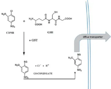
Figure 3 – Detoxification scheme for glutathione
conjugation. Scheme of detoxification of 1-chloro-2, 4-dinitrobenze through catalytic thioether formation. GST – Glutathione S-Transferase; GSH – Glutathione. Tew and Townsend, 2012 (adapted).
23 3.1. Glutathione S-transferase Pi
GSTP is one of the most extensively studied GSTs and has been implicated in the protection of cells from ROS-inducing agents due to its ability to alter levels of cellular glutathione in response to production of ROS, ameliorating the oxidative milieu (Tew and Ronai, 1999). Mainly found in the cytoplasm and widely distributed in a range of tissues (Suzuki et al., 1987), GSTP is induced by exposure to electrophiles. At the transcriptional level GSTP is regulated mainly by the GSTP enhancer 1 element which is recognized by Nrf2 (Suzuki et al., 2005). In contrast to the mouse that has two pi class GST genes (Bammler et al., 1994), humans have a single functional GSTP gene termed GSTP1 located in chromosome 11q13 (Board et al., 1989). Single nucleotide polymorphisms (SNPs) in the GSTP1 gene have been found and correlated with several malignancies such, as the development and progression of Hodgkin’s and non-Hodgkin’s lymphoma, and cancer drug response (Lourenço et al., 2009). These polymorphisms cause a steric change at the substrate binding site of the enzyme without affecting the GSH binding affinity (Ali-Osman et al., 1997).
GSTP has also been shown to protect cells from ROS by modulating S-glutathionylation of proteins following oxidative and nitrosative stress, a post-translational modification that consists of the addition of GSH to low pKa cysteine residues of target proteins (Tew 2007). Protein cysteines are evolutionarily highly conserved and sparingly used throughout the proteome, suggesting a tight control of cysteine-dependent biological functions. They have a wide-ranging reactivity towards oxidants, which depends on their pKa (Dalle-Donne et al., 2007; Rutkevich and Williams, 2012). It is currently believed that the type of cysteine oxidation controls the biological response as well as the fate of the oxidized protein (Finkel, 2011). Certain types of cysteine oxidation may occur preferentially in subcellular compartments where the redox environment may be facilitating these oxidations, such as the ER (Chakravarthi et al., 2006). S-glutathionylation generally occurs when a cysteine within the protein forms a disulfide bond with GS-,which can occur either in response to
24
endogenous oxidative or nitrosative stress mediated signaling events or from exposure to external environment drug treatments (Townsend, 2007). A wide range of chemicals can induce S-glutathionylation such as hydrogen peroxide, glutathione disulfide, diamide and various nitric oxid donors (Townsend, 2007; Townsend et al., 2008). S-glutathionylation has been associated with the stabilization and protection of proteins against irreversible oxidation of critical cysteine residues, and with the regulation of protein functions and transcription. These proteins include enzymes with catalytically important cysteines, particularly those involved in protein folding and stability, nitric oxide regulation and redox homeostasis, such as kinases, phosphatases, heat shock proteins, proteins involved in energy metabolism and transcription factors (Tew et al., 2012).
It has been shown that Keap1 is modified by S-glutathionylation (Zhang et al 2010). This protein has high cysteine content, making it an excellent candidate for this post-translational modification. Interestingly, GSTP itself is subject to S-glutathionylation, which reduces its enzymatic activity against chemical substrates and promotes its multimerization (Dalle-Donne et al 2007). Altered levels of S-glutathionylation in some proteins have been associated with numerous pathologies, many of which linked to stress within the ER (Janssen-Heininger et al 2013).
Besides being involved in cell redox homeostasis, GSTP also has a number of other functions such as catalyzing degradation of nitro-compounds (Lo Bello et al., 2001), and participating in reactions involving stress kinases (Adler et al., 1999). In regards to the latter, it has been shown that GSTP1 monomer can act as a ligand-binding protein controlling the catalytic activity of JNK (Adler et al., 1999; Castro-Caldas et al., 2012), an interaction that involves the C-terminal region of GSTP and of JNK (Monaco et al., 1999), preventing c-Jun phosphorylation and inhibiting the subsequent trigger of the cell death cascade. Under conditions of oxidative stress,
GSTP dissociates from JNK, which may then be phosphorylated and consequently phosphorylate its downstream substrates (Yin et al., 2000). It has also been shown that after systemic administration of MPTP, GSTP expression
25
is significantly increased in glial cells in the vicinity of dopaminergic neurons cell bodies and fibers (Castro-Caldas et al., 2009).
The creation of two GSTP KO strains has been reported, one involving mouse GSTP1 (GSTP1-1) and the second inactivating both genes (Henderson et al., 1998), and all studies reported so far involved the double KO mouse strain (GSTP1/2 null mice). These mice do not present striking or lethal phenotypes and have been mainly used to explore the role of GSTP in tumourigenesis (Board, 2007). The results suggest that the contribution of GSTP to tumourigenesis is multifaceted; GSTP1/2 null mice are more sensitive to a two-stage skin tumourigenesis model (Henderson et al., 1998) showing a detoxification role for GSTP.
Furthermore, it has been shown that GSTP KO mice are more susceptible to the neurotoxic effects of MPTP than their wild type counterparts. Administration of MPTP induced a demise of nigral dopaminergic neurons together with the degeneration of striatal fibers at an earlier timepoint in the GSTP KO mice when compared to the wild type counterparts. It has also been shown that in vivo GSTP can act as an endogenous regulator of the MPTP-induced cellular stress by controlling JNK activity through protein-protein interactions (Castro-Caldas et al., 2012). The various contributions of GSTP to carcinogenesis and other biological processes reflect the mounting evidence that some GSTs participate in cell signaling pathways that are independent of their drug and xenobiotic detoxification roles.
3.1.1. GSTP in PD
There have been multiple reports concerning a role for GSTP in PD, both in cellular and animal models. It was demonstrated that over-expression of wild-type GSTP1 before treatment with rotenone effectively protected Neuro2A cells by reducing oxidative stress, cell death, neurite loss and attenuating PERK activation and CHOP induction, all of which are important components of neurodegeneration in PD progression (Oakes and Papa, 2015; Shi et al., 2009), and GSTP1 mutants with low catalytic activity had a diminished effect, providing compelling evidence that GSTP’s biological functions effectively reduced
26
oxidative stress and the associated ER stress (Shi et al., 2009). Also, using primary cultured dopaminergic cells harvested from the SN of MPTP resistant mice (Hamre et al., 1999), Smeyne and collaborators have shown that by inhibiting GSTP there was an increase in the amount of MPP+-induced neuronal death (Smeyne et al., 2007). Administration of MPTP to mice lacking GSTP also showed altered protein ubiquitination and increased susceptibility to UPS damage and inactivation (Carvalho et al., 2012). Furthermore, reports have shown that by mutating the parkin gene in Drosophila and deleting the GSTS1 gene, the loss of dopaminergic neurons is enhanced and overexpression of GSTS1 ameliorates this neurodegeneration (Whitworth et al., 2005).
The analysis of the SN in the post-mortem brain of PD patients has revealed a substantial reduction of GSH levels and low activity of enzymes related to the de novo synthesis of GSH that seems to be specific to this pathology since changes in GSH were not detected in other neurodegenerative diseases (Di Monte et al., 1992; Sian et al., 1994; Pearce et al., 1997). Another study that compared ventricular cerebrospinal fluid from PD and normal control subjects, has shown differences in proteins expression in PD individuals, namely in GSTP1-1 (Maarouf et al., 2012). Epidemiological studies have shown that decreased GSTP expression is a significant risk factor in PD and that GSTP wild type allele is an individual protective genetic trait in idiopathic PD (Golbe et al., 2007). Furthermore, it has been found an association between the A313G polymorphism in GSTP1 and sporadic PD (Kelada et al., 2003; Vilar et al., 2007), suggesting that the decreased conjugation of some GSTP substrates may be relevant to the etiology of PD.
4. TUDCA as a therapeutic approach
4.1 UDCA and TUDCA: endogenous functions and therapeutic properties
Bile acids are detergent molecules synthesized in the liver from neutral sterols (Russell and Setchell, 1992). In most animals (including humans) bile acids are produced mainly from the cholesterol metabolic pathway, and