UNIVERSIDADE DO ALGARVE
FACULDADE DE CIÊNCIAS E TECNOLOGIA
T
T
H
H
E
E
R
R
OL
O
LE
E
O
OF
F
V
V
IT
I
TA
AM
M
IN
I
N
K
K
I
IN
N
O
OS
ST
TE
EO
OA
A
RT
R
TH
HR
R
IT
I
TI
IS
S
Carla Margarida da Silva Pereira
Dissertação para obtenção do Grau de Mestre em Ciências Farmacêuticas
Trabalho efetuado sob a orientação da Professora Doutora Dina Simes
2016UNIVERSIDADE DO ALGARVE
FACULDADE DE CIÊNCIAS E TECNOLOGIA
T
T
H
H
E
E
R
R
OL
O
LE
E
O
OF
F
V
V
IT
I
TA
AM
M
IN
I
N
K
K
I
IN
N
O
OS
ST
TE
EO
OA
A
RT
R
TH
HR
R
IT
I
TI
IS
S
Carla Margarida da Silva Pereira
Dissertação para obtenção do Grau de Mestre em Ciências Farmacêuticas
Trabalho efetuado sob a orientação da Professora Doutora Dina Simes
2016MICF Carla Margarida da Silva Pereira
T
T
H
H
E
E
R
R
O
O
L
L
E
E
O
O
F
F
V
V
I
I
T
T
A
A
M
M
I
I
N
N
K
K
I
I
N
N
O
O
S
S
T
T
E
E
O
O
A
A
R
R
T
T
H
H
R
R
I
I
T
T
I
I
S
S
Declaração de autoria de trabalho
Declaro ser a autora deste trabalho, que é original e inédito. Autores e trabalhos consultados estão devidamente citados no texto e constam da listagem de referências incluída.
(Carla Margarida da Silva Pereira)
Copyright © 2016 Carla Margarida da Silva Pereira.
A Universidade do Algarve tem o direito, perpétuo e sem limites geográficos, de arquivar e publicitar este trabalho através de exemplares impressos reproduzidos em papel ou de forma digital, ou por qualquer outro meio conhecido, ou que venha a ser inventado, de o divulgar através de repositórios científicos e de admitir a sua cópia e distribuição com objetivos educacionais ou de investigação, não comerciais, desde que seja dado crédito ao autor e editor.
MICF Carla Margarida da Silva Pereira
“O conhecimento torna a alma jovem e diminui a amargura da velhice. Colhe, pois, a sabedoria.Armazena suavidade para o amanhã.”
MICF Carla Margarida da Silva Pereira
Agradecimentos
Ao longo da elaboração desta monografia contei sempre com várias pessoas, a quem pretendo agradecer, por facilitarem este percurso e me ajudarem a atingir o objetivo.
Em primeiro lugar, agradeço imenso à Professora Doutora Dina Simes, que me orientou ao longo de toda esta etapa. Muito obrigada por toda a bibliografia cedida, pela sua amabilidade, solicitude, incentivo e por partilhar comigo algum do seu conhecimento, através de inúmeros conselhos e ensinamentos.
Quero agradecer à minha amiga (“irmã”) do coração, por toda a ternura e apoio constante, por escutar os meus desabafos e por me fortalecer ao acreditar tanto em mim. Obrigada por todos os bons momentos e sorrisos partilhados.
Agradeço aos meus patrões pela oportunidade concedida, pelo voto de confiança, por toda a compreensão e auxílio. Fico também muito grata às minhas queridas colegas da parafarmácia e da farmácia, por me acolherem tão bem, pela paciência, pela cooperação, pela partilha de experiências e por toda a amizade.
Um agradecimento muito especial ao meu companheiro, pelo bom humor, pela ajuda, pela compreensão, por me perdoar as múltiplas ausências, por me ouvir, por respeitar as minhas opções, por ser o meu porto de abrigo, pelo carinho e por todos os “mimos extra” sempre gentilmente enviados pela mãe.
Por fim, um eterno agradecimento à minha Estrelinha. Eu sei que estás orgulhosa e que continuas no céu sempre a olhar e a zelar por mim…
MICF Carla Margarida da Silva Pereira
Abbreviations
ACR - American College of Rheumatology
ADAMTS - A Disintegrin And Metalloproteinase with Thrombospondin motifs
AGEs - Advanced Glycation End-products
ANK - Ankylosis
AP - Alkaline Phosphatase
ASU - Avocado Soybean Unsaponifiables
BCP - Basic Calcium Phosphate
BMI - Body Mass Index
BM - Bone Mass
BMLs - Bone Marrow Lesions
BMPs - Bone Morphogenetic Proteins
BSP - Bone Sialoprotein
Ca - Calcium
CM - Chylomicrons
COMP - Cartilage Oligomeric Matrix Protein
COX - Cyclooxygenase
CPPD - Calcium Pyrophosphate Dehydrated
CR - Chylomicrons Remnants
CT - Computed Tomography
DMDs - Disease Modifying Drugs
DMOAD - Disease Modifying Osteoarthritic Drug
DRIs - Dietary Reference Intakes
ECM - Extracellular Matrix
EIA - Enzyme Immunoassay
ELISAs - Enzyme Linked Immunosorbent Assays
EMA - European Medicines Agency
EULAR - European League Against the Rheumatism
F IX - Factor IX
F VII - Factor VII
F X - Factor X
FDA - Food and Drug Administration
FGFs - Fibroblast Growth Factors
GGCX - Gamma Glutamyl Carboxylase
MICF Carla Margarida da Silva Pereira
Glu – Glutamate
GRP - Gla-Rich Protein
HDL - High Density Lipoprotein
IGF-1 - Insulin-like Growth Factor 1
IUPAC - International Union of Pure and Applied Chemistry
HA - Hyaluronic Acid
HIF - Hypoxia Inducible Factor
HPLC - High Performance Liquid Chromatography
Ihh - Indian hedgehog
ILs – Interleukins
JSN - Joint Space Narrowing
JSW - Joint Space With
KO - Vitamin K 2,3-epoxide
LC-MS - Liquid Chromatography-Mass Spectrometry
MAPK - Mitogen Activated Protein Kinases
MGP - Matrix Gla Protein
MMPs - Matrix Metalloproteinases
MRI - Magnetic Resonance Imaging
MVs - Matrix Vesicles
NF-kB - Nuclear Factor Kappa light chain enhancer of activated B cells
NIH - National Institutes of Health
NLRP - Nucleotide-binding oligomerization domain Like Receptors
NO - Nitric Oxide
NPP1 - Nucleotide Pyrophosphatase Phosphodiesterase 1
NSAID - Non Steroidal Anti-Inflammatory Drugs
OA - Osteoarthritis
OARSI - Osteoarthritis Research Society International
OC - Osteocalcin ON - Osteonectin OP – Osteoporosis OPN – Osteopontin PC - Protein C PGE2 - Prostaglandin E2 Pi - Inorganic Phosphate
MICF Carla Margarida da Silva Pereira
PPi - Inorganic Pyrophosphate
PRGP1 - Proline-Rich Gla Proteins-1
PRGP2 - Proline-Rich Gla Proteins-2
PS - Protein S
PTHrP - Parathyroid Hormone-related Peptide
PZ - Protein Z
RA - Rheumatoid Arthritis
RANKL - Receptor Activator of Nuclear factor κB Ligand
RIA - Radioimmunoassay
ROS - Reactive Oxygen Species
Runx2 - Runt-related transcription factor 2
SBCs - Subchondral Bone Cysts
SNRI - Serotonin and Norepinephrine Reuptake Inhibitor
Sox9 - SRY-type high-mobility-group box transcription factor-9
TG - Triglyceride
TGF-β - Transforming Growth Factor Beta
TIMPs - Tissue Inhibitors of Metalloproteinases
TMG3 - Transmembrane Gla Proteins-3
TMG4 - Transmembrane Gla Proteins-4
TNAP - Tissue Nonspecific Alkaline Phosphatase
TNF-α - Tumor Necrosis Factor Alpha
tPA - tissue Plaminogen Activator
TLRs - Toll-Like Receptors
UNO - United Nations Organization
US - Ultrasound
uPA - urokinase Plaminogen Activator
VEGF - Vascular Endothelium Growth Factor
VKAs - Vitamin K Antagonists
VKDP - Vitamin K Dependent Proteins
VKOR - Vitamin K epoxide Reductase
VKR - Vitamin K Reductase
VSMCs - Vascular Smooth Muscle Cells
MICF Carla Margarida da Silva Pereira i
Table of Contents
Index ... iIndex of Figures ... iii
Index of Tables ... iv Abstract ... v Resumo ... vi 1. Introduction ... 1 2. Osteoarthritis ... 4 2.1. Definition ... 4 2.2. Epidemiology ... 4 2.3. Risk Factors ... 5
2.3.1. Systemic Risk Factors ... 5
2.3.1.1. Non Modifiable Systemic Risk Factors ... 5
2.3.1.2. Modifiable Systemic Risk Factors... 6
2.3.2. Local Risk Factors ... 7
2.4. Physiopathology ... 9
2.4.1. Physiology of the Synovial Joints ... 9
2.4.2. Pathological Changes in the Synovial Joints Structures ... 10
2.4.2.1. Articular Cartilage ... 10
2.4.2.2. Synovium ... 13
2.4.2.3. Subchondral Bone ... 15
2.4.3. Pathological Calcification ... 19
2.4.4. Concluding Remarks in the Description of the Disease ... 25
2.5. Diagnosis ... 26
2.5.1. Imaging Analysis ... 27
2.5.1.1. Imaging Biomarkers ... 27
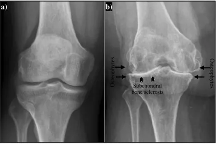
2.5.1.2. Radiography ... 27
2.5.1.3. Magnetic Resonance Imaging ... 29
2.5.1.4. Ultrasound ... 29 2.5.1.5. Computed Tomography ... 30 2.5.1.6. Nuclear Medicine ... 30 2.5.2. Laboratory Analysis ... 32 2.5.2.1. Biochemical Biomarkers ... 32 2.5.2.2. Assessment Methods ... 33 2.5.2.3. Crystals Examination... 33
MICF Carla Margarida da Silva Pereira ii 2.6. Pharmacological Therapies ... 34 2.6.1. Symptomatic Treatments ... 34 2.6.1.1. Oral Drugs ... 35 2.6.1.2. Topical Drugs ... 36 2.6.1.3. Injectable Drugs... 36
2.6.1.4. Slow-acting Symptomatic Drugs ... 37
2.6.2. Structure Modifying Treatments ... 38
2.6.2.1. Disease Modifying Osteoarthritis Drugs Targeting Cartilage ... 39
2.6.2.2. Disease Modifying Osteoarthritis Drugs Targeting Subchondral Bone ... 42
3. Vitamin K ... 44 3.1. Discovery ... 44 3.2. Characterization ... 44 3.3. Sources ... 46 3.3.1. Dietary ... 46 3.3.2. Non Dietary ... 47 3.4. Metabolic Processes... 48
3.4.1. Absorption and Distribution ... 48
3.4.2. Metabolism and Excretion ... 49
4. Vitamin K Dependent Proteins ... 50
4.1. Hemostatic Vitamin K Dependent Proteins ... 52
4.2. Vitamin K Dependent Proteins Associated with Calcification ... 52
5. The Role of Vitamin K and VKDPs in Osteoarthritis ... 55
6. Conclusion ... 58
MICF Carla Margarida da Silva Pereira iii
Index of Figures
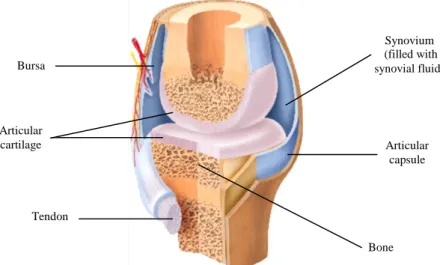
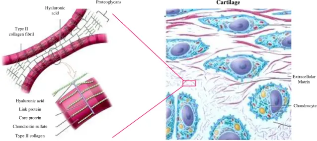
Figure 2.1- Structure of synovial joint ... 9Figure 2.2- Principal molecular components of the articular cartilage ... 11
Figure 2.3- Interaction between inflammation, angiogenesis and cartilage degradation in OA ... 14
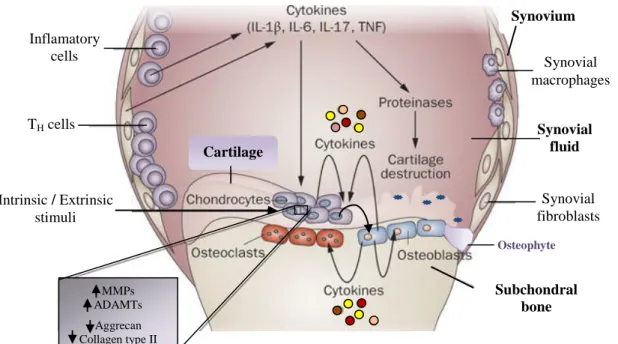
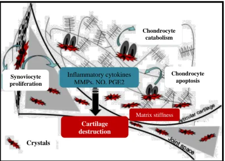
Figure 2.4- Joint intertwined pathways in the pathophysiology of OA ... 17
Figure 2.5- Changes across cartilage zones in OA pathological calcification ... 20
Figure 2.6- Schematic representation of crystal formation in articular cartilage ... 22
Figure 2.7- Model of microcrystal stress in OA joint ... 24
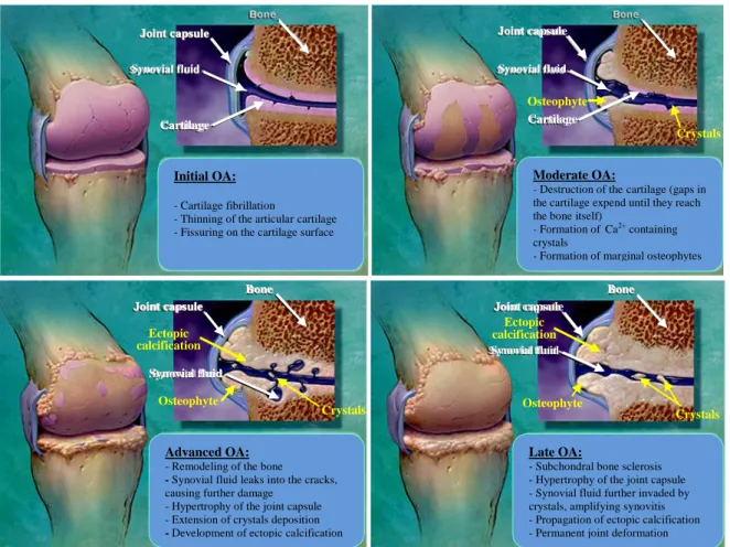
Figure 2.8- Progressive features along the OA illness ... 25
Figure 2.9- X-ray comparison of: a) normal knee; b) end-stage OA knee ... 28
Figure 3.1- Schematic representation of metabolism and excretion of vitamin K ... 49
Figure 4.1- Post-translational enzymatic modification of Vitamin K Dependent Proteins ... 51
Figure 4.2- Amino acid structure of mature human osteocalcin ... 53
Figure 4.3- Amino acid structure of mature human matrix Gla protein ... 53
MICF Carla Margarida da Silva Pereira iv
Index of Tables
Table 2.1- Principal proteolytic enzymes involved in cartilage tissues degradation ... 12Table 2.2- Principal non-collagenous proteins involved in cartilage mineralization regulation ... 22
Table 2.3- The ACR criteria for OA diagnosis of the hand, hip and knee ... 26
Table 2.4- Kellgren-Lawrence radiographic grading system for OA diagnosis ... 28
Table 2.5- Principal biochemical markers of OA ... 31
Table 2.6- Analytical methods applied in BCP detection ... 33
Table 2.7- Pharmacological symptomatic treatment options for OA management ... 34
Table 2.8- Prospective Disease Modifying Osteoarthritis Drugs (DMODs) under investigation ... 39
Table 3.1- Classification of the principal forms of vitamin K ... 45
Table 3.2- Phylloquinone content of selected common food ... 46
Table 3.3- Menaquinone content of selected common food ... 47
Table 3.4- Adequate intakes of vitamin K for the different age groups ... 47
MICF Carla Margarida da Silva Pereira v
Abstract
Osteoarthritis (OA) is the most frequent chronic rheumatic disease, affecting approximately 15% of the population, with a higher prevalence among the elderly; occurring in synovial joints such as the hips, knees and the ankle.
This condition develops when the joint organ homeostasis is affected, causing abnormal remodeling of the articular tissues, leading to degradation of the cartilage, thickening of the subchondral bone, formation of osteophytes and variable degrees of inflammation.
The burden of OA clinically characterized by chronic pain and significant disability is high, and only few nonpharmacologic and pharmacologic treatment options are available, mostly focused on providing symptomatic relief and showing limited efficacy and several side effects. The research on this disease in need for novel therapeutic alternatives has increased and lately is becoming fully recognized that joint’s calcification and the crosstalk with inflammation should be considered as an OA therapeutic target. In this context, vitamin K has been recognized as playing multifunctional roles that may modulate the pathogenesis of the disease.
Vitamin K acts as an essential coenzyme in the post-translational modification of specific glutamic acid residues (Glu) into γ-carboxyglutamic acid residues (Gla) in target proteins, known as vitamin K-dependent proteins (VKDPs), to make them biologically active. Mineral-related Gla proteins, have been proposed as regulators of cell differentiation and inhibitors of mineralization in articular systems, so impairment in their γ-carboxylation status should have an impact in joint’s health, showing a plausible rationale for the connection of vitamin K through the OA stages.
This old vitamin is now presented in a new perspective, with emerged value in human’s health, crucial in the prevention of pathological calcification and an important protective tool against inflammation and oxidative stress; revealing a promising potential as a prophylactic and therapeutic agent in OA.
Keywords: Osteoarthritis; Vitamin K; Gla Proteins; Pathological Calcification; Inflammation.
MICF Carla Margarida da Silva Pereira vi
Resumo
A osteoartrite (OA) é considerada a doença reumática crónica mais frequente, atingindo cerca de 15% da população e afetando pessoas de todas as idades, embora com uma maior predominância entre os idosos. A maioria dos indivíduos com idade superior a 65 anos apresenta evidências radiográficas e/ou clínicas desta patologia.
Esta doença representa um problema de saúde pública crescente, estimando-se que em 2020 seja a quarta principal causa de incapacidade motora. Apesar de não ser uma consequência inevitável do envelhecimento, mantém uma relação estreita e direta com a idade, em termos de incidência e prevalência. Assim, num futuro próximo, um aumento na esperança de vida conduzirá provavelmente a um crescimento do seu número de casos.
Estudos epidemiológicos mostram que esta é uma doença multifactorial muito complexa, dependente da associação de fatores genéticos e epigenéticos, sexo, etnia e idade, estando também associada com outras comorbidades, sedentarismo, lesões desportivas e fatores nutricionais.
Apresenta uma maior incidência de desenvolvimento em articulações que suportam o peso, tais como os joelhos, as ancas e os tornozelos, embora possa surgir em qualquer articulação sinovial, individualmente ou em simultâneo e com variáveis níveis de severidade.
A nível fisiológico, esta condição desenvolve-se quando a homeostasia da articulação é afetada, causando uma remodelação anormal dos tecidos articulares. As principais alterações patológicas comumente observadas em articulações atingidas por OA incluem a degradação irreversível da cartilagem articular, o espessamento do osso subcondral, formação de osteófitos, graus variáveis de inflamação, nomeadamente a nível da membrana sinovial e a presença de focos de mineralização ao nível da matriz extra celular.
O fardo da OA é elevado, sendo uma patologia caracterizada clinicamente por dor crónica, redução gradual do espaço interarticular e da mobilidade articular, podendo evoluir até causar uma incapacidade significativa. As opções de tratamento não farmacológico e farmacológico atualmente disponíveis são poucas e principalmente focadas em fornecer alívio sintomático, revelando uma eficácia limitada e apresentando vários efeitos colaterais.
A pesquisa sobre esta doença, com necessidade de novas terapias alternativas tem aumentado e, ultimamente, já se reconhece que a interrelação entre a calcificação patológica da articulação e a inflamação deve ser considerada um alvo terapêutico na OA. Por outro lado, o conhecimento sobre mecanismos moleculares da OA é ainda muito limitado e o seu
MICF Carla Margarida da Silva Pereira vii
diagnóstico tardio. Neste contexto e dada a falta de medicamentos eficazes para o tratamento da doença, é essencial a descoberta de novos alvos moleculares e biomarcadores que beneficiem a patologia. Neste âmbito, a vitamina K tem sido reconhecida como um alvo muito interessante a explorar pelo seu papel multifuncional que pode ser modulador da patogénese da doença e contribuir para o seu tratamento e prevenção.
A vitamina K corresponde a uma família que inclui uma série de compostos essenciais e lipossolúveis. O termo vitamina K é usado de forma genérica para os compostos estruturalmente relacionados, que possuem um núcleo 2-metil-1,4-naftoquinona comum, mas diferem na composição de uma cadeia lateral na posição 3. As três diferentes formas de vitaminas K são classificadas de acordo com a estrutura química dessa cadeia lateral: K1-
Filoquinona, K2-Menaquinonas e K3- Menadiona.
A fonte dietética predominante de vitamina K é a filoquinona, obtida a partir de plantas, sendo as suas maiores concentrações encontradas em vegetais de folhas verdes, óleos vegetais, frutas e grãos. Relativamente às menaquinonas, para além da sua presença no fígado de animais, podem ser também encontradas em alimentos fermentados por bactérias, que nas dietas ocidentais são tipicamente representados por queijos e no Japão pelo natto, que é um alimento feito de soja fermentada.
O papel metabólico da vitamina K é atuar como cofator numa reação de carboxilação específica que transforma determinados resíduos de glutamato (Glu) em gamacarboxiglutamato (Gla), tendo um papel essencial para a função de proteínas dependentes da vitamina K (VKDP). Este processo de carboxilação ocorre no retículo endoplasmático e é catalisado pela enzima gamaglutamil carboxilase (GGCX), utilizando como cofator ativo, a forma reduzida de vitamina K (KH2). Em simultâneo com a conversão
Glu, KH2 é oxidado a vitamina K 2,3-epóxido (KO), numa reação catalisada pela enzima
redutase VKOR e convertido de volta para KH2, num processo de reciclagem que é crucial
para a função da vitamina K e para manutenção de níveis adequados de biodisponibilidade desta vitamina no organismo. Esta capacidade de reciclagem pode explicar a necessidade diária muito baixa de vitamina K, quando comparado com outras vitaminas e cofatores.
Este processo de gamacarboxilação resulta na formação de proteínas Gla, biologicamente ativas, capazes de ligar o cálcio e minerais de fosfato de cálcio. No entanto, na ausência de vitamina K, ou da presença de inibidores de VKOR (fármacos anticoagulantes, como por exemplo, 4-hidroxicumarinas: varfarina), a carboxilação das proteínas VKD é
MICF Carla Margarida da Silva Pereira viii
incompleta e as proteínas são secretadas em várias formas menos funcionais, em todos os tecidos.
Durante as últimas décadas, a vitamina K inicialmente descoberta e associada a uma função hemostática, considerada como necessária para a correta função dos fatores de coagulação do sangue produzidos no fígado, para uma vitamina mais abrangente em termos funcionais, com a descoberta de outras VKDPs extra-hepáticas e associadas á calcificação como a osteocalcina (OC), a proteína Gla da matriz (MGP) e a proteína rica em Glas (GRP); Estas proteínas são caracterizadas por uma distribuição tecidual muito generalizada e um amplo impacto fisiológico com um papel em vários processos biológicos e fisiológicos, ao nível do osso, tecido vascular, pele e cartilagem.
Este grupo de VKDPs extra-hepáticas associadas á calcificação foi descrito como tendo um papel crucial na saúde, principalmente devido à sua função na regulação do cálcio e deposição e mineral nomeadamente patológicos de calcificação em tecidos moles.
Na última década, vários investigadores desenvolveram um conjunto de estudos genéticos e farmacológicos para adquirir mais informação sobre o papel das VKDPs extra-hepáticas, OC, MGP e GRP no processo de calcificação. Na verdade, OC e MGP são descritas como estando implicadas na regulação da calcificação endocondral. A MGP é considerada um poderoso inibidor da calcificação vascular. Mais recentemente, foi descrita uma associação direta das formas não funcionais da MGP e GRP á doença osteoartrítica.
A população em geral apresenta níveis de carboxilação baixos (10-40%) das proteínas VKDPs extra-hepáticas, pelo que, a atividade biológica destas proteínas em circulação pode ser considerada insuficiente. Esta baixa funcionalidade das VKDPs, não é essencial para a sobrevivência a curto prazo, o que sugere que a biodisponibilidade de vitamina K nos tecidos extra-hepáticos seja inferior ao necessário para assegurar a correta gamacarboxilação das proteínas. Em consequência da restrição da vitamina K, aumenta a vulnerabilidade a doenças associadas com o envelhecimento, com implicações importantes no osso e cartilagem, como é o caso da OA.
Uma vez que a calcificação e inflamação são processos comuns e bastante interligados em OA, a importância da vitamina K através da ação de OC, MGP e da GRP, abre novas perspetivas sobre o potencial da vitamina K como novo alvo terapêutico da OA.
Com o conhecimento emergente do seu valor na modulação da patogénese da OA esta vitamina é agora apresentada com uma nova perspetiva. A gama de ação da vitamina K provou ser crucial na prevenção da calcificação patológica, nomeadamente a nível vascular e
MICF Carla Margarida da Silva Pereira ix
da pele, assim como ser essencial na proteção contra a inflamação e o stress oxidativo. Globalmente a informação disponível permite concluir que esta vitamina é uma promissora candidata a ser um potencial agente profilático e terapêutico na OA.
Palavras-chave: Osteoartrite; Vitamina K; Proteínas Gla; Calcificação Patológica; Inflamação.
MICF Carla Margarida da Silva Pereira 1
1. Introduction
In our society, musculoskeletal conditions pose a huge burden on individuals, health systems and social care organisms; accounting for a significant economic impact. They represent the major cause of severe long-term pain and physical disability, affecting hundreds of millions of people in all continents, leading to more functional limitations in the adult population and reduction of the quality of life than any other series of disorders [1-3].
These conditions comprise a wide diversity in terms of pathophysiology, but share common anatomic features and are associated with pain and impaired physical function. They include a wide spectrum of situations, from acute onset and short duration, to lifelong disorders that include osteoarthritis (OA), rheumatoid arthritis (RA) and osteoporosis (OP) [1].
OA is the most frequent form of arthritis, affecting all the populations and ethnic groups investigated thus far [3-6].It should not be thought of as a single disease, but rather as the pathologic endpoint of a variety of events that conspire to incite a cascade of events within the whole joint, that perpetuate its progressive degeneration and eventual failure [7, 8].
The OA disease occurs when the dynamic steady state between destructive forces and repair mechanisms destabilises the joint organ homeostasis. This imbalance is thought to be the driving force for this irreversible and debilitating condition, which slowly progresses in a cyclic interaction between systemic and local factors. Over the years, this interplay between biological, structural and mechanical changes, compromises the articular cartilage and causes disturbances in the underlying bone, soft tissues of the joint and surrounding muscles [4-6, 9-14].
Commonly, OA develops in weight bearing joints such as the hips, knees and the ankle, but it can occur in any synovial joint of the body, individually or simultaneously, with variable degree intensities [9]. The disease is clinically characterized by joint pain, tenderness, crepitus, stiffness and limitation of movement with occasional effusion and unpredictable levels of local inflammation[13, 14].
From prehistoric times to the present day, OA has proven to be a challenging disease, found in nearly every period and civilization. The disease history is rich and ancient, since it can be traced back in time, with historical depictions and paleopathological findings in Neanderthal and Cro-Magnon skeletal remains and Egyptian mummies. The pathological changes observed in a 100 million year old bone and in a contemporary bone are remarkably similar, suggesting that OA has not changed much throughout evolution [15].
Despite the nearly ubiquitous presence of the pathology, OA was not recognized until the late 18th century, possibly due to its few obvious clinical signs [15].
MICF Carla Margarida da Silva Pereira 2
Further a nomenclature confusion delayed its identification, because OA and RA were for some time considered to be the same entity, known as arthritis deformans. Only in 1859, Sir Alfred Baring Garrod proposed a clinical distinction between these two diseases [15].
Historically the disease knowledge development and treatment innovation in OA has been considered slow, dependent upon the leisurely evolution of the understanding of the elaborate nature of joint tissue biology. OA is an extraordinarily intricate pathology with marked heterogeneity in onset, clinical presentation, rate of disease progression, pattern of joint involvement and synovial tissue structures affected [5].
Nowadays, OA is considered the most frequent chronic rheumatic disease. Several data indicate that affects people of all ages, with higher predominance among the elderly. A majority of individuals over the age of 65 have radiographic and/or clinical evidences of that condition [4, 5, 16, 17].Although not an inevitable consequence of aging, OA maintains a close relationship with age, its incidence and prevalence grows with it, thus a longer life expectancy will lead to a possible increase in the number of cases in a near future [1, 4, 17-20].
In recent decades, there was a clear process of aging and the period between 1975 and 2025 was considered by the United Nations Organization (UNO) as “The Era of the Aging“. In developing countries, this population aging is even more significant and accelerated. Due to the demographic changes OA is a growing public health problem across the globe, estimated to be the fourth leading cause of disability by the year 2020 [1-5, 20]. A rather worrying situation, aggravated by the fact that this is a condition with still no known disease modifying drugs (DMDs) available for treatment.
Some epidemiological studies tend to present different prevalence numbers for OA, according to geographical regions. Possible explanations for these differences range from genetic, socio-economic conditions, health-care access, environmental factors or different lifestyles. Indicating that predisposition and susceptibility, for this old disease that affects humankind, depends on the association of various risk factors [4, 22, 23].
Thanks to the increasing research and the use of molecular biology and proteomic tools, OA is being defined as a very complex, multifactorial disease. Although the disease can be dependent on genetic and epigenetic factors, sex, ethnicity and age, it is also associated with obesity, sedentary lifestyle, sport injuries and dietary factors [1, 2, 4, 19, 20, 24].
The requirements of this society andincreased costs of living, force more and more people to acquire wrong routines, reducing the time that they used to dedicate to themselves; as a consequence, people devote less time to sports activity and choose fast food instead of healthy meals [4].
MICF Carla Margarida da Silva Pereira 3
Our body is in an extremely controlled balance, where every little change may be responsible for several alterations. An equilibrated diet, including all required nutritional factors and moderate physical activity are necessary to maintain the body equilibrium and contribute to avoid pathological modifications in the joints [4].
Several studies have shown that insufficient intake of vitamin K, over long periods of time, is a risk factor for development of a wide range of diseases, including OA, OP, vascular calcification and cardiovascular disease, and even some types of cancer [18, 25-29].Some clinical studies have demonstrated that the nutritional intake and concentration of vitamin K in circulation decreases with age [30, 31].This suggests that the increase in vitamin K consumption can be made, either by using fortified foods or food supplements and might therefore represent a potential modifiable risk factor for several health disorders, including OA [25, 31].
Unfortunately, there are serious limitations for OA management, since it lacks specific and sensitive biomarkers for early diagnosis, prognosis and therapeutic monitoring [5, 8, 24]. Furthermore, there are few nonpharmacologic and pharmacologic treatment options, mostly focused on providing general symptomatic relief, although showing a quite limited efficacy and several side effects, prompting the need for novel therapeutic alternatives [18, 32-34].
Till date, there are no therapies approved by regulatory authorities to modify the onset or arrest the progression of OA structural damages. So, these limitations of pharmacological approaches, with no treatments available to prevent or halt the development of the illness, hasten the increased need of joint replacement surgeries. Consequently, there is an urgent need to identify novel prophylactic and therapeutic agents, which prove to be safe for clinical use and efficacy to prevent the initiation or slow the progression of OA [5, 34, 35].
In this context, vitamin K is a potential OA target and an interesting candidate for a disease modifying osteoarthritis drug (DMOAD), due to its multifunctional roles in health. Vitamin K has been described to have a determinant role in regulating bone and cartilage mineralization metabolisms, preventing soft tissue mineralization and further suggested as a protective agent against inflammation, suppressing the signaling of inflammatory mediators which have been reported to be involved in OA pathological pathways. More recently, a low plasma vitamin K has been suggested to be associated with higher prevalence and progression of knee OA features [18, 27, 29, 31, 36]. Altogether the information available strongly indicates that vitamin K should represent a promising tool that might help to alter the course of this pervasive disease.
MICF Carla Margarida da Silva Pereira 4
2. Osteoarthritis
2.1. Definition
The Osteoarthritis Research Society International (OARSI) Disease State Working Group defined OA as “progressive disease representing the failed repair of joint damage that, in the preponderance of cases, has been triggered by abnormal intra-articular cell stress”[37, 38].
Presently, there is a consensus that OA represents a heterogeneous and complex group of conditions difficult to define. The process involves all the tissues of the movable joints affected, starting first as a molecular disturbance, followed by anatomic, and/or physiologic derangements that can culminate in illness. This degenerative syndrome, complicated by inflammatory reactions, leads to eventual failure of one or more joints of the body, causing chronic pain and significant disability [4, 9, 39-41].
2.2. Epidemiology
OA is the most common form of arthritis, affecting every population and ethnic group investigated thus far. This chronic rheumatic pathology represents a huge cause of morbidity, activity limitation, physical disability and excess health care utilization, especially in people aged 45 years and above [1, 3, 42-46].
Worldwide estimates show that this musculoskeletal condition affects approximately 15% of the population, with an incidence of about 100.000 new cases per year [1, 9, 12, 44].In our country, general data presented by the Portuguese League Against Rheumatic Diseases revealed that around 6% of the Portuguese population has the disease [13].
Most of the OA burden is attributable to the involvement of the hip and the knee joints and women have higher rates than man, especially after the age of 50. The World Health Organization’s Scientific Group on Rheumatic Diseases estimates that 18.0% of women and 9.6% of men aged 60 or older suffer from these disorders [1, 46-49].
Due to demographic changes, the ageing of population and rising obesity, it is anticipated that the impact of OA will grow and become a major problem, in the near future; with its prevalence projected to double by the year 2020 [1, 5, 43, 47].
Depending on the geographical regions, the epidemiological studies tend to report different numbers for OA. In general, this condition is more prevalent in Europe and the UnitedStates of America than in other parts of the world [46]. Possible explanations for these variances range from genetics, socio-economic conditions, environmental characteristics or different lifestyles;indicating that predisposition and susceptibility to the development of OA depends on the association of several factors [4, 38, 48].
MICF Carla Margarida da Silva Pereira 5
A major goal of epidemiologic studies is to evaluate the disease risk factors. For OA, understanding whether and why certain individuals or groups are at high risk providesinsights into disease biology andleads to novel opportunitiesfor its prevention or treatment[41].
2.3. Risk Factors
OA is recognized as a multifactorial disorder that can be considered the product of a complex interplay between systemic and local factors [48-51].
2.3.1. Systemic Risk Factors
The systemic factors increase the individual predisposition to develop OA. This category of factors may act by raising the susceptibility of the joints to injury, by direct damage to joint tissues or by impairing the process of repair in damaged joint tissues [12, 48].
2.3.1.1. Non Modifiable Systemic Risk Factors
Some specific non modifiable systemic risk factors have been established.
OA in all its heterogeneous forms is, to a large extent, genetically determined. Evidences of the genetic influence come from a number of sources, including epidemiological studies of family history and family clustering, twin studies and exploration of rare genetic disorders related to OA. Taken together, these estimates suggest a heritability of 50% or more
[51, 52]
. The OA genetic architecture is similar to other complex polygenic diseases with contributions of several or even perhaps hundreds of genes, most of them affecting the occurrence in many joints, although there may be specific genes for specific sites, with some having small effects and a few having large effects [13, 51]. Overall findings indicate that half the variation in predisposition and susceptibility to OA in the population is explained by inherited components but, we need to keep in mind that, environmental and lifestyle factors are key modulators of gene expression [15, 48].
Many studies support the role of ethnicity in the development of OA based on variations among ethnic and racial groups [12, 15].The better characterized data of racial and ethnic differences in OA patterns of joint involvement come from large database studies, but the evidences are conflicting. The relative contributions of biological, lifestyle, and socioeconomic factors to ethnic differences in OA are still unclear [50, 51].
Even though aging is not sufficient for the development of OA, it is known as one of the strongest risk factor for the disease in all joints [15, 20]. Several studies and evidences confirm the high correlation between aging and OA, but the real mechanism behind this strongest predictor to the condition is still poorly understood [4, 43].
MICF Carla Margarida da Silva Pereira 6
The increased incidence and prevalence of OA on the elderly, probably, is a consequence of cumulative exposure to various risk factors and biologic changes that occur with aging and can turn a joint less able to cope with adversity [12, 20]. The basic cellular mechanisms that maintain tissue homeostasis decline with age, leading to an inadequate response to stress or injury, resulting in joint tissue destruction [12, 48].
Several epidemiologic studies of OA suggest the relevant difference between gender in the onset and development of this disease in males and females. Before the age of 50, males are reported to have a higher prevalence of OA, but after this age it is higher in females and with an enlarged severity [4, 15]. In addition, the definite increase in OA in women following menopause has led investigators to hypothesize that hormonal factors may play a role in the onset of OA. However, the results on effect of estrogen, either endogenous or exogenous, on OA from observational studies have been inconsistent. Gender disparities may also be caused by differences in anatomy, mechanical alignment, bone strength and neuromuscular strength, pregnancy, etc. [53, 54].
2.3.1.2. Modifiable Systemic Risk Factors
Numerous studies have shown a strong positive association between body mass index (BMI) and OA. Situations of overweight or obesity could precede the development and increase the progression of OA in weight-bearing joints, such as the hip and knee, as well as in non-weight-bearing joints, like the hand [55, 56]. Prospective studies indicate that obesity increases the relative risk of developing knee OA by two to tenfold [57, 58]. The pathogenesis of obesity-associated OA is not completely understood, but recent studies indicate that adipose tissue, and in particular infrapatellar fat, is a local source of pro-inflammatory mediators that are augmented with obesity and have been shown to increase cartilage degradation in cell and tissue culture models [13, 56]. Adipose tissue, once considered a passive storage portal of energy, is now recognized as a highly metabolic endocrine organ, with its adipocytes implicated in the secretion of active agents, including adipocytokines that play pleiotropic functions in bone, cartilage and others tissues of the joint formation [8, 56-58].
Emerging studies have also suggested clustering between OA and comorbidities, vascular health and cardiovascular risk factors, such as hypertension, hypercholesterolemia and diabetes [48, 49, 59]. It has been reported that approximately 55% of knee OA patients over 65 years old have hypertension and 13% diabetes type 2 [60]. The association of OA with diabetes is based on the suggestion that high glucose concentration produces reactive oxygen
MICF Carla Margarida da Silva Pereira 7
species (ROS) and advanced glycation end-products (AGEs), which are proteins or lipids that become glycated after exposure to sugars, accumulate in aged cartilage causing its degradation. In addition, long-term insulin therapy often needed to treat diabetes may overload tissues such as cartilage. Indeed, joint damage severity is higher in diabetic patients [4, 61].
The role of diet and nutrition in OA has been a recent area of research. There is evidence, from longitudinal studies, that low dietary intake and serum levels of vitamin D may be associated with the risk of knee and hip OA development. Without sufficient vitamin D, bones became thin, brittle or misshapen [15, 23].
Since antioxidants provide defense against tissue injury, high dietary intake of antioxidant vitamins could be postulated to protect against OA. For example, high vitamin C intake has been shown to reduce the progression of radiographic knee OA threefold as well as reducing the risk of developing knee pain [43, 51].
Several studies indicate that lack of adequate vitamin K may be a significant risk factor, for OA condition. Vitamin K has several potential effects that may modulate the development of OA. Some investigators show that a poor intake of vitamin K can result in abnormal bone and cartilage mineralization. Research demonstrated that high levels of vitamin K are associated with low OA prevalence ratios [18, 27, 29, 36]. Moreover, a recent study reported the beneficial effect of the assumption of extra virgin olive oil, rich in antioxidants such as vitamin A, E and K, in rats suffering from OA, underlining its possible application as preventive agent [62].
A few studies have identified high bone mass (BM) as a potential OA risk factor. Cross-sectional studies, in a variety of populations, have demonstrated associations between increased BM and radiographic OA in the large joints of the hip and knee. Longitudinal studies have also associated high BM with incident knee and hip OA [63, 64]. Like OA, OP is a common age-related skeletal disorder and the preponderant evidence suggests an inverse relationship between those two diseases [12, 51].
2.3.2. Local Risk Factors
Local factors are most commonly biomechanical affecting adversely the forces applied to the joint. Abnormal joint biomechanics, whether from trauma or other cause, may be the initiator of changes in the local joint environment that can, if ignored, result in OA development [12, 48].These factors have the potential for intervention and risk modification [50].
MICF Carla Margarida da Silva Pereira 8
Numerous cases prove that acute injury to the structures of the joint increase the risk for OA development and musculoskeletal symptomatology; especially in the knees, but nearly all joints can be affected. It is reported that knee injuryistheleadingmodifiableriskfactorin menandthesecondinwomen [48, 49]. Individuals with a history of joint injury are three to six times more likely to develop OA and are diagnosed approximately ten years earlier than individuals without any joint trauma. It is concerning that certain types of injuries may be associated with a rapid cascade towards joint failure in less than one year [44, 65].
The professional occupation is another appointed risk factor for OA onset. It appears that people who are exposed to certain physically demanding activities in their jobs, which involve repetitive use of joints during their tasks, are at increased risk of developing localized
OA [47, 51]. Workers whose occupations demand squatting or kneeling have twice the risk for
knee OA, prolonged standing and lifting might cause hip OA and work that require increased manual dexterity have been associated with features of hand OA [20, 44].
Participation in certain competitive sports, such that done by elite level athletes, that demand high intensity, torsion, direct impact as a result of contact with others participants, playing surfaces or equipment, increases OA risk too [50, 51].
The relationship between muscle strength and OA is complex and may vary, depending on specific muscles and joints examined. Recent reviews conclude that muscle weakness and atrophy may predispose for knee OA onset and progression. Sarcopenia is common in patients with knee OA and can occur as a consequence of OA related to disuse due to pain avoidance [23, 48]. Greater muscle strength is not, however, always protective as it corresponds to higher forces and thus increased joint loading during activity [12, 48].
A concerning factor is joint alignment, because any shift from a neutral or collinear alignment of the hip, knee and ankle will alter load distribution across the articular surfaces of the knees. A systematic review confirmed that knee malalignment is one of the strongest predictors of knee OA progression. Abnormal increases in compartmental loading are thought to increase stress on the articular cartilage, and other joint structures, subsequently leading to degenerative change. Medial progression of knee OA was four times more likely in individuals with varus deformity, whilst lateral progression was five times more likely in individuals with valgus alignment [12, 44].
Relative importance of the risk factors may vary in the different joints and according to the stages of the disease. The multifactorial etiology, with such a different set of intrinsic and extrinsic factors, turns it difficult to make a distinction between single and clustered factors. Each aspect is thought to be a risk not just because of one of the abovementioned reasons, but as a combination of them, presenting a synergistic or cumulative effect [13].
MICF Carla Margarida da Silva Pereira 9
2.4. Physiopathology
2.4.1. Physiology of the Synovial Joints
Humans have the capacity to maintain shape and produce a great variety of postures and movements that require that the musculoskeletal system of the body both generate and respond to forces that produce and control movement at the body’s joints. These joints, or articulations, are the points where two or more adjacent bones of the skeleton come together to form a connection and articulate with each other [66, 67].
The three major kinds of joints can be classified structurally as: fibrous, cartilaginous, and synovial. In this classification scheme, joints are categorized according to the major connective tissue type that covers the surfaces of the adjacent bones and whether they are strongly anchored or not to each other [66, 67].
In the human body, synovial joints are the most common. They are formed by articular cartilage of dense irregular connective tissue (2-5 mm thick) and present a joint cavity, surrounded by walls of articular capsule, filled with a lubricating fluid. These joints fundamental structural characteristics (figure 2.1) allow the bones to move smoothly, providing great mobility. However, since the bones are free to move in relation to each other, being only held together by ligaments, the synovial joints are less stable and more vulnerable.
Figure 2.1- Structure of synovial joint (Adapted from reference 67).
These diarthrodial joints are quite important for normal human body functions and autonomy. Fortunately, they are self maintaining, but damage, disorder or disease of a synovial joint, like OA, can lead to huge difficulty in movement and chronic pain, hampering the quality of life.
Bursa Bone Articular capsule Synovium (filled with synovial fluid) Articular cartilage Tendon
MICF Carla Margarida da Silva Pereira 10
2.4.2. Pathological Changes in the Synovial Joints Structures
Findings of detrimental pathologic changes in all of the synovial joint tissues lead to consider OA as a disease of the whole joint as an organ, being difficult to understand when and where the changes begin [6, 68-72].
OA is not a uniform disease, but a complex network of processes that occur when the dynamic steady state between destructive and repair mechanisms destabilises the joint homeostasis, adversely affecting the multiple articular components, including cartilage, subchondral bone, synovium, ligaments and neuromuscular support structures [68-71, 73, 74].
In the long-term, is reasonable to conceive that the pathologic bioreactivity of the individual tissues and the crosstalk between them likely contribute to the disease perpetuation, underlining that the pathological pathways are, in many respects, intertwined [70, 74-76].
2.4.2.1. Articular Cartilage
Adult articular cartilage is a hyaline cartilage, avascular, aliphatic and aneural structure subjected to a harsh biomechanical environment. This highly specialized tissue is designed for efficient gliding motion, with a limited capacity for intrinsic repair so, in this regard, its preservation is vital for the synovial joints health [77, 78].
The resilience, integrity and function of articular cartilage are highly dependent on the specialized mesenchymal cells, the chondrocytes, the sole cellular component found in this tissue, responsible for the production and maintenance of its surrounding extracellular matrix (ECM) infrastructure [79]. This abundant ECM, that comprises 95% or more of the cartilage volume, is composed of a big amount of water with dissolved inorganic ions (calcium, sodium, potassium) and organic macromolecules, mainly collagens, proteoglycans and small amounts of other noncollagenous proteins, glycoproteins and lipids [77-80].
Normal structure of the articular cartilage ECM is made of highly crosslinked fibrils, mostly of type II collagen molecules and, in a minor proportion, of type I, III, VI, IX, X, XI, XII and XIV; intertwined with two major classes of proteoglycans (figure 2.2), large monomers or aggrecans and small proteoglycans including decorin, biglycan, fibromodulin and lumican [74, 77-81].
The aggrecan is composed of a core protein with covalently attached glycosaminoglycans side chains of chondroitin and keratin sulfates that interact with hyaluronic acid (HA) (figure 2.2), which creates an hydrophilic environment, providing cartilage osmotic properties that are crucial to give tensile strength and withstand compressive forces [77-79].
MICF Carla Margarida da Silva Pereira 11
Figure 2.2- Principal molecular components of the articular cartilage (Adapted from reference 77).
This specific organization of articular cartilage results from the joints embryogenesis, in a process called endochondral ossification, comprising four steps: chondrogenesis from mesenchymal condensations, chondrocyte differentiation and hypertrophy, mineralization of the matrix with invasion of bone cells and the definitive formation of bone [74].
Histologically, cartilage tissues can be divided into a thick nonmineralized layer composed by superficial, transitional and radial zones, separated by a tidemark to a thin calcified deeper area. All of them characterized by a different phenotypic and gene expression patternsoftheresident chondrocytes,as well as distinctECM compositionand organization[80]. In healthy adult cartilage in a non-stressed steady state, the chondrocytes are quiescent cells and the articular cartilage equilibrium is regulated by a complex interplay between the anabolic growth factors, including insulin-like growth factor 1 (IGF-1), transforming growth factor beta (TGF-β), bone morphogenetic proteins (BMPs) and fibroblast growth factors (FGFs) and the catabolic proinflamatory factors, particularly some interleukins (ILs), tumor necrosis factor alpha (TNF-α), prostaglandin E2 (PGE2) and nitric oxide (NO) [74].
Much work has been devoted to the knowledge of how the homeostatic balance of the cartilage is perturbed at molecular level and how this leads to disease. Changes in chondrocytes activity are pivotal for OA development, through mechanisms that are not completely understood. It is highly feasible that normal cartilage, in spite of the low cellular activity and matrix turnover, possesses a strong metabolic potential. Activation of chondrocytes is driven by a cascade of signals, particularly by nuclear factor kappa light chain enhancer of activated B cells (NF-kB) and mitogen activated protein kinases (MAPK) pathways, which are critical in cell survival, differentiation and chondrogenesis [6, 72, 81, 82].
Type II collagen fibril Hyaluronic acid Hyaluronic acid Link protein Core protein Chondroitin sulfate Type II collagen Cartilage Extracellular Matrix Chondrocyte
MICF Carla Margarida da Silva Pereira 12
Disruption of the chondrocytes resting state may be viewed as an active response, with the recapitulation of developmental programs of the matrix substitution. A disturbed cell-matrix relationship lies at the centre of OA pathogenesis, promoted by chondrocytes phenotypic shift, rising cell proliferation and production of diverse cytokines, chemokines and matrix proteinases, either as initiating or as feedback amplification events [6, 72, 81, 82].
Notably, there appear to be significant differences between the individual zones of OA cartilage. In the upper zones, expression of collagen type II is significantly suppressed while collagen type III, fibronectin and cartilage oligomeric matrix protein (COMP) are upregulated during the progression of matrix destruction. Thus, chondrocytes produce a different kind of matrix structure, which is more susceptible to erosion. In contrast, the middle and deeper zones exhibit normal metabolism producing predominantly cartilaginous molecules [10, 74].
The involvement of several proteolytic enzymes is well established in the degradation of cartilage proteoglycans and collagens (table 2.1). Members of both matrix metalloproteinases (MMPs) and a disintegrin and metalloproteinase with thrombospondin motifs (ADAMTS) families are important mediators of proteoglycan degradation, while collagen catabolism is majorly attributed to the action of MMPs [14, 83, 84]. Initial OA changes may be due to MMP-3 and ADAMTS-5 that degrade aggrecan, followed by MMP-13, which is highly efficient in degradation of type II collagen. At some point, the biosynthetic anabolism is unable to keep pace with the catabolic activity and, once the collagen network is broken, the process is irreversible [6, 72, 83-85].
Table 2.1- Principal proteolytic enzymes involved in cartilage tissues degradation [14, 79, 83, 84].
Metalloproteinases Aggrecanases Other Proteinases
Collagenases: MMP-1, MMP-8, MMP-13 ADAMTS-4 Cathepsins K,B,D,G,L
Stromelysins: MMP-3, MMP-10, MMP-11 ADAMTS-5 Tissue Plaminogen activator (tPA) Gelatinases: MMP-2, MMP-9 Urokinase Plaminogen activator (uPA)
In OA advanced stage, the cartilage becomes hypocellular, often accompanied by lacunar emptying, which has been considered as evidence that chondrocyte death is a feature in OA progression. Most eukaryotic cells attach to neighboring cells or to the surrounding matrix for survival, a phenomenon called anchorage dependence. Indeed, chondrocyte survival is mediated by integrins that are responsible for the connection of ECM components to various intracellular cytoskeletal proteins. So, it is plausible that chondrocyte anchorage to the ECM is disturbed to a significant degree that leads to cellular death [4, 86].
MICF Carla Margarida da Silva Pereira 13
Additionally, OA cartilage produces endogenously NO and ROS, causing oxidative stress and mitochondrial dysfunction [10, 87]. The excess production of NO in OA tissues has been linked with chondrocyte apoptosis both in vitro and in vivo. The activation of the caspase cascade seems to play an essential role, along with another possible mechanism that has recently been identified. In the superficial layer where most of the apoptotic cells are located, a subpopulation of OA chondrocytes expresses the Fas antigen, which upon ligand binding could induce cell apoptosis [86, 88].
However, the relative contribution of chondrocyte apoptosis in the OA pathogenesis is difficult to ascertain. It has been suggested that matrix loss and chondrocyte death may form a vicious cycle with chondrocyte apoptosis being an inducer of cartilage degeneration and at the same time, a byproduct of cartilage destruction. The release of intracellular contents from apoptotic cells, as well as local production of inflammatory mediators might also play a further role in the disease progression [4, 86, 89].
2.4.2.2. Synovium
Synovium structure consists of a soft tissue covering the spaces of diarthrodial joints, tendon sheaths and bursae. Under normal physiological conditions it includes a thin surface layer, named intima, of macrophages and fibroblasts surrounded by an amorphous fine fibrillar ultrastructure of collagens type I, III, IV, V and VI, with laminin, fibronectin and chondroitin-6-sulfate-rich proteoglycan and an underlying subintima, containing scattered blood and lymphatic vessels, fibroblasts and fat cells in a collagenous matrix [70, 90, 91].
The synovium is an important source of synovial fluid components, which provide the major route of nutrition and help to modulate chondrocyte activity, while contribute to maintain the integrity and functional properties of the articular cartilage. Two of those essential synovium secreted molecules are HA and lubricin, which together reduce friction, providing boundary lubrication at the articular joint and lubricin reducing the pathologic deposition of proteins at the cartilage surface [70, 90-92].
As adult articular cartilage has no intrinsic vasculature it relies on the adjacent tissues, including the synovium, for removal of products of the chondrocytic metabolism and the matrix turnover. The synovium acts as a semipermeable membrane, controlling molecular traffic in the joint cavity, maintaining the composition of the synovial fluid and preserving the physiologic state. Therefore, alterations in the synovial membrane can result in decreased concentrationsofcartilageprotectingfactorsandincreasedproductionof degradationfactors[91].
MICF Carla Margarida da Silva Pereira 14
The synovium produces some of the chemokines and metalloproteinases that degrade cartilage (figure 2.3), even though the cartilage itself produces most of these destructive molecules in a vicious autocrine and paracrine manner. In turn, cartilage breakdown products can provoke the release of collagenases and other hydrolytic enzymes from synovial cells and lead to synovial hypertrophy and vascular hyperplasia, with an increased number of lining cells, often accompanied by infiltration of the sublining tissue with scattered foci of lymphocytes [8, 69, 75, 89].
In the past, OA was categorized as a non-inflammatory condition because of the neutrophils absence in the synovial fluid and the lack of systemic manifestations of inflammation. Recent studies have been proving that, even in early OA, some degrees of synovitis may be observed. The synovium tissue is the most inflammatory on molecular level (figure 2.3) and provides the best evidence of a coordinated biochemical process along with cartilage and subchondral bone. However, in contrast to RA, synovial inflammation in OA is mostly confined to adjacent areas of pathologically damaged cartilage and bone [8, 75, 76].
Figure 2.3- Interaction between inflammation, angiogenesis and cartilage degradation in OA (Adapted from
reference 92). HIF- hypoxia-induced factor; IL- interleukin; MMP- matrix metalloprotease; NF-κB- nuclear factor-κB;
NO- nitric oxide; PGE2- prostaglandin E2; ROS- reactive oxygen species; TNF-α- tumor necrosis factor alpha.
Sy n o v ial m em b ran e Sy n o v ial flu id Ar ticu lar ca rtilag e Angiogenesis Cartilage breakdown products and microcrystals
IL-1β, IL-6, IL-8, TNF-α, PGE2, MMP, ROS
MICF Carla Margarida da Silva Pereira 15
Innate immunity is the first level of immune system activation in response to inflammatory challenges. The available information suggest that OA synovitis may be established through at least two mechanisms, via stimulation of cell receptors known as toll-like receptors (TLRs) and the activation of the complement cascade (figure 2.3) [72, 81, 91, 93].
A consequence of the low-grade inflammatory processes is the induction of synovial IL-1β and TNF-α, which are active contributors to the degradative cascade. There are also reports of increased numbers of immune cells in synovial tissue, such as activated B cells and T lymphocytes, including evidence for a clonally expanded, antigen-driven B-cell response that may contribute to the development of the disease [8, 89, 91].
Local hypoxia is a major feature of the inflammatory tissue that also triggers angiogenesis. Hypoxia stimulates the expression of hypoxia inducible factor (HIF) which acts predominantly via upregulation of the vascular endothelium growth factor (VEGF). Angiogenic factors produced by various cell types in the synovium activate local endothelial cells which, in turn, release proteolytic enzymes that degrade the endothelial basement membrane and the perivascular ECM. Endothelial cells then proliferate and migrate into the interstitial tissue forming a ‘primary sprout’. The lumen formation within these sprouts leads to the formation of ‘capillary loops’ followed by synthesis of a novel basement membrane and ultimately capillary formation (figure 2.3) [92]. Then, the blood vessel permeability and upregulation of adhesion molecules that are seen as part of angiogenesis potentiates the inflammatory response. A new road map is created to perpetuate the transport of these inflammatory cells and nutrients to the sites of inflammation, in a vicious way [71, 76].
Pathologic studies describe that patterns of synovial change in OA are diverse, varying with the stage of the disease. Nonetheless there is a common agreement that persistence of synovitis is correlated with symptom severity, faster rates of cartilage erosion and osteophytosis [6, 16, 90-93].
2.4.2.3. Subchondral Bone
Despite the focus on the contribution of subchondral bone to the pathogenesis of OA, there remains a controversy whether the alterations within subchondral bone are a driving force or a consequence of cartilage breakdown, being nowadays still a matter of debate if bone changes are preceding, concurring with or following cartilage degradation [71, 94, 95].
Subchondral bone refers to the zone of epiphyseal bone just underneath the articular cartilage and includes two distinct anatomic entities, the subchondral bone plate and the
MICF Carla Margarida da Silva Pereira 16
underlying trabecular bone. The subchondral bone plate is a thin cortical lamella, lying beneath the calcified cartilage and separated from the deeper calcified cartilage by a sharp borderline, called the cement line. This cortical endplate is a slightly porous structure with channels that provide a direct connection between cartilage and the subchondral trabecular bone that is more porous and metabolically active, containing blood vessels, sensory nerves, and bone marrow [70].
Evidently, a close contact exists between the deeper layer of non-calcified cartilage, the tidemark, calcified cartilage, cement line and the subchondral bone forming a strictly functional unit called the osteochondral junction. There is intensive biomechanical and biochemical cross-talk across this region that plays a role in maintenance of the joint [96, 97].
Subchondral bone is a dynamic specialized connective tissue made up of several components including the specific cells osteoblasts, osteocytes and osteoclasts, inorganic non-collagenic substances such as proteoglycans and a non-collagenic component, majorly of collagen type I [69, 71]. Its integrity is maintained by a distinct equilibrium through processes of bone remodeling and modeling, which are regulated by the osteocytes that are the major source of the osteoclast differentiating cytokine, receptor activator of nuclear factor κB ligand (RANKL). Control of skeletal patterning, tissue remodelling and cell development involves a complex network interaction of signaling molecules including hormones and local growth factors, like IGF-1,TGF-β, BMPs, MAPK and wingless type (Wnt) proteins [97, 98].
The detection of bone changes in OA prior to the appearance of detectable changes in the cartilage can be attributed, in part, to the marked differential capacity of adaptation to altered mechanical loads and damage. Bone can rapidly alter its architecture and structure via the cell-mediated processes of modeling and remodeling. In contrast, the capacity of chondrocytes to repair and modify their surrounding matrix is relatively limited [69, 71]. Under physiologicalconditions,boneandcartilageturnoverare coupled.Along the progression of OA, subchondral bone turnover appears tobe20fold increasedcomparedtonormal turnover [94,99].
In OA, the fissuring and flanking along with the augmented vascularization across the osteochondral junction seem to operate as instigators of the bone remodeling process and provide a mean of direct passage of the catabolic mediators secreted from cartilage to bone and vice versa (figure 2.4), affecting the homeostasis of all neighboring tissues. The osteochondral junction zone undergoes uncontrolled catabolic and anabolic remodeling processes to adapt to local biochemical and biological signals. The changes in cartilage and bone are not merely secondary manifestations of OA, they are active components of the disease, contributing to its severity [70, 96, 97].
![Table 2.1- Principal proteolytic enzymes involved in cartilage tissues degradation [14, 79, 83, 84]](https://thumb-eu.123doks.com/thumbv2/123dok_br/18936093.939089/29.892.129.777.763.890/table-principal-proteolytic-enzymes-involved-cartilage-tissues-degradation.webp)


![Table 2.3- The ACR criteria for OA diagnosis of the hand, hip and knee [13, 15, 140]](https://thumb-eu.123doks.com/thumbv2/123dok_br/18936093.939089/43.892.105.798.470.937/table-acr-criteria-oa-diagnosis-hand-hip-knee.webp)