This article was published in Future Microbiology, 9 (10), 1131–1142, 2014
http://dx.doi.org/10.2217/fmb.14.68
A new colorimetric peptide nucleic acid-based assay for the specific
detection of bacteria
Carina Almeida1,2, Diogo Constante1, André Ferreira1, Laura Cerqueira2, Maria J Vieira2 & Nuno F Azevedo*,1
1LEPABE, Department of Chemical Engineering, Faculty of Engineering, University of Porto, Porto, Portugal
2Centre of Biological Engineering, Universidade do Minho, Campus de Gualtar 4710–057, Braga, Portugal
ABSTRACT Aim: Developments on synthetic molecules, such as peptide nucleic acid (PNA),
make FISH procedures more robust for microbial identification. Fluorochromes use might hinder a broader implementation of PNA-FISH, but colorimetric applications are inexistent so far. Methods: A biotin-labeled eubacteria probe was used to develop a colorimetric PNA-in situ hybridization (ISH) assay. An enzymatic-conjugate, targeting biotin, was introduced. The procedure was optimized and evaluated regarding sensitivity, specificity and detection limit.
Results: Results have shown strong ISH signals. The method was specific, but permeabilization
problems were observed for Gram-positive bacteria. Detection limit was 5 × 10⁷ CFU/ml, limiting current applications to pre-enriched samples. Conclusion: The PNA-ISH procedure described here is a simple alternative to other detection methods, and is also the base for the development of other PNA colorimetric systems.
The identification of pathogenic microorganisms at an early stage is critical to improve the clinical outcome of a patient and to prevent the release of contaminated food products. However, rapid detection is still difficult to achieve, especially using conventional techniques. It has been demonstrated that pathogenic microorganisms could be detectable with in situ hybridization (ISH) and that this method is useful for the accurate and rapid diagnosis of infectious agents, especially in blood samples [1,2].
A number of labeled DNA or RNA probes and hybridization protocols are available to detect target nucleic acids of pathogens, especially in clinical specimens, but these techniques are usually laborious [3]. The need for more expedite and sensitive detection of target nucleic acids prompted researchers to explore the hybridization characteristics of nucleic acid mimics, synthetic molecules that mimic DNA or RNA [3,4]. One widely studied DNA mimic is peptide nucleic acid (PNA), which is known to bind to complementary nucleic acid sequences with much stronger affinity and with more stable binding than DNA or RNA molecules [5].
PNA molecules have an achiral, neutral polyamide backbone formed by repetitive units of N-(2-aminoethyl) glycine. They present a quicker and stronger binding, attributed to the lack of charge repulsion between the neutral PNA strand and the complementary DNA or RNA strand
instance, a typical 15-mer PNA/DNA melts at 69°C, whereas the corresponding DNA/DNA duplex melts at 54°C [6]. Because of this, probes used for PNA-FISH are shorter, usually consisting of 15 bases, than conventional DNA probes (usually contain 20–24 bp). Also, the melting temperature difference between a perfectly matched target and a mismatched target is substantially higher than that observed when a DNA oligonucleotide is used, which allow an easier discrimination of single mismatches [6,7]. More precisely, the melting temperature changes caused by single base mismatch averaged 15 and 11°C, respectively, in PNA/DNA and DNA/DNA duplexes for a 15-mer oligo.
Another important advantage is that PNA probes are better for targeting highly structured nucleic acids, such as rRNAs, since hybridization with PNA probes can be performed efficiently under low salt concentrations. These conditions destabilize the secondary structures of the rRNA, resulting in an improved access to the target sequences. Other advantages frequently reported include a faster hybridization time, resistance to nuclease and protease attack and higher specificity and sensitivity [4].
Several hybridization procedures using PNA probes have been described in recent years, but most of them are based on fluorescently-labeled probes [4,5]. This limits the application of this technique to laboratories that either have an epi- fluorescent microscope or a flow cytometer in their facilities. Nonetheless, some recent works have already used biotinylated PNA probes to allow the simple incorporation of streptavidin/ avidin-enzyme conjugates, able to generate colorimetric signals. Some examples of the useful application of biotinylated PNA probes to ISH protocol include. The detection of virus; the localization of probes within human tis- sues and the development of nanocarriers for drug delivery [3,8–10]. These PNA-ISH assays are still very scarce and the utility of this technique as a microbial identification tool has not been successfully attempted so far. However, the potential of PNA to increase general sensitivity and specificity of ISH and to accelerate the diagnostic is promising.
In this study, the application of the PNA-ISH method for microbial identification was evaluated. For this, a probe targeting all eubacteria was used to detect a broad spectrum of bacteria (both Gram-negatives and Gram-positives) and the PNA-ISH procedure was optimized to achieve the best signal to noise (S/N) ratio Determination of the method specificity, sensitivity and LOD, was also addressed.
Materials & methods
● Microorganisms & culture maintenance
For the different optimization steps, Escherichia coli CECT 434 was used. For further specificity tests, other eubacteria, with different cell envelope properties, have been selected. A set of Gram-negative (E. coli CECT 434, Pseudomonas fluorescens ATCC 13525 and Delftia tsuruhatensis BM90) and Gram-positive bacteria (Listeria innocua CECT 910, Staphylococcus aureus and Bacillus cereus) was selected. For the yeast Saccharomyces cerevisiae PYCC 3507 and the archaea Methanobacterium formicicum DSM 1535, were also included in the specificity test. The Gram-negative Proteus hauseri strain NCTC 4175 (accession number DQ885262), which presents two mismatch positions with probe EUB 338, was used as a negative control for the probe specificity test.
All eubacteria were maintained on tryptic soy agar plates at 37°C, except for P. fluorescens which was incubated at 30°C. S. cerevisiae was maintained in yeast extract peptone dextrose medium (1% yeast extract, 2% peptone, 2% glucose and 2% agar) at 37°C. All these microorganisms were incubated overnight and subcultured every 24 h. For M. formicicum, a
bicarbonate buffer mineral salt medium was used [11]. The medium was displaced in bottles and sealed with butyl rubber septa and aluminum screw caps. The bottles were flushed and pressurized with a mixture of H2/CO2 (80:20 [volume/volume (vol/vol)], 1.7 × 10⁵ Pa) and autoclaved for 20 min at 121°C. Before inoculation, the medium was reduced with 0.8 mM sodium sulfide and supplemented with bicarbonate and salts plus vitamins solutions [11]. Inoculation of M. formicicum as well as the addition of solutions was performed aseptically using sterile syringes and needles. The bottles were incubated at 37°C, statically and in the dark. After M. formicicum growth, the headspace of the bottles was flushed under sterile conditions with N /CO (80:20 vol/vol, 1.7 × 10⁵ Pa).
● Probe specificity/sensitivity test
In order to access the probe specificity/sensitivity without the interference of the changes implemented in the ISH protocol proposed, a standard PNA-FISH protocol was performed as previously described in Almeida et al. [12]. For this, a universal probe targeting most eubacteria, EUB 338 (5’–TGC CTC CCG TAG GA–3’) [13], was labeled at the N terminus with AlexaFluor® 488 (Life technologies, CA, USA) via a double 8-amino-3,6-dioxaoctanoic acid (AEEA) linker. All the microorganisms referred above, were used.
Briefly, smears of each strain were prepared in coated microscope slide (Marienfeld) and then immersed in 4% (wt/vol) paraformaldehyde for 10 min. Next, the excess of paraformaldehyde was removed using absorbent paper, and 50% (vol/vol) of ethanol was added for 10 min. The slides were allowed to air dry and then the smears were covered with 20 ul of hybridization solution (10% [wt/vol] dextran sulfate, 10 mM NaCl, 30% [vol/vol] formamide, 0.1% [wt/vol] sodium pyrophosphate, 0.2% [wt/vol] polyvinylpyrrolidone, 0.2% [wt/vol] Ficoll® [Sigma-Aldrich, MO, USA], 5 mM disodium ethylenediamine- tetraacetic acid (EDTA), 0.1% [vol/vol] Triton X™100 [Sigma-Aldrich, MO, USA], 50 mM Tris-HCl [pH 7.5] and 200 nM of the EUB 338 PNA probe). The slide wells were covered with coverslips (20 × 20 mm, Marienfeld), placed in moist chambers protected from the light and incubated for 45 min at 57°C. After hybridization, the coverslips were removed and the slides were immersed in a prewarmed (59°C) washing solution (15 mM NaCl [Sigma], 1% [vol/vol] Triton X-100 and 5 mM Tris base [pH 10; Sigma]) for 30 min. The slides were allowed to air dry before microscopic visualization.
The samples were mounted with a drop of nonfluorescent immersion oil (Merck) and covered with coverslips. The samples were analyzed in a fluorescence microscope (Leica DM LB2) equipped with a Leica DFC300 FX camera.
Colorimetric in situ hybridization assays For these assays, the same EUB338 probe was
used, but this time it was labeled, at the N terminus, with the biotin molecules via AEEA linker (Panagene) [13]. A loop of biomass was taken and dissolved in sterile water. Optical density (OD, 600 nm) was adjusted to obtain approximately 1 × 10⁸ CFU/ml. Thirty micro- liter of suspension were pipetted into each wells of a 96-well plate (untreated polystyrene plates, Scientific Orange) and inoculums were dried in an incubator at approximately 60°C (Venticell). Then 30 l of 4% (wt/vol) paraformaldehyde were added into each well and allowed to act for 10 min. The paraformaldehyde was removed by inverting the plate on absorbent paper, and 30 l of lysozyme 10 mg/ml (Thermo Fisher), diluted in phosphate buffered saline (PBS), were added into each well. The plate was incubated at 37°C for 1 h. The lysozyme was removed by inverting
the plate and the wells were washed with 100 l of PBS. Twenty l of hybridization solution were added to each well (as a control, hybridization solution without probe was used) and the plate was incubated for 15, 30, 45 or 60 min at 57°C. Next, the hybridization solution was removed, 200 l of wash solution were added and the plate was incubated again at 57°C for 30 min. The wash solution was removed and 40 l of a 2.5 g/ml work solution of horseradish peroxidase–streptavidin (HRP–S) conjugate (prepared with and without 1% [vol/vol] blocking reagent [BR] [Invitrogen], see below for more details) were added to each well and incubated at room temperature for 30 min. The solution was removed and the wells washed twice with 100 l of PBS. For the visualization step, 200 l of a ready-to-use 3,3’,5,5’-tetramethylbenzidine (TMB, HRP substrate) solution (Invitrogen) was added to each well and allowed to act for 5 min. One hundred microliters of the sample were transferred into a new plate and 100 l of sulfuric acid (0.5 M) were added to stop the reaction. OD values were obtained using a spectrophotometer (SpectraMax® M2 [Molecular Devices, CA, USA]) at a wavelength of 450 nm. Concentration of HRP–S and incubation time on TMB, were further optimized as described below.
● Effect of the blocking reagent
In an attempt to increase the S/N of the test, the inclusion of a commercial blocking reagent (BR) (Invitrogen) specific for biotin/streptavidin detection systems, was tested. BRs are typically used to block the nonspecific biding of antibodies to nontarget antigens. For this, a step of 1% (vol/vol) BR solution was included in two different steps of the ISH protocol. prior to hybridization and after the washing solution step. For both cases the BR was allowed to react for 10 min and after that the wells were washed with 100 l of PBS. The procedure was then performed as described above.
● Effect of HRP conjugate concentration & detection step
Other parameters that may interfere with the S/N are the HRP–S conjugate concentration and the incubation period in the HRP substrate (TMB). Both excessive concentrations of HRP–S or extended incubation periods in the TMB, may result in strong background signals. As such, minimum concentrations and periods, that do not compromise signal intensity, should be selected. For this, ISH protocol was repeated changing the HRP–S conjugate concentration (1, 2.5 and 5 g/ml) and the incubation period in TMB (5, 15 and 30 min).
● Detection limit of the ISH procedure
The detection limit (DL) is an important feature of any detection system as it refers to the lowest amount of analyte in a sample which can be detected as an exact value (i.e., minimum concentration of the target microorganism needed to produce a clear positive result). The DL was carried out in E. coli cells. The test was performed with different inoculum concentrations, ranging from 1.0 × 10⁴ to 109 CFU/ml. Then, the DL was estimated based on the SD of the response (OD values) and the slope, as previously described [14]:
where SD is the standard deviation of the response and S the slope of the calibration curve. The slope S was estimated from a calibration curve (CFU/ml vs OD values), prepared with
the E. coli concentration presented above.
● Specificity/sensitivity of the ISH procedure All microorganisms mentioned above
were used to assess the specificity and sensitivity of ISH, using the ISH procedure here described. A hybridization period of 45 min, a HRP–S conjugate concentration of 1 g/ml and an incubation period of 15 min in TMB, were used.
● Statistical analysis
Results were compared using a t-test (two sample assuming unequal variances), using Microsoft Office Excel. All tests were performed with a confidence level of 95%. Differences between samples were considered statistically different when p-values were lower than 0.05.
Results & discussion
● Optimization of the PNA-ISH procedure
ISH is traditionally based on the use of DNA or RNA probes to detect, usually on tissue sections, the presence of complementary mRNA and rRNA sequences. Applications are very diverse including the analysis of gene expression; the study of molecular mechanisms implicated in certain diseases; or the detection of pathogenic agents [15–17]. Those application for the detection of fungus or bacteria are usually based on fluorescence (FISH), by using fluorochromes as reporter molecules [15]. Despite scarce, colorimetric ISH methods for detecting virus, bacteria or molds are also available [1–2,18–21]. These colorimetric strategies overcome the autofluorescence problems, simplify the results analyses and are easier to use [21,22]. These properties explain the growing popularity of these techniques.
Chromogenic visualization (colorimetric method) is typically based on enzyme-conjugated antibodies that recognize the targeted probe. Then, the addition of the correct substrate to the enzyme used leads to chromogenic precipitates. There are no colorimetric PNA-ISH methods described with applications to microbial identification and, thus, this work intends not only to evaluate PNA-ISH performance and potential, but also to be the base for future works and developments on this technology.
Before conducting the optimization of an alternative ISH approach, the PNA probe selected for this study was evaluated in terms of specificity and sensitivity, using a standard FISH procedure. This would allow us to compare the effect of the new protocol on these parameters. A general probe, EUB338, that targets all eubacteria, was selected, and a set of bacterial species with different properties were used [13]. Since PNA molecules present a high thermal stability and then a higher melting temperature than DNA molecules, the probe sequence was adapted to PNA by reducing the original sequence. This reduction does not compromise the specificity and sensitivity of the probe (Supplementary Table 1; for the theoretical evaluation of the probe, see online at www.future- medicine.com/doi/suppl/10.2217/fmb.14.68) ), which was determined using the Probe Match at the Ribosomal Database Project [23]. In fact, in opposition to the DNA probes, it has been shown that PNA probes can maintain sequence discrimination up to the level of a single mismatch [7,24]. The short size of PNA probes assures that single mismatches have a higher impact on the heteroduplex stability.
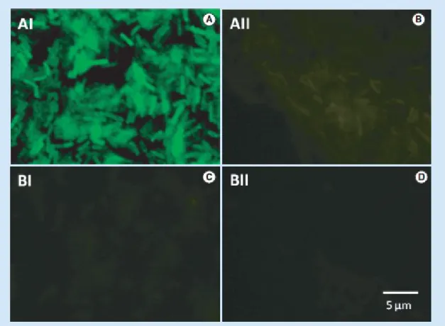
The PNA-FISH test with the EUB338 probe performed as expected. The probe was able to hybridize with eubacteria, while no cross- hybridization was observed for the S. cerevisiae and M. furmicium strains (see Figure 1 for some examples).
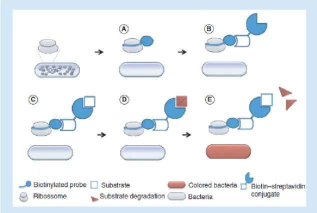
For the ISH-PNA experiments the EUB338 probe was labeled with a biotin molecule in order to allow the introduction of a streptavin enzymatic conjugate. Streptavin has high affinity to biotin and the biotin–streptavidin complex is very resistant to extreme conditions [25]. After hybridization with the biotin-labeled PNA probe, the streptavidin is added and the binding detected by an enzyme conjugated to streptavidin (Figure 2), following a similar strategy to the one described in Bonvisini et al., 2007 [26]. The enzyme will then generate a color change when incubated in the presence of the respective substrate. A typical option is the enzyme HRP because, comparing with the most popular alternatives (such as alkaline phosphatase), the enzyme is smaller, more stable, less expensive and able to generate strong signals in short periods [27]. Thus, an HRP–S conjugate was selected as a reporter molecule.
After selecting the detection strategy, the PNA-ISH procedure was optimized by testing the effect of hybridization time, HRP–S conjugate concentration, incubation period in TMB and the inclusion of BR.
Firstly, we intended to determine the mini- mum hybridization time that allows a strong ISH signal. It was observed that shorter periods allowed an easy discrimination of a positive signal from the background noise, but the highest S/N was obtained for 45 min of hybridization (Figure 3). This hybridization period is actually very short comparing to hybridization times used for other ISH procedures using EUB338 or other DNA probes, which usually take between 6 and 18 h [2,28].
In an attempt to reduce the background noise observed, the inclusion of a BR was tested. BRs are used to increase the specificity of the hybridization procedures. Their use is common on solid phase immunoassays, ISH or western blot procedures, to avoid the nonspecific to nontarget antigens/molecules. They act by blocking active binding sites that may be the source for cross- reactivity [29]. In here, the procedure steps more susceptible to cross-reactivity are the hybridization (probe binding to the rRNA) and the addition of the HRP–S conjugate, where the binding between biotin and streptavidin takes place. As such, the inclusion of BR immediately before these steps would probably increase the reaction stringency. Results revealed no statistic difference (p > 0.05) between the inclusion or not of the BR (Figure 3). The noise observed was not dependent on the nonspecific biding of the probe, since the noise was observed even when no probe was added. It seems a result of some residual nonspecific binding of the conjugate, which was not solved by the addition of BR.
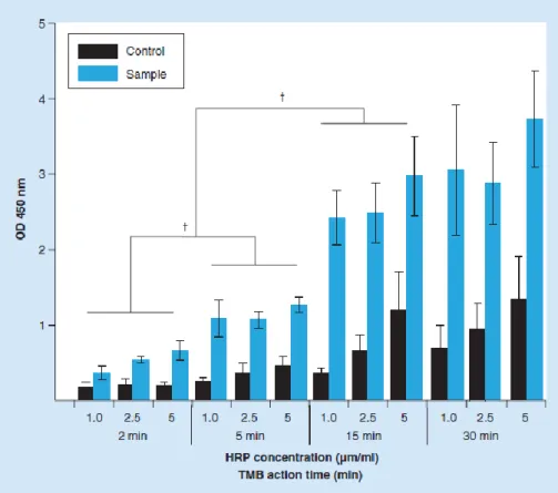
Another factor that can influence the S/N of this procedure is the HRP–S conjugate concentration and the incubation period in the substrate (time that the enzyme HRP is allowed to act). Both high concentration of conjugate and extended incubation periods in the TMB solution might increase the reaction background and, consequently, hinder the discrimination between positive and negative samples. As such, three concentrations of HRP– S conjugate and three incubation periods (in the TMB solution), were tested (Figure 4). An important observation is the lack of statistical significant differences (p > 0.05) between positive samples, exposed to different concentration of conjugate, but with the same incubation period in the TMB solution. This means that, for positive samples, increased concentration of conjugate do not have influence on the PNA-ISH result. On the other hand, the average noise obtained for controls increased with the conjugate concentration. This behavior is particularly noticed for the longer incubation periods (15 and 30 min). This means that high concentrations of conjugate will result in high residual amounts of free conjugate, even after careful washing. Thus controls noise will be further amplified with longer incubation periods. Regarding the positive samples, 15 and 30 min provided the strongest signal and
allowed for a clear discrimination between the positive samples and the controls. However, there is no need to extend the incubation period after 15 min, since no statistically significant difference (p > 0.05) was observed between 15 and 30 min. Taking into account these results, the combination that allowed a better discrimination between the positive samples and the controls, with a reduced background level, in a shorter period of time; was 1 g/ml of conjugate and 15 min in the TMB solution. These were the conditions used in all subsequent tests.
● Specificity/sensitivity of the PNA-ISH procedure
The specificity/sensitivity test allowed not only to verify the performance of the PNA-ISH protocol, but rather to ascertain whether the designed protocol would reveal the same behavior in bacteria with different cell envelope properties.
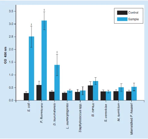
The results (Figure 5) showed that this protocol was effective for Gram-negative eubacteria strains, with visible and statistically significant (p < 0.05) differences between samples and controls on E. coli, P. fluorescens and D. tsuruhatensis. The same was not found in the Gram-positive bacteria used in this study, L. monocytogenes,
S. aureus and B. cereus. Regarding the noneubacteria microorganisms (S. cerevisiae and M. for- micicum) and the mismatched P. hauseri strain, as expected, negative outcomes were observed. As such, this alternative procedure revealed a similar pattern to that of standard PNA- FISH probe specificity/sensitivity test (section ‘Optimization of the PNA-ISH procedure’); but it was unable to correctly identify Gram-positive bacteria.
The unsuccessful detection of the Gram-positive bacteria is probably related to the permeabilization step. Permeabilization of cells for standard PNA-FISH procedures is not usually a problem since molecular weights of fluorochromes are generally 500–1000 Da [30]. For ISH procedures, however, it is necessary to introduce enzymatic conjugates with a large size [31], a fact that can affect the specificity/sensitivity results of the ISH protocol. For instance, the HRP used here is approximately 5–6 nm with a molecular weight of approximately 40 kDa [30]. Thus, this large molecule penetrates fixed cells very poorly compared with fluorescently labeled probes [30].
The major structural difference of Gram- positive bacteria is the presence of a thicker peptidoglycan wall: 20–80 nm on Gram-positive in contrast to 2–3 nm on Gram-negative [32]. It was precisely because of this fact that the lysozyme has been included into the permeabilization step. Lysozyme hydrolyzes the -1, 4 linkages between the peptidoglycan disaccharide subunits; N-acetylglucosamine and N-acetylmuramic acid [33]; but, in here, lysozyme was unable to permeabilize Gram-positive bacteria. However, this failure does not mean that the lysozyme was not able to digest the peptidoglycan layer. Probably the time used (30 min) for the digestion to take place was not enough to allow the permeabilization of such thick layer. Some studies demonstrated that that lysozyme only partly digests the murein multilayers of fixed Gram-positive cells [34,35] and that, for some types of bacteria, combination with other enzyme or solvents might be necessary [36,37].
The problem of large molecular weight molecules diffusion into whole fixed cells, was already described in other studies [30–31,37–38]. Permeabilization protocols must be optimized for each group of microorganism of interest. This is often laborious because the balance between the accessibility of target molecules and the loss of target molecules or complete cell lysis is very difficult to achieve.
● Detection limit
Determining the DL for a new procedure is a key step for its characterization. The DL refers to the minimum amount of cells, which can be detected but not necessarily quantitated. A good DL will thus prevent the occurrence of false negative results. As it is possible to observe in Figure 6A, the statistically significant differences between controls and samples started at 2.5 × 10⁷ CFU/ml, which can also be confirmed at naked eye (Figure 6B). For concentrations higher than 5 × 10⁸ CFU/ml, samples seem to suffer a saturation effect, shown by the small difference in absorbance between this and the 109 CFU/ml.
However, the DL should establish a minimum concentration that can be reliably detected, but at which detection is feasible [14]. As such, the DL was determined based on the slope of the regression line (obtained from the CFU vs OD values, see Supplementary Figure 1) and the SD of low, but known, cellular concentrations [14]. This way rational data is used to determine conclusively what cellular concentration is necessary to distinguish the bacterium presence from its absence [39]. The DL obtained was 5 × 10⁷ CFU/ml.
It would be expected that this assay presented DL similar to those reported for ELISAs, due to the similarity of the strategies. However, these results do not appear to be very favorable when compared with the DLs of others methods based on ELISA, which are usually between 105 and 106 CFU/ml [40–43]; or even PNA-FISH, which are approximately 105 CFU/ml [44–46]. However, the estimation of the DL does not typically take into account the SDs of the measurements, which, when considered, has a negative impact on the DL. It should be noticed that, despite the use of the same concentrations, the amounts of cells used in the experiments change significantly from one study to another. More precisely, we have used 30 l of each inoculum (which for the DL of the PNA-ISH method – 5 × 10⁷ CFU/ml – represents 1.5 × 10⁶ CFU per well); while other authors have used, for instance, 100 l of the innoculum [40,42]. Also, if a centrifugation step is introduced in the procedure, DL may increase significantly. Nonetheless, it should be noticed that the DL here determined only applies to similar culture condition to the ones used in this study.
The DL obtained implies that a suitable enrichment step associated to the detection procedures is needed, in order to ensure that the tar- get bacterium concentration exceeds the DL. In fact, pre-enrichments are used in all technologies (traditional of molecular) applied to microbial identification and are essential to prevent the occurrence of false negatives [47].
Conclusion
Here, an alternative ISH procedure for microbiologic detection, using a biotinylated PNA probe and an enzymatic complex as a reporter molecule, was used. The procedure was optimized and evaluated regarding sensitivity, specificity and DL.
Briefly, the use of a BR was not effective to reduce the noise of the PNA-ISH signal. Increasing concentration of enzymatic conjugate does not improve the signal obtained, regard less the exposure period to the substrate. The specificity/sensitivity test has shown that the procedure is specific, but unable to detect Gram- positive eubacteria, a problem probably related to their thick peptidoglycan layer. The protocol tested achieved a DL of 5 × 10⁷ CFU/ml, which will always imply the inclusion of a suitable pre-enrichment step.
In its current state it is expected that the described method could be used to analyze the presence of Gram-negative bacteria, using a specific probe for that purpose. Nonetheless, future
improvements are necessary in order to solve the permeabilization issues and improve the DL of the procedure.
Future perspective
Microbial identification is facing a huge evolution due to the introduction of nucleic acid mimics, such as PNA or locked nucleic acid, into molecular techniques. New techniques are arising and future developments will allow their introduction in the routine procedures. The method here described intends to be the base for the development of other PNA-based colorimetric systems.
Financial & competing interests disclosure
The authors have no relevant affiliations or financial involvement with any organization or entity with a financial interest in or financial conflict with the subject matter or materials discussed in the manuscript. This includes employment, consultancies, honoraria, stock ownership or options, expert testimony, grants or patents received or pending or royalties.
No writing assistance was utilized in the production of this manuscript.
References
Papers of special note have been highlighted as:
• of interest; •• of considerable interest
1 Enomoto H, Inoue S, Matsuhisa A et al. Development of a new in situ hybridization
method for the detection of global bacterial DNA to provide early evidence of a bacterial infection in spontaneous bacterial peritonitis. J. Hepatol. 56(1), 85–94 (2012).
• Shows the potential of an in situ hybridization (ISH) procedure in the detection of an
infectious disease.
2 Kudo M, Matsuo Y, Nakasendo A et al. Potential clinical benefit of the in situ hybridization
method for the diagnosis of sepsis. J. Infect. Chemother. 15(1), 23–26 (2009).
• Shows the clinical benefit of the ISH method for the diagnosis of sepsis.
3 Bonvicini F, Filippone C, Manaresi E et al. Peptide nucleic acid-based in situ hybridization
assay for detection of parvovirus B19 nucleic acids. Clin. Chem. 52(6), 973–978 (2006).
4 Cerqueira L, Azevedo NF, Almeida C, Jardim T, Keevil CW, Vieira MJ. DNA mimics
for the rapid identification of microorganisms by fluorescence in situ hybridization (FISH). Int. J. Mol. Sci. 9(10), 1944–1960 (2008).
• Overview of nucleic acid mimics applications to microbial identification.
5 Stender H. PNA FISH: an intelligent stain for rapid diagnosis of infectious diseases. Expert
Rev. Mol. Diagn. 3(5), 649–655 (2003).
•• A review about peptide nucleic acid (PNA)-FISH, its characteristics and
potential for the rapid diagnosis of infectious diseases.
6 Egholm M, Buchardt O, Christensen L et al. PNA hybridizes to complementary
oligonucleotides obeying the Watson–Crick hydrogen-bonding rules. Nature 365(6446), 566– 568 (1993).
7 Jensen KK, Orum H, Nielsen PE, Norden B. Kinetics for hybridization of peptide
nucleic acids (PNA) with DNA and RNA studied with the BIAcore technique. Biochemistry 36(16), 5072–5077 (1997).
8 Chinnery PF, Taylor RW, Diekert K, Lill R, Turnbull DM, Lightowlers RN. Peptide
9 Coester C, Kreuter J, Von Briesen H, Langer K. Preparation of avidin-labelled gelatin
nanoparticles as carriers for biotinylated peptide nucleic acid (PNA). Int. J. Pharm. 196(2), 147– 149 (2000).
10 Langer K, Coester C, Weber C, Von Briesen H, Kreuter J. Preparation of avidin-labeled
protein nanoparticles as carriers for biotinylated peptide nucleic acid. Eur. J. Pharm. Biopharm. 49(3), 303–307 (2000).
11 Stams AJ, Van Dijk JB, Dijkema C, Plugge CM. Growth of syntrophic
propionate-oxidizing bacteria with fumarate in the absence of methanogenic bacteria. Appl. Environ. Microbiol. 59(4), 1114–1119
(1993).
12 Almeida C, Azevedo NF, Fernandes RM, Keevil CW, Vieira MJ. Fluorescence in situ
hybridization method using a peptide nucleic acid probe for identification of Salmonella spp. in a broad spectrum of samples. Appl. Environ. Microbiol. 76(13), 4476–4485 (2010).
13 Amann RI, Krumholz L, Stahl DA. Fluorescent-oligonucleotide probing of whole cells for
determinative, phylogenetic, and environmental studies in microbiology. J. Bacteriol. 172(2), 762–770 (1990).
14 Validation of analytical procedures. Text and methodology. International Conference on
Harmonization (ICH) of Technical Requirements for the Registration of Pharmaceuticals for Human Use, ICH–Q2B. www.ema.europa.eu
15 Bouvier T, Del Giorgio PA. Factors influencing the detection of bacterial cells using
fluorescence in situ hybridization (FISH): a quantitative review of published reports. FEMS Microbiol. Ecol. 44(1), 3–15 (2003).
• Review focusing on factors influencing FISH.
16 Carter BS, Fletcher JS, Thompson RC. Analysis of messenger RNA expression by in situ
hybridization using RNA probes synthesized via in vitro transcription. Methods 52(4), 322– 331 (2010).
17 Stylianopoulou E, Lykidis D, Ypsilantis P, Simopoulos C, Skavdis G, Grigoriou M. A rapid and highly sensitive method of non-radioactive colorimetric in situ
hybridization for the detection of mRNA on tissue sections. PLoS ONE 7(3), e33898 (2012).
18 Birner P, Bachtiary B, Dreier B et al. Signal-amplified colorimetric in situ hybridization for
assessment of human papillomavirus infection in cervical lesions. Mod. Pathol. 14(7), 702–709 (2001).
19 Montone KT. Differentiation of Fusarium from Aspergillus species by colorimetric in situ
hybridization in formalin-fixed, paraffin- embedded tissue sections using dual fluorogenic-labeled LNA probes. Am. J. Clin. Pathol. 132(6), 866–870 (2009).
20 Montone KT, Feldman MD. In situ detection of Aspergillus 18s ribosomal RNA sequences
using a terminally biotinylated locked nucleic acid (LNA) probe. Diagn. Mol. Pathol. 18(4), 239– 242 (2009).
•• Article describing an ISH approach, using locked nucleic acid molecules, for the
detection of Aspergillus.
21 Schroder S, Hain M, Sterflinger K. Colorimetric in situ hybridization (CISH) with
digoxigenin-labeled oligonucleotide probes in autofluorescent hyphomycetes. Int. Microbiol. 3(3), 183–186 (2000).
22 Tanner M, Gancberg D, Di Leo A et al. Chromogenic in situ hybridization: a practical
archival breast cancer samples. Am. J. Pathol. 157(5), 1467–1472 (2000).
•• Article describing a colorimetric ISH approach for the detection of an oncogene.
23 Cole JR, Chai B, Farris RJ et al. The Ribosomal Database Project (RDP-II): sequences and
tools for high-throughput rRNA analysis. Nucleic Acids Res. 33, D294–D296 (2005).
24 Ray A, Norden B. Peptide nucleic acid (PNA): its medical and biotechnical applications and
promise for the future. FASEB J. 14(9), 1041–1060 (2000).
25 Gonzalez M, Bagatolli LA, Echabe I et al. Interaction of biotin with streptavidin.
Thermostability and conformational changes upon binding. J. Biol. Chem. 272(17), 11288– 11294 (1997).
26 Bonvicini F, Mirasoli M, Gallinella G, Zerbini M, Musiani M, Roda A. PNA-based
probe for quantitative chemiluminescent in situ hybridisation imaging of cellular parvovirus B19 replication kinetics. Analyst 132(6), 519–523 (2007).
27 Carlsson GH, Nicholls P, Svistunenko D, Berglund GI, Hajdu J. Complexes of horseradish
peroxidase with formate, acetate, and carbon monoxide. Biochemistry 44(2), 635–642 (2005).
28 St Amand AL, Frank DN, De Groote MA, Basaraba RJ, Orme IM, Pace NR. Use of specific
rRNA oligonucleotide probes for microscopic detection of Mycobacterium tuberculosis in culture and tissue specimens. J. Clin. Microbiol. 43(10), 5369–5371 (2005).
29 Tate J, Ward G. Interferences in immunoassay. Clin. Biochem. Rev. 25(2), 105–120 (2004). 30 Kubota K. CARD-FISH for environmental microorganisms: technical advancement and
future applications. Microbes Environ. 28(1), 3–12 (2012).
31 Bidnenko E, Mercier C, Tremblay J, Tailliez P, Kulakauskas S. Estimation of the state of
the bacterial cell wall by fluorescent in situ hybridization. Appl. Environ. Microbiol. 64(8), 3059– 3062 (1998).
32 Seltmann G, Holst O. The Bacterial Cell Wall. Springer, Berlin, Germany (2002).
33 Deckers D, Vanlint D, Callewaert L, Aertsen A, Michiels CW. Role of the lysozyme
inhibitor Ivy in growth or survival of Escherichia coli and Pseudomonas aeruginosa bacteria in hen egg white and in human saliva and breast milk. Appl. Environ. Microbiol. 74(14), 4434–4439 (2008).
34 Dellinger M, Geze M, Santus R et al. Imaging of cells by autofluorescence: a new tool in
the probing of biopharmaceutical effects at the intracellular level. Biotechnol. Appl. Biochem. 28(Pt 1), 25–32 (1998).
35 Wendeberg A. Fluorescence in situ hybridization for the identification of environmental
microbes. Cold Spring Harb. Protoc. 2010(1), pdb.prot5366 (2010).
36 Sekar R, Pernthaler A, Pernthaler J, Warnecke F, Posch T, Amann R. An improved protocol
for quantification of freshwater Actinobacteria by fluorescence in situ hybridization. Appl. Environ. Microbiol. 69(5), 2928–2935 (2003).
37 St Amand AL, Frank DN, De Groote MA, Pace NR. Use of specific rRNA oligonucleotide
probes for microscopic detection of Mycobacterium avium complex organisms in tissue. J. Clin. Microbiol. 43(4), 1505–1514 (2005).
38 Schonhuber W, Fuchs B, Juretschko S, Amann R. Improved sensitivity of whole-cell
hybridization by the combination of horseradish peroxidase-labeled oligonucleotides and tyramide signal amplification. Appl. Environ. Microbiol. 63(8), 3268–3273 (1997).
39 Armbruster DA, Tillman MD, Hubbs LM. Limit of detection (LQD)/limit of quantitation
(LOQ): comparison of the empirical and the statistical methods exemplified with GC-MS assays of abused drugs. Clin. Chem. 40(7 Pt 1), 1233–1238 (1994).
separation and ELISA end-detection procedure. Lett. Appl. Microbiol. 31(4), 279–283 (2000).
41 Padhye NV, Doyle MP. Rapid procedure for detecting enterohemorrhagic Escherichia coli
O157:H7 in food. Appl. Environ. Microbiol. 57(9), 2693–2698 (1991).
42 Jones JB, Somodi GC, Scott JW. Increased ELISA sensitivity using a modified extraction
buffer for detection of Xanthomonas campestris pv. Vesicatoria in leaf tissue. J. Appl. Microbiol. 83(4), 397–401 (1997).
43 Tan S, Gyles CL, Wilkie BN. Comparison of an LPS-specific competitive ELISA with
a motility enrichment culture method (MSRV) for detection of Salmonella typhimurium and S. enteritidis in chickens. Vet. Microbiol. 56(1–2), 79–86 (1997).
44 Almeida C, Azevedo NF, Iversen C, Fanning S, Keevil CW, Vieira MJ. Development and
application of a novel peptide nucleic acid probe for the specific detection of Cronobacter genomospecies (Enterobacter sakazakii) in powdered infant formula. Appl. Environ. Microbiol. 75(9), 2925–2930 (2009).
45 Peleg AY, Tilahun Y, Fiandaca MJ et al. Utility of peptide nucleic acid fluorescence in situ
hybridization for rapid detection of Acinetobacter spp. and Pseudomonas aeruginosa. J. Clin. Microbiol. 47(3), 830–832 (2009).
•• Article describing the potential of PNA-FISH in the clinical diagnostics.
46 Selvaraju SB, Kapoor R, Yadav JS. Peptide nucleic acid-fluorescence in situ hybridization
(PNA-FISH) assay for specific detection of Mycobacterium immunogenum and DNA- FISH assay for analysis of pseudomonads in metal working fluids and sputum. Mol. Cell. Probes 22(5–6), 273– 280 (2008).
47 Ponsard I, Soumillion P. New immunoenzymatic strategy for rapid and selective growth
Figure 1. Peptide nucleic acid-FISH results for the EUB338 peptide nucleic acid probe.
Bacillus cereus subjected to a standard FISH protocol with (positive control [A]) and without PNA probe (no probe control [B]). An Archaea (non-target strain) Methanobacterium formicicum DSM 1535, also subjected to the FISH protocol with (C) and without PNA probe (D). Images were obtained with equal exposure times, and with a magnification of ×1000. PNA: Peptide nucleic acid.
Figure 2. In situ hybridization procedure used in this work. A biotinylated PNA probe,
targeting rRNA conserved regions of the target bacterium, is added (A). Then, an HRP–S conjugate that binds specifically the biotin molecule is provided (B). When incubated with the substrate – TMB (C), the enzyme will degrade it (D & E), producing a colorimetric compound
(E). HRP–S: Horseradish peroxidase–streptavidin; PNA: Peptide nucleic acid; TMB:
5’-tetramethylbenzidine.
Figure 3. Effect of the hybridization time and blocking reagent in the peptide nucleic acid-in situ hybridization signal obtained for E. coli CECT 434. The optimization of the
hybridization time (A) has shown a clear increase of the fluorescence up to 45 min. Blocking reagent (B) was introduced in two different steps: before the addition of HRP–S conjugate and before the hybridization step. Controls were subjected to the same procedure, but no probe was added to the hybridization solution. BR: Blocking reagent; HRP–S: Horseradish peroxidase– streptavidin; OD: Optical denisty.
Figure 4. Peptide nucleic acid-in situ hybridization outcome for different concentrations of horseradish peroxidase–streptavidin conjugate (1, 2.5, 5 g/ml) and incubation periods in 5’-tetramethylbenzidine (2, 5, 15 and 30 min). Samples and controls
refer to tests with and without probe. †Show significant differences between samples (p < 0.05). HRP: Horseradish peroxidase; OD: Optical density; TMB: 5’-tetramethylbenzidine.
Figure 5. Results for the specificity/sensitivity test of peptide nucleic acid-in situ hybridization procedure. †Show significant differences between samples and controls (p <
0.05). OD: Optical density.
Figure 6. Detection limit for the peptide nucleic acid-in situ hybridization assay. Peptide
nucleic acid-ISH signal for different cellular concentrations (A) and visual aspect of a representative area of the 96-well plate (B). †Show significant differences between samples and controls (p < 0.05). ISH: In situ hybridization; OD: Optical density.