UNIVERSIDADE DA BEIRA INTERIOR
Ciências
Improvement of STEAP 1 Biosynthesis from Pichia
pastoris X33 cells under an optimized feeding
strategy
Diana Rute Tavares Duarte
Dissertação para obtenção do Grau de Mestre em
Biotecnologia
(2º ciclo de estudos)
Orientador: Prof. Doutor Luís António Paulino Passarinha
Coorientador: Prof. Doutor Cláudio Jorge Maia Baptista
iii
Acknowledgments
Firstly, I would like to make special thanks to my supervisors, Professor Luís Passarinha and Professor Claúdio Maia for the opportunity to let me develop this work, for helping and guiding me through this year.
To my colleagues and friends at CICS, the biggest thanks for all the patience, friendship, laughs, support, and encouragement. Special thanks to Margarida Gonçalves, Fátima Santos, and Augusto Pedro for the biggest help, companionship, and teachings during the past year.
To my friends that have always been with me and believe in me. I thank you for your friendship and kind words that make me forget a bad day and give me hope in the future.
My forever thankfulness goes to my dad and my little brother Ivo, for your support, love and believing. To my mom special thanks, I hope that you are proud of me. I can never repay what you have done for me.
v
Resumo
O cancro da próstata é uma das patologias com maior incidência em todo o mundo. Atualmente, os meios de diagnóstico e terapêuticos existentes são invasivos e apresentam uma eficácia limitada sobretudo em estágios mais avançados da doença. Desta forma, torna-se necessário o estudo de proteínas específicas cuja expressão esteja relacionada com o seu desenvolvimento e progressão. Diversos estudos têm sugerido a proteína Six-transmembrane Epithelial Antigen of the Prostate 1 (STEAP1) como um possível biomarcador e/ou alvo imunoterapêutico para o cancro da próstata. A STEAP1 é constituída por 6 domínios transmembranares e encontra-se presente na membrana plasmática das células epiteliais, nomeadamente nas junções que promovem a comunicação célula-célula. Alguns estudos suportam a hipótese que a STEAP 1 assume um papel preponderante na comunicação entre células tumorais, estando desta forma envolvida na progressão do cancro. No entanto, estudos complementares são ainda necessários para resolver a sua estrutura tridimensional de forma a melhor compreender as suas funções na carcinogénese assim como delinear novas estratégias terapêuticas. Deste modo, elevadas quantidades de STEAP1 são requeridas a partir de tecnologias emergentes de DNA recombinante. Nestes domínios, a levedura Pichia pastoris tem-se revelado um hospedeiro adequado na expressão de proteínas recombinantes. Em particular, a sua capacidade para realizar modificações pós-tradução torna-a num sistema microbiano ideal para a produção recombinante de proteínas membranares. Assim, os principais objetivos do presente trabalho são: 1) Aumentar a escala de produção da proteína STEAP1 para bioreator em culturas de Pichia pastoris X33 testando diferentes feeds de glicerol e metanol; 2) Avaliar a adição de diferentes chaperones químicos que contribuam para a estabilização conformacional da STEAP1; 3) Estudar a influência dos diferentes feeds de glicerol em eventuais processos de N-glicosilação. Os resultados obtidos demonstraram que - através um feed por gradiente de glicerol e constante de metanol - houve um aumento da produção de STEAP1, para cerca do dobro, aquando a suplementação do meio com 1M de Prolina. Observa-se que na aplicação de um feed exponencial de glicerol e constante de metanol, a quantidade de STEAP1 produzida encontra-se no peso molecular correto (~35kDa), embora encontra-se tivesencontra-se verificado níveis de produção reduzidos. Adicionalmente, denotou-se através da digestão dos lisados com a enzima PNGase F que um feed constante de glicerol e metanol parece produzir STEAP1 com N-glicosilação. Como trabalho futuro serão desenvolvidas estratégias de purificação da proteína STEAP1.
Palavras-chave:
vii
Resumo alargado
O cancro da próstata é uma das patologias com maior incidência nas sociedades modernas, principalmente em homens com idade superior a 50 anos. Os meios de diagnóstico e de terapia existentes são invasivos e com eficácia limitada. Assim, é necessário encontrar e estudar genes específicos que codifiquem proteínas específicas neste grupo de patologias. Ao longo dos últimos anos, inúmeras biomoléculas têm sido identificadas como biomarcadores para o cancro da próstata, dos quais a proteína Six-transmembrane epithelial antigen of the prostate 1 (STEAP1). Esta encontra-se maioritariamente presente na membrana plasmática das células epiteliais, nomeadamente nas junções que promovem a comunicação célula-célula. É constituída por seis domínios transmembranares interligados por três loops extracelulares e 2 intracelulares. De acordo com a literatura, é sugerido o seu papel na comunicação entre células tumorais, estando envolvida em processos de carcinogénese e invasão tumoral. No entanto, estudos adicionais são necessários para resolver a sua estrutura 3D de forma a compreender o papel da STEAP1 no desenvolvimento e progressão tumoral assim como desenvolver moléculas terapêuticas que diminuam a sua função oncogénica. De uma forma geral para a realização de estudos cristalográficos são requeridas elevadas quantidades de proteína alvo. Nas últimas décadas a tecnologia de DNA recombinante emergiu e otimizou exponencialmente a obtenção de níveis de expressão, consideráveis, de proteínas membranares para o desenvolvimento de estudos de bio-interação e estruturais. Neste domínio cientifico, a levedura Pichia pastoris tem-se revelado o hospedeiro ideal na expressão recombinante de proteínas membranares, destacando-se a sua capacidade de realizar modificações pós-tradução similares às identificadas em eucariotas superiores. Assim, os principais objetivos do presente trabalho são: 1) Aumentar a escala de produção da proteína STEAP1 em biorreator com culturas de Pichia pastoris X33 testando diferentes feeds de glicerol e metanol; 2) Avaliar a adição de diferentes chaperones químicos que contribuam para a estabilização conformacional da STEAP1; 3) Estudar a influência dos diferentes feeds de glicerol em eventuais processos de N-glicosilação. Relativamente à estratégia desenvolvida, o processo fermentativo compreendeu três fases: o batch em glicerol, fed-batch (2 horas de feed de glicerol e 1 hora de mistura glicerol/metanol) e por último a etapa de indução com metanol. Tendo como primeiro objetivo o incremento da biossíntese recombinante da proteína STEAP1 foram testados diferentes feeds de glicerol e metanol, durante a fase de fed-batch e de indução, respetivamente. Especificamente, analisaram-se três feeds de glicerol distintos - constante, gradiente e exponencial - e dois feeds de metanol - constante e exponencial. As concentrações de glicerol e metanol no meio extracelular foram quantificadas ao longo do processo fermentativo por HPLC com um índice de refração acoplado. Devido à existência de dímeros e/ou bandas de maior peso molecular em análises efetuadas por Western blot (WB), comparando a densitometria das bandas, usou-se a enzima PNGase F para compreender se as alterações de peso molecular observadas seriam
viii
devido a diferentes padrões de N-glicosilação da proteína em estudo. Deste modo, obteve-se para um feed constante de glicerol e metanol um pico de produção de STEAP1 às 2 horas de indução, sendo produzida com um peso molecular superior (~48KDa). A realização de ensaios complementares com a enzima PNGase F demonstraram que as diferenças no peso molecular podem estar relacionadas com a existência de N-glicosilações na estrutura 3D da biomolécula alvo. Relativamente ao feed de glicerol por gradiente e constante de metanol obteve-se um pico de produção às 10 horas de indução. No entanto, por WB denotou-se a existência de bandas com elevado peso molecular (~63 kDa), sugerindo a formação de agregados proteicos: dímeros. Consequentemente, testaram-se diferentes concentrações de prolina (0,2, 0,5 e 1M), trealose (0,1, 0,25 e 0,5 M) e histidina (0,04 e 0,08 mg/mL) no meio de cultura, de forma a avaliar se as moléculas adicionadas poderiam funcionar como chaperones químicos, promovendo a consequente estabilização da STEAP1 durante a sua expressão e diminuindo a concentração de dímeros. Os resultados obtidos demonstraram que - através um feed por gradiente de glicerol e constante de metanol - houve um aumento da produção da STEAP1, para cerca do dobro, aquando a suplementação do meio com 1M de Prolina. Do tratamento dos lisados obtidos, nesta estratégia com a enzima PNGase F, observa-se que não há alteração nos padrões de glicosilação, mesmo para diferentes concentrações de enzima durante os vários tempos de reação testados. Para complemento dos resultados obtidos, efetuaram-se ensaios adicionais por eletroforese bidimensional, de forma a averiguar se os diferentes padrões de N-glicosilação conduzem a uma alteração do ponto isoelétrico da STEAP1 (pI teórico ~ 9,28). Os resultados obtidos reforçam que os dímeros de STEAP1 formados durante o feed de glicerol por gradiente e constante de metanol, podem não estar relacionados com interações que originem modificações pós-tradução. Adicionalmente, denota-se que a aplicação do feed exponencial de glicerol e constante de metanol, embora decresça a quantidade de STEAP1 obtida, toda a proteína produzida encontra-se no peso molecular correto e descrito na literatura (~35kDa). Observou-se também que os níveis de metanol (< 8.3 g/L nas primeiras horas de indução) e glicerol (entre 0.7- 7.0 g/L) permanecem em níveis tolerantes e inócuos para as culturas de P. pastoris, não impedindo desta forma a expressão da STEAP1. Finalmente foi ainda testado um feed exponencial de metanol durante a etapa de indução tendo por base os feed exponencial e gradiente de glicerol anteriormente referenciados. Com a estratégia descrita obteve-se um incremento basal na biossíntese da STEAP1, prevalecendo a formação de dímeros e degradações de baixo peso molecular. Concluindo, foi otimizada com sucesso uma nova estratégia que visa ao aumento da biossíntese da proteína STEAP1 de forma recombinante em biorreactor. Este incremento foi alcançado pela combinação de diferentes perfis de alimentação de fonte de carbono e de indutor com o uso de diferentes chaperones químicos. Em suma, a estratégia ideal é através da aplicação de um feed de glicerol por gradiente combinado um feed constante de metanol durante 10 horas e suplementado com 1M de Prolina. Como trabalho futuro serão desenvolvidas novas estratégias de purificação da proteína STEAP1.
ix
Abstract
Prostate cancer (PCa) is the most common type of cancer in aged men. Actually, the main problem arises from the fact that both PCa diagnosis and therapy are still invasive and limited in advanced stages of this disease. Thus, it is necessary to identify, study and characterize specific proteins whose expression correlates with these pathologies. Concerning this, it has been suggested that the Six-transmembrane epithelial antigen of the prostate 1 (STEAP1) protein as a good biomarker and/or immunotherapeutic target for PCa. It is located in the plasma membrane of epithelial cells, in both tight and gap junctions. STEAP1 is composed of six transmembrane domains, connected by three extracellular and two intracellular loops. Therefore, it has been suggested that this protein plays an important role in intracellular communication between cancer cells, contributing to the cancer process and tumor invasiveness. The characterization of STEAP1 structure and function might allow the development of specific inhibitors, envisaging a decrease of its oncogenic role. However, the techniques used for protein structural and functional characterization demand for high quantities of the target protein, which may be achieved through the recombinant DNA technology. Therefore, the aim of this work was to improve STEAP1 biosynthesis from mini-bioreactor Pichia pastoris X33 methanol induced cultures. This was achieved through the study of different glycerol and methanol feeding profiles during the fed-batch phases. Briefly, the medium supplementation with Proline 1M in a gradient glycerol and constant methanol feed, leads to high quantities of STEAP1 (increase for the double). An exponential glycerol and constant methanol feed produces fewer amounts of the protein but in the correct molecular weight (~35kDa). The influence of the fermentation conditions on STEAP1 molecular weight and N-glycosylation was studied using the enzyme PNGase F. The results showed that a constant glycerol feed seems to produce STEAP1 with N-glycosylation. However, the dimers produced in the gradient glycerol feed are not due N-glycosylation process. Two-dimensional electrophoresis proves this, and it was demonstrated that they correspond to different N-glycosylation patterns. Overall, it was successfully optimized a new strategy for recombinant STEAP1 biosynthesis, through the study of different feeding profiles. Future work encompassing will be developed an alternative strategy to perform the purification on the target protein.
Keywords:
xi
Table of Contents
Chapter I – Introduction ... 1
1. Human Prostate ... 1
Anatomy and Physiology ... 1
1.2. Prostate Cancer ... 2
2. General overview of STEAP family ... 6
Structure and Function of STEAP1 ... 8
STEAP 1 as an immunotherapeutic target ... 9
3. Recombinant protein biosynthesis ... 11
Host strain and promoter selection ... 11
Fermentation strategies and main conditions ... 15
Chapter II – Aims ... 21
Chapter III – Materials and Methods ... 23
1. Materials ... 23
2. Methods ... 23
2.1. Strain, plasmids, and media ... 23
STEAP1 biosynthesis ... 24
Cell lysis and Protein Recovery ... 25
Total protein quantification ... 26
SDS-PAGE and Western Blotting ... 26
Dry Pichia pastoris weight assessment ... 27
Glycerol and Methanol assessment ... 28
Evaluation of N-glycosylation ... 29
STEAP1 Quantification ... 30
Chapter IV – Results and Discussion ... 33
1. Setting up the batch phase ... 33
2. Optimization of the Fed-batch phases ... 35
xii
Exponential feed profile ... 37
Gradient feed profile ... 39
Comparison of the three feeds ... 41
3. Optimization of fermentation conditions ... 45
Chemical chaperones ... 46
Exponential Methanol Feeding ... 51
4. Evaluation of N-glycosylation ... 54
Chapter V – Conclusions and Future Perspectives ... 59
Chapter VI – Bibliography ... 61
xiii
List of Figures
Figure 1 – General anatomical and microscopical structure of the prostate, from [1,2].
Figure 2 - Cellular and molecular model of early prostate neoplasia progression. Formation of a) PIA lesions; b) PIN lesions; c) Prostate carcinoma; and d) Metastic carcinoma, adapted from [31].
Figure 3 - Schematic illustration of the domain organization of STEAPs family, from [60]. Figure 4 - Schematic STEAP1 protein structure, cellular localization, and physiologic functions, adapted from [49].
Figure 5 - Prediction of the several N-glycosylation, glycation, phosphorylation and O-β-GlcNAc sites of STEAP1 using: A) NetNGlyc 1.0; B) NetGlycate 1.0; C) NetPhos 2.0 and D) YinOyang 1.2, respectively, adapted from [78].
Figure 6 – pPICZα (A, B, C) expression vector (Retrieved from Invitrogen, EasySelect™ Pichia Expression Kit no. 25, 2010).
Figure 7 - Methanol/Glycerol pathway in Pichia pastoris metabolism [100].
Figure 8 - Specific AOX activity during the transition phase in P. pastoris X33 high cell density fed-batch cultivation, from [109].
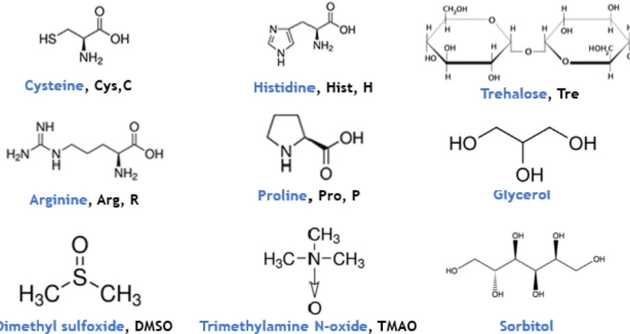
Figure 9 - Chemical structure of several molecules used as chaperones.
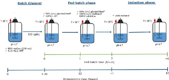
Figure 10 - Structure of the production process implemented and developed for recombinant STEAP1 biosynthesis in P. pastoris bioreactor cultures, adapted from [121].
Figure 11 – BSA calibration curve for total protein quantification (µg/mL) ranged between 25 – 2000 μg/mL.
Figure 12- Relationship between the OD600nm and the P. pastoris dry weight (g/L).
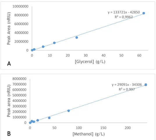
Figure 13 – Calibration curves measured by HPLC-RID for A) Glycerol ranged between 0.125-62.5g/L; B) Methanol ranged between 0.395- 237 g/L.
Figure 14- Typical standard ELISA curve, ranged between 0.39 ng/mL to 25 ng/mL.
Figure 15 – Residual glycerol concentration (g/L) measured by HLPC- RID during the batch fermentation (hours).
xiv
Figure 17 - Glycerol concentration (g/L) and biomass levels (g/L) over 60 hours of methanol induction in the constant glycerol feed.
Figure 18 – Western blot analysis of STEAP1 expression during a constant glycerol feed with 60 hours of methanol induction.
Figure 19 - Biomass levels (g/L) for the reactor 1 and 2, over 60 hours of methanol induction for a typical exponential glycerol feed.
Figure 20 – Glycerol concentration (g/L) in reactor 1 and 2, over 60 hours of methanol induction for the exponential glycerol feed.
Figure 21 – Western blot analysis of STEAP1 expression during an exponential glycerol feed with 60 hours of methanol induction.
Figure 22 – Biomass levels (g/L) over 60 hours of methanol induction for the gradient glycerol feed in reactor 1 and 2.
Figure 23 – Glycerol concentration (g/L) in reactor 1 and 2, over 60 hours of methanol induction for a typical gradient glycerol feed.
Figure 24 – Western blot analysis of STEAP1 expression during a gradient glycerol feed with 60 hours of methanol induction.
Figure 25 – Analysis of the spikes of STEAP1 production with a glycerol constant (2h), exponential (5h) and gradient (10h) feed: A) Western blot, B) SDS-PAGE.
Figure 26 – Residual glycerol concentration (g/L) in the different glycerol feeding tested. Figure 27 – Residual methanol concentration (g/L) in the different glycerol feeding tested during the methanol induction.
Figure 28 – Western blot of the different proline concentrations tested: 0.1 M, 0.5 M, and 1 M, respectively.
Figure 29 - Western blot of the several histidine concentrations tested: A) 0.04mg/mL; B1 and B2) Comparation between 0.04 mg/mL and 0.08 mg/mL histidine, respectively.
Figure 30 – Western blot of the three trehalose concentrations tested: 0.1 M, 0.25 M, and 0.5M, respectively.
Figure 31 – Comparison of WBs obtained in the original glycerol gradient feed and glycerol gradient feed supplemented with different concentrations of chaperones: Proline, Trehalose, and Histidine.
xv
Figure 32 – Comparison by SDS-PAGE of the original glycerol gradient feed and glycerol gradient feed supplemented with distinct concentrations of chaperones: Proline, Trehalose, and Histidine.
Figure 33 – STEAP1 relative quantification for the original gradient glycerol feed and additional feeds supplemented with proline, trehalose, and histidine.
Figure 34 – Western blot analysis of an exponential methanol feed and the original glycerol gradient feed (with a methanol constant flow-rate during the induction).
Figure 35 – Western blot analysis of an exponential methanol feed and the original glycerol exponential feed (with a methanol constant flow-rate during the induction).
Figure 36 - Residual methanol concentration (g/L) for the exponential and gradient glycerol feed in an exponential methanol feed during the induction.
Figure 37– Western blot analysis of a cell lysate obtained from a glycerol gradient feed, non-treated (A) and non-treated (B) with 3 µL of PNGase F for 1 hour.
Figure 38 – SDS-PAGE analysis of the cell lysate treated with the enzyme PNGase F, obtained from the original glycerol gradient feed supplemented with chaperones (0.04 mg/mL histidine, 0.2M proline, and 0.1 M trehalose).
Figure 39 – SDS-PAGE and Western blot analysis of the cell lysate treated with the enzyme PNGase F, obtained from the glycerol gradient feed supplemented with 0.1M of trehalose. A) original Trehalose 0.1M without PNGase F treatment; Trehalose 0.1M treated with B) 3 µL PNGase F for 1 hour; C) 5 µL PNGase F for 1 hour; D) 3 µL PNGase F for 2 hours.
Figure 40 – Description of Pichia pastoris X33 proteome by bi-dimensional electrophoresis obtained in a gradient glycerol and constant methanol feed during 10 hours of methanol induction.
Figure 41 – Western blot analysis of the cell lysate obtained from a constant glycerol and methanol feed, non-treated (A) and treated (B) with 3 µL PNGase F for 1 hour.
xvii
List of Tables
Table 1 – Specific growth rate (µ, h-1) of each glycerol feed during the glycerol fed-batch and methanol phase.
Table 1 - The relationship between DO600nm and the different chaperone tested.
Table 2 - The relationship between DO600nm and the exponential methanol feed during the induction phase.
xix
List of Acronyms
2-DE Two-dimensional electrophoresis µ Specific growth rate (h-1)
AOX1 Alcohol Oxidase
AR Androgen Receptor
BMGH Buffered Minimal Medium Containing Glycerol BSA Bovine Serum Albumin
BSM Basal Salt Medium
DMSO Dimethyl Sulfoxide
DO Dissolved oxygen
DTT Dithiothreitol
E. coli Escherichia coli
HPLC High-performance liquid chromatography OD600nm Optical Density at 600nm
PCa Prostate Cancer
pI Isoelectric point
PIA Proliferative Inflammatory Atrophy
P. pastoris Pichia pastoris X33
PSA Prostate Specific Antigen SDS Sodium Dodecyl Sulphate
SDS-PAGE Sodium dodecyl sulphate-polyacrylamide gel electrophoresis STEAP Six-Transmembrane epithelial antigen of the prostate
WB Western Blot
xxi
Work presented in this thesis has resulted in:
Oral communication in the XII Annual CICS-UBI Symposium, Covilhã (2017): Duarte DR, Pedro AQ, Maia CJ, Passarinha LA, Improvement of STEAP1 biosynthesis from mini-bioreactors Pichia pastoris X33 cultures.
Poster presentation at the II International Congress on Health Sciences Research towards Innovation and entrepreneurship: Trends in Biotechnology for Biomedical Application, Covilhã (2017): Duarte DR, Pedro AQ, Maia CJ, Passarinha LA, Improvement of STEAP1 biosynthesis from Pichia Pastoris X33 cells under an optimized strategy.
Poster presentation at the 9th Conference on Recombinant Protein Production, Croatia (2017): Barroca-Ferreira J, Pais JP, Santos MM, Duarte DR, Pedro AQ, Maia CJ, Passarinha LA, Evaluation of Escherichia coli and Pichia pastoris host in the biosynthesis of STEAP1: a membrane therapeutic target for prostate cancer.
1
Chapter I – Introduction
1. Human Prostate
Anatomy and Physiology
The prostate is the main gland in male reproductive and urinary system. It is oval shaped with a rounded tip. The size varies from man to man, but generally, it has approximately 4 cm wide and 3 cm thickness. It possesses two lobes that bring urine from the bladder, through the prostate, to the penis. The main function is the production of the fluid that protects and nourish the sperm in the semen [1–3]. It is composed of two main compartments, the stroma and the epithelium. Both structures influence each other reciprocally via different signaling pathways to the normal prostate development and homeostasis. According to the general anatomical and microscopical structure of the prostate depicted in Figure 1, the prostate gland is covered by a layer of connective tissue called the prostatic capsule. Anatomically, it is composed of three main zones: the central, the transition, and the peripheral zone [2,4]. The peripheral zone is the predominant zone, comprising almost 70% of glandular tissue and covers the posterior and lateral features of the gland. This peripheral zone is the most associated with adenocarcinoma development. The transition and the central zone accounts for 5-10% and 20-25% of the total glandular tissue, respectively. The transition zone has an important clinical significance once it is the main region where benign prostatic hyperplasia (BPH) are diagnosed [1,2].
2
In relation to the microscopic structure, the prostate is composed by a glandular epithelium embedded in a fibromuscular stroma. The main function of the stromal compartment is to maintain the correct microenvironment of the epithelial cells. Also, it helps to maintain or restore the homeostasis of the prostate [1,5,6]. The epithelial compartment has the main glandular function, once it secrets the prostatic fluid that contributes to the most volume of the entire ejaculate. Some factors control the ejaculation and regulate the proteins involved in sperm maturation, they have mechanistically and functionally linked to each other. The accumulation of zinc (Zn2+) and citrate play an important physiological role in prostate. Also, the loss of this ability may be related to the development and progression of prostate malignancy [5,7–9].The epithelium is divided into several types of cells such as columnar luminal, basal, and neuroendocrine cells. The main function of the columnar cells is the production of prostatic secretions. These cells express high levels of androgen receptors (AR) and require androgens for its survival and secretory activity. On the other hand, the basal cells are undifferentiated cells, with a distinct morphology, that can originate all types of epithelial cells. Their differentiation and growth only occur in the presence of androgens, despite its survival and maintenance are androgen independent [1,4,10,11]. The neuroendocrine cells have regulatory functions that seem to be involved in the proliferation of adjacent cells. They produce neuropeptides such as, chromogranin A (Cg A), neuron-specific enolase (NSE), somatostatin or calcitonin, which has been described as biological markers for cancer [12,13]. Prostate development and function are dependent on AR signaling, particularly from 5dihydrotestosterone (DHT). The intracellular reduction of testosterone into DHT by 5- α-reductase in the prostatic epithelium is necessary to complete prostate morphogenesis. After the development of the prostate, androgens are involved in the survival promotion of the secretory epithelium. Besides that, it is described that the AR is differentially expressed in the stroma and epithelium compartments, with a paracrine and autocrine control, respectively. Alterations in these pathways may promote tumorigenesis [5,11,14].
1.2. Prostate Cancer
Carcinogenesis is a process characterized by changes in the cellular phenotype of some cells, that are based on genome changes [15,16]. Typically, the cancer cells have the ability to grow into several environments and fail the response to the usual controls on such proliferation. Contrary to normal cells, cancer cells replication is not limited beyond the limits imposed by telomere length [16,17].
They also present the ability to stimulate new blood vessel formation, thus ensuring the oxygen and nutrients required for their survival and proliferation. Also promote angiogenesis, vessel co-option, and vascular mimicry to create an extracellular matrix rich in growth factors with a
3
specific pH that makes difficult the anticancer-drug proliferation [16–19]. Moreover, the cancer cells display specific characteristics, namely: unlimited proliferation, self-sufficiency in growth signals, resistance to antiproliferative and apoptotic stimuli, tissue invasion and metastasis [15,20]. Typically, the cancer environment is composed of stromal cells, acting as support cells for the tumor itself, which is responsible to attract new blood vessels to bring nutrients and oxygen, invade detection, and metastasizing to distal organs. The major problem in the currently available treatments is the lack of specificity of the drugs, once they should present a large therapeutic window to kill tumor cells while sparing normal cells [20,21].
From all the cancers studied in the literature, this work will focus on the prostate cancer (PCa). Concerning this, the PCa is one of the most frequently diagnosed cancer, more than 3.3 million men living with this pathology in the United States. It is the second leading cause of male cancer-related death in North America and the third one in Portugal [22–25]. The PCa incidence in Portugal has been increasing since 1998, and it was the most frequent cancer among men in 2009, with 5433 new cases. It is predicted the existence of new 8600 incident cases and 1700 deaths caused by PCa in 2020 [26].
Typically, it tends to develop in older men, aged 50 and over. In many clinical cases, PCa develops slowly, although in some patients it can be aggressive and metastasize to other parts of the body. The risk factors associated with the appearance of PCa include aging and ethnicity, family history and genetic factors, diet and lifestyle, hormonal levels, and also environmental factors [27–30].
The mechanisms involved in the prostate carcinoma are not well understood but, it seems that the PCa progression is down-regulated by androgen-responsive genes [31,32]. These critical factor contributes to the development of prostate tumors, through the inhibition of apoptosis rather than an enhanced cellular proliferation [33]. Furthermore, there is evidence for the formation of pre-cancerous lesions initiated by an inflammatory process that occurs during tissue injury. Indeed, it was described the existence of three different stages involved in PCa development and progression, namely prostatic intraepithelial neoplasia (PIN), proliferative inflammatory atrophy (PIA) and prostatic and metastatic carcinoma, as shown in Figure 2 [2,31,34].
4
PIA seems to be a precursor of PCa, and these lesions occur mainly in stromal and epithelial cells, as described in Figure 2a. The PIN lesions are described as the most likely precursor to PCa progress, and involve an abnormal proliferation of basal and luminal cells, showing a dysplastic behavior, according to Figure 2b. When the PCa cells reach the blood, they progress through the bloodstream and metastasize into different organs, including liver, lung, and bones( Figure 2c and d) [3,10,34]. Changes of epithelial and stromal cells genes expression during the different development stages of PCa notably contribute to the enhancement of tumor growth, survival, migration, and invasiveness [3,35]. The PCa is androgen-dependent at initial stages. In the primary PCa development, only a few AR genes have been mutated and the androgen ablation therapies reveal that the tumor is in regression. However, in advanced stages of PCa, the prostate cells became AR-independent, as they are able to survive and proliferate without circulating androgens, thus restricting the use of androgen ablation therapies [36,37].
1.2.1. Diagnosis and Treatment
Current diagnosis of PCa can be achieved by prostate-specific antigen (PSA) test, digital rectal examination, and biopsies for histopathological staging [38–41]. Early detection of PCa has increased dramatically with a serum test for the PSA, which is a serine protease secreted by epithelial cells of the prostate and has an important role in seminal fluids liquefaction [38,41]. However, the PSA test may not distinguish PCa from benign disease such as BPH and prostatitis, leading to the detection of false positives [40,42]. Thus, the best way to perform a correct PCa diagnosis is a biopsy of the respective tissue, that is currently collected by ultrasound transrectal sampling. The main disadvantages of this procedure are the fact that it is an invasive test and offer a significant risk of posterior infection [39,40].
Figure 2. Cellular and molecular model of early prostate neoplasia progression. Formation of a) PIA lesions ; b) PIN lesions; c) Prostate carcinoma ; and d) Metastic carcinoma, adapted from [31], .
5
Thereby, the Gleason score grading is the method used to unify the PCa progression and aggressiveness. With this grading, it is possible to characterize and distinguish the different stages of PCa. This system was created by Dr. Donald F. Gleason and is based on the histological pattern of carcinoma cells in the prostatic tissue. The Gleason-score is ranged between 2 and 10, in agreement with the severity of the disease. An increasing of Gleason grade is directly related to tumor size and invasiveness [43,44]. Treatments for PCa depend on the stage of cancer and the age of the patient. For example, in men with the low-Gleason score (2-4), the main action is through the regular measurement of serum PSA levels and prostate biopsies as monitoring. In aggressive cancers, related with high-grade Gleason score (9-10) the most common treatment is the androgen receptor ablation therapy, radiation, prostatectomy, or a combination of both. Nevertheless, these therapies may be violent and can diminish the life quality of the patients [25,32,43,45]. As mentioned, therapies through the androgen receptor ablation have been reported as a possibility, but the loss of androgen-dependence for advanced cancer stages may preclude the application of these treatments. Most androgen-independent PCa still express the androgen receptor protein, suggesting the importance for androgen-refractory PCa [35,46,47]. Despite the many advances in PCa diagnosis and treatments, it is crucial to identify of novel markers and therapeutic targets to improve the diagnostic and treatment specificity.
1.2.2. Immunotherapeutic targets
Nowadays, there are several biomolecules that may be used as cancer biomarkers and allow a specificity both in the diagnosis and treatment. Proteins that are over-expressed in PCa can be considered as an ideal immunotherapeutic target. In general, these proteins are highly expressed in cancer disease, in comparison with normal cells and are accessible to therapeutic modalities at the cell surface [27,41]. The main biomarkers of PCa are the prostatic acid phosphatase (PAP), prostate-specific membrane antigen (PSMA), prostate stem cell antigen (PSCA), Gg A, NSE and, the six-transmembrane epithelial antigen of the prostate I (STEAP1), as will be described below [12,38,39,41,48,49] .
The PAP is a dimeric glycoprotein produced predominantly by the prostate, in spite of being identified in several organs, such as the liver, brain, and lungs. It is used as a serum biomarker for metastatic PCa detection. However, PAP reveals a low sensitivity to detect the local of the disease [38,41].
The PSMA is a transmembrane glycoprotein expressed on the surface of prostatic epithelial cells. It was identified in several prostate tissues, being suggested that is upregulated in carcinomas when compared with the benign tissue. Additionally, it was well described the correlation between high PSMA levels, high Gleason score values and a PCa invasive and aggressiveness stage [38,41,48].
6
The PSCA is a glycoprotein expressed on cell-surface of the prostate basal cells and detected in prostate tissues. Several studies suggest it might play an important role in carcinogenesis, being involved in cell adhesion, signaling, and prevention of apoptosis. Indeed, an increased PSCA expression is correlated with higher Gleason score values and consequently advanced PCa stage and metastasis [39,48,50,51].
As mentioned behind, CgA and NSE could be used as a PCa biomarker. Several studies suggest that some neuroendocrine peptides may increase the invasive potential of PCa cells. Thereby, it may lead to a rapid progression and aggressiveness of tumors rich in neuroendocrine elements [52–54]. Also, it was described the association between NSE expression with other biomarkers, namely CgA and PSA. Therefore, an increased expression of these biomolecules might be associated with the presence of a mixed epithelial-neuroendocrine tumor cell population [12,55,56]. The CgA is a peptide produced by neuroendocrine cells. It is commonly used to detect neuroendocrine features on tissues or serum. While its functions are still unknown, it is believed that CgA is involved in the regulation of protein secretion. Several studies suggest that CgA plays an important role in the initial PCa detection, eventually combined with free total PSA test [12,13,39,46]. The NSE is known to be a cell specific isoenzyme of glycolytic enzyme enolase. It is a specific marker for neuroendocrine prostatic cells and is associated with tumor differentiation and invasiveness. So, NSE can be a biomarker for the diagnosis, staging, and treatment of related neuroendocrine prostatic tumors [52,53,56].
Finally, STEAP1 is a transmembrane protein mostly expressed in the plasmatic membrane of epithelial prostate cells. Several studies in the literature suggest its relevance in cell-cell communication and tumor invasiveness, being associated with high Gleason score and Ewing tumors. Indeed, STEAP1 is pointed as a promising immunotherapeutic target as long as over-expressed in all stages of Pca in comparison with normal prostate cells [22,49,57].
2. General overview of STEAP family
The human Six-transmembrane epithelial antigen of the prostate (STEAP) proteins family are found in mammals and comprises at least five homologous members - STEAP1 to 4 and STEAP1B [49,58]. Typically, they act as metalloreductases, suggesting their role in metal homeostasis. Still, STEAP proteins family have great importance in responses to inflammation, oxidative stress response, cell-cell communication, proliferation and tumor invasiveness, fatty-acids and glucose metabolism as well as endoplasmic reticulum stress [22,49,58–61].
Regarding on the amino acids sequence of the STEAP1 proteins family, they share at least 60% similarity. It was described their capacity to form homo or hetero-oligomers each other’s, although they seem to have different function and location in the cell [58,61,62].
7
All members of the STEAP proteins family, unless STEAP1B, have six transmembrane domains and an intracellular N- and C-terminal groups [61]. Their N-terminal group has a structural GXGXXGA/A motif (Rossman-fold), which is typically found in proteins that bind nucleotides [58,61,63]. Also, their C-terminal group, share a great homology with FRE family of metalorredutases in yeasts. As suggested in Figure 3, all proteins, except STEAP1B, have an heme binding histidine group close to the transmembrane domain 3 and 5 [49,58,61].
The first member of this family identified was STEAP1, whose gene is located on the chromosome 7q21.13 near to STEAP1B gene located on 7q15.3. Also, the genes encoding STEAP2 (7q21.13) and STEAP4 (7q21.12) appears at the same place, just like other genes predicted to encode membrane proteins. Only the STEAP3 gene is located on chromosome 2q14.2 [49,58,61]. STEAP1B gene is transcribed into a mRNA of 1.2 kb that origins a protein of 245 amino acids, with 28.8 kDa. It shares 88% amino acids similarity to STEAP1 but, does not have the last two transmembrane domains. These two proteins lacks the FNO-like domain and the Rossman fold and therefore seem do not have the capacity to reduce metals [61].
STEAP2 is encoded by a mRNA of 2.2 kb and produces a protein of 490 amino acids, with 56.1 kDa. This protein is associated with the plasma membrane and Golgi complex, suggesting that it may be involved in the partial induction of cell cycle, as a receptor to bind exogenous and endogenous ligands [49,58,61,64,65].
In relation to STEAP3, this protein is weakly expressed in most human tissues, although it seems to be highly expressed in bone marrow and liver [61,66]. Encodes a mRNA of 4.3 kb which gives rise to a protein with 488 amino acids, 54.6 kDa and with a preferential localization in plasma and endosomal membranes. It was related the role of STEAP3 in secretory pathways as supporting important physiologic functions in iron metabolism [49,61,67].
Figure 3. Schematic illustration of the domain organization of STEAPs proteins family, adapted from [60].
8
Finally, STEAP4 is encoded by a mRNA of 4.5 kb and a protein with 459 amino acids, 52.0 kDa. It appears to be involved in secretory and endocytic pathways such as STEAP2 and 3 [61]. Therefore, its major location is in the plasma membrane, close to the Golgi complex and vesicular-tubular structures of the cytosol [49,68]. STEAP4 has abundant expression and a relevant physiological function into various organs such as placenta, heart, and lung [49,58,61].
Structure and Function of STEAP1
The STEAP1 protein was identified in 1999 by Hubert and coworkers as a novel marker and therapeutic target for PCa [22,49]. The STEAP1 gene is located close to the telomeric region of the chromosome and the size comprises 10.4 Kb, including 5 exons and 4 introns. It gene transcription gives rise to two different mRNA of 1.4 kb and 4.0 kb, which only the first is processed into a mature protein. The protein produced has 339 amino acids with a predicted molecular weight of 39 kDa [22,61]. STEAP1 predicted secondary structure has six transmembrane domains, an intracellular C- and N-terminal, three extracellular and two intracellular loops [49,58,61,64]. The STEAP1 is present in the plasma membrane of the epithelial cells, with lesser intensity on the cytoplasm. Thus, due to its location, several studies suggested the relevance of STEAP1 as an important play in cancer development and progression [49,58]. The predicted structure and location at cell-cell junctions suggest that STEAP1 could take part in intracellular cell communication and cell adhesion [69]. Indeed, it appears to act as an ion channel, by mediating the transport of some proteins in both tight and gap junctions. Also, it was reported that STEAP1 promote cell growth by raising the intracellular level of reactive oxygen species (ROS) as shown in Figure 4 [49,57,61,70].
Figure 4. Schematic STEAP1 protein structure, cellular localization, and physiologic functions, adapted from [49].
9
Several research groups comproves that STEAP1 may promote cancer cell proliferation and invasion, perhaps through modulation of sodium (Na+), calcium (Ca2+), potassium (K+) and also small molecules [49,71]. Interestingly, it was described that higher levels of Na+ promote an invasive phenotype in PCa cells [72]. Also, the modulation of Ca2+ and K+ seems to have a prevalent role in PCa progression. The presence of these channels appears to be linked with the loss of androgen receptor expression and function, and consequently, the cancer development [22,61,71,73]. In addition, STEAP1 is overexpressed in PCa but it was also detected in other human cancer cell lines, such as pancreas, colon, breast, prostate, testicular, cervical, bladder and ovarian carcinoma, severe lymphocytic leukemia, and Ewing sarcoma [22,57,70,73].
Gomes and coworkers reported the positive association between STEAP1 expression and, high Gleason score, suggesting its role in PCa progression and aggressiveness. Concerning its role as a biomarker, STEAP1 seems to be highly reliable for distinguishing malignant PCa from BPH [73]. In another study, it was reported the correlation between STEAP1 expression, the invasive behavior, and the oxidative stress of Ewing tumors. This comprises the second most common type of bone-associated cancer in children and is characterized by the fusion of oncogenic proteins and early metastasis [57]. Also, it was denoted by RT-PCR the presence of high quantities of STEAP1 in solid tumors compared with normal tissues. This finding provides evidence that this gene is associated with the deregulation of normal cell growth [70]. It was reported that 17b-Estradiol (E2) downregulates STEAP1 gene regulation in both rat mammary glands and MCF-7 breast cancer cells [74]. Another study related that STEAP1 is down regulated by sex hormones. This group describes that neoplastic prostate cells (LNCaP) treated with DHT, and E2 for different periods induces a down-regulation of STEAP1 expression. Additionally, using inhibitors of androgen and estrogen receptor (AR and ER) it was showed that the down-regulation of STEAP1 is AR-dependent, but ER-independent [74,75].
STEAP 1 as an immunotherapeutic target
Nowadays, STEAP1 is a promising candidate to be imposed as a therapeutic and diagnosis target, due its location and over-expression in several cancers. Hereupon, during the last years several strategies have been developed for targeting STEAP1, including antibody-drug conjugates, DNA cancer-vaccines, small-molecule therapy [22,61,69,76,77].
In the last years it has become recurrent the use and commercialization of antibodies as treatment for several pathologies. Owing to its specificity, has highlighting the need to identification novel cell surface targets suitable for cancer therapies. Furthermore, it was described the use of monoclonal antibodies (mAb) that have a higher specificity to bind STEAP1
10
extracellular loops leading to a decrease of it oncogenic function following the mAb administration [69]. Recently, some studies have shown STEAP1 as a suitable antigen for T-cell-based immunotherapy in prostate, colon, pancreas, bladder, Ewing sarcoma, breast, testicular cancer. The results show that STEAP186–94 and STEAP1262–270 are specific stimulators for CD8+T cells acting as HLA-A*0201 restricted epitopes [59,67]. Also, the vaccination with STEAP1262–270 peptides encapsulated into PLGA microspheres in HLA-A*0201 transgenic mice, revealing a new approach in PCa immunotherapy [76].
The deep knowledge of STEAP1 structure and function is crucial for the development of new molecules with therapeutic and clinical applications. Thereby, several modifications in the STEAP1 structure were reported and supported through in silico analysis, such as N-glycosylation, N-Glycation, Phosphorylation and O-linked β-N-acetyl glucosamine [78]. So, different mechanisms of post-translational modification (PTM) could be involved in the differential expression of STEAP1 between non-neoplastic (PNT1A) and LNCaP cells. It was denoted the presence of two consensus motifs Asn-X-Thr/Ser (X may be any amino acid except proline) in protein sequence, but just a single potential N-glycosylation site was identified in Asn143. The potential score of another Asparagine was below the threshold line, as shown in Figure 5A. Based on the potential score, several Lysines are suggested as potential glycation sites, at positions Lys5, 15, 17, 30, 108, 148, 149, 156, depicted in Figure 5B. Some amino acids could be phosphorylated, such as Serine, Threonine, and Tyrosine. It was predicted that four Serines (Ser3, 187, 240 and 244), two Threonines (Thr160 and 246) and four Tyrosines (Tyr27, 147, 219 and 252) are potentially phosphorylated on STEAP1, as showed in Figure 5C. O-linked β-N-acetyl glucosamine (GlcNAcylation) is usually associated with phosphorylation. The potential O-β-GlcNAc anchor sites can be found on Thr236 and 333, Ser237 and 242 of the STEAP1 sequence, much close to phosphorylation predicted sites, as shown in Figure 5D [62,78– 80].
Figure 5. Prediction of the several N-glycosylation, glycation, phosphorylation and O-β-GlcNAc sites of STEAP1 using: A) NetNGlyc 1.0; B) NetGlycate 1.0; C) NetPhos 2.0 and D) YinOyang 1.2, respectively, adapted from [78].
11
Curiously it was demonstrated that STEAP1 mRNA and the native protein are highly stable in LNCaP cells in comparison with the PNT1A cells. So, the differences reported in PTM could contribute for STEAP1 overexpression and stabilization in PCa cells [78] . It is well documented that these modifications tend to confer higher stability to proteins and are often implicated in the development and pathogenesis of several diseases, including cancer [81]. Furthermore, certain PTM’s may even be used as diagnostic targets and are known to enhance tumor cell proliferation and invasion, as in PCa. Hence, more specific studies related with these particular PTM are required to evaluate the role of these modifications in tumorigenesis [62,78,81–83].
3. Recombinant protein biosynthesis
The major bottleneck for the characterization of membrane proteins is the production of sufficient amounts of high-quality samples. For this reason, the use of recombinant technology has revolutionized the strategies for protein production. It is possible to obtain high quantities of different human recombinant proteins when compared with the natural source. The selection of an ideal expression system, as well as the appropriated growth conditions, the characteristics of the target protein and its posterior application, are specific examples of factors to be considered when producing proteins.
Host strain and promoter selection
There are several systems that allow overproduction of recombinant proteins such as prokaryotic and eukaryotic. The ideal expression system should combine a wide range of features, namely low-cost, safety, rapid growth and if necessary the capacity to perform secretion-linked protein modification steps, as glycosylation [84,85]. As mentioned below, the most widely system used to express recombinant proteins, both on a laboratory and industrial scale, is the Prokaryotic system. While, if the target protein needs some posttranslational modifications the Eukaryotic cells are preferable [85,86].
3.1.1. Prokaryotic system
Prokaryotic cell cultures are cheap and allow the production of large amounts of recombinant proteins in a short period of time. Due to the well-known transcription and translational mechanism, bacterial cells are frequently used to produce heterologous proteins. Noteworthy
12
is the wide availability of promoters and mutant host strains. However, the disadvantages of bacteria cells are the inability to perform PTM in target synthesized protein [85,86].
Escherichia coli (E. coli) is a Gram-negative bacterium and a useful system for recombinant protein expression. The ease to perform genetic manipulation and the well-characterized genome are the greatest advantage of its use. Moreover, these bacteria are capable grow rapidly in order to achieve high cell densities onto fermentation media with inexpensive substrates. A great disadvantage of E. coli is the inability to perform post-translational modifications, as well the lack of efficient secretion systems into the extracellular medium. An additional problem is the presence of recombinant lipopolysaccharide (LPS) anchored into the bacteria membrane surface.For therapeutic proteins, it is necessary the complete removal of LPS, once they can promote, in humans, a strong immune responses [85–88].
Besides, others prokaryotic systems can be used in the upstream step, such as the gram-positive Bacillus spp. The most commonly used are Bacillus megatherium, Bacillus subtitles and Bacillus brevis. The bacillus strain used in biotechnology are not pathogenic and do not secrete toxic secondary metabolites. Their greatest advantage is the absence of LPS in the membrane and the efficient secretory system of heterologous proteins, which greatly facilitates their isolation and purification. However, the main disadvantages of these hosts are the requirement of complex media, high endogenous protease activity, structural instability of the plasmid and production of lower levels of heterologous protein when compared with E.coli [85,89,90].
3.1.2. Eukaryotic System
Distinct eukaryotic systems such as mammalian and insect cells, yeast, filamentous fungus, and microalgae are an interesting alternative for recombinant protein biosynthesis. Mammalian and insect cells allow obtaining catalytical active biomolecules, with the correct PTM [85,91–93] However, the culture of these cells is time-consuming, expensive and the amount of target product is usually low. Unlike, the yeast cells culture receives special attention. The hands-on is facilitated and produces large quantities of the recombinant protein in a short period of time. The common yeasts applied to biosynthesized heterologous protein are Saccharomyces cerevisiae (S. cerevisiae) and Pichia pastoris (P.pastoris) [85,94,95].
Saccharomyces cerevisiae was the first eukaryote to have its complete genome sequenced. Being commonly associated with the pharmaceutical, brewing and baking industries. It is the most widely employed species in the laboratory for the production of membrane proteins [94,96]. It might grow in an aerobic or anaerobic medium, in the presence of several carbon sources, such as glucose or glycerol. The switch from a respiratory to a fermentative metabolism occurs in response to a change in the external concentration of a carbon source
13
easily metabolized. The main advantages of S. cerevisiae are the tolerance to low pH and the ability to perform posttranslational modifications [94,97]. The major difference between S. cerevisiae and P. pastoris is the N-glycosylation performed. In P. pastoris, the oligosaccharides are a shorter chain length, no more than 20 residues, compared with 50-150 residues in S. cerevisiae. In addition, P. pastoris lacks the mannosyl-transferase which yields immunogenic α -1, 3-linked mannosyl terminal linkages in S. cerevisiae [94,98].
3.1.2.1. Pichia pastoris
Since the 1980 Pichia pastoris (P. pastoris) has been used as a host for heterologous protein expression, being described for the first time by Guillirmond in 1919. It is a methylotrophic yeast, this means that it is capable of metabolizing methanol as its sole carbon and energy source [84,99]. The most advantage of its application is the ability to perform the correct protein folding, the post-translational modifications inside the cell and, forward the heterologous protein into the extracellular medium. In addition, it has the genome completely sequenced which facilitate its manipulation in the laboratory [100–102].
P. pastoris can have different promotors consonant the product to express and the resources to be used, like the inducer. The promoter mostly used is AOX1, although there are others, GAP, FLDI, PEX8 or YPT7. In relation to the plasmids, the most used belongs to pPICZα (A, B, or C) plasmids family, that confer zeocinTM resistance.
This vector has multiple cloning sites and its pUC origin allows replication and maintenance of the plasmid in E. coli cells before P. pastoris transformation. The fragment 5’AOX1, containing the AOX1 promoter, permits methanol inducible high-level expression in P. pastoris cells [103– 106].
Figure 6. pPICZα (A, B, C) expression vector (Retrieved from Invitrogen, EasySelect™ Pichia pastoris Expression Kit no. 25, 2010).
14
Normally, the heterologous protein expression in P. pastoris is regulated using the strong and inducible AOX1 promoter, which synthesizes the enzyme alcohol oxidase 1 (AOX1), allowing to control the expression of genes required for methanol metabolism [100,107]. Depending on the ability to metabolize methanol, there are several phenotypes of P. pastoris, including MutS (methanol utilization slow), Mut (methanol utilization) and Mut+ (methanol utilization plus) [101,104,108]. The main difference between each phenotype is the expression of the AOX1 or AOX2 proteins. The strain Mut+ (e.g., P. pastoris X33 or GS115) express both AOX1 and AOX2, whereas MutS (e.g., P. pastoris KM71H or MC 100-3) only express the AOX2. Typically, the AOX1 gene is more strongly transcribed than AOX2 [102,104,108–110].
The most relevant metabolic pathways in P. pastoris are glycolysis, pentose phosphate pathway, citric acid cycle, methanol metabolism, gluconeogenesis, anapleurotic reactions depending on carbon sources [84,107]. All operational conditions, such as a carbon source, flow rates, pH, temperature, dissolved oxygen, might regulate the expression of genes involved in the metabolic pathways mentioned above, and consequently, the quantity and quality of the target protein may change [84,101,107]. The first reaction of methanol metabolism occurs in peroxisomes and is the oxidation of methanol to formaldehyde using molecular oxygen by AOX1 and AOX2 proteins. This reaction generates formaldehyde and hydrogen peroxide that are toxic to the cells. Its reduction to water and oxygen is made by the enzyme catalase in the peroxisomes. The followings metabolites will then go to the cytoplasm for more reaction, as demonstrated in Figure 7 [84,100,107].
It is well known that, P. pastoris can grow on several carbon and energy sources, where glycerol, glucose, and methanol are the most commonly used. Once the AOX1 expression is repressed by glycerol and glucose, is important to switch to less repressive carbon sources, such as sorbitol, trehalose, rhamnose and some amino acids [101,111]. Also, it was described that
15
mixed feeds often generate high cell expression profiles which can increase specific production rates, optical densities and decrease the induction time period [99,110,111].
One of the major inconvenient in the application of P. pastoris as an expression host, is the high-level expression of proteases during the fermentation and the hiper-glycosylation pattern of the mature protein [103,109,112]. The glycosylation is a very common post-translational modification (PTM) that occurs in the endoplasmic reticulum. P. pastoris can add both N and O-Glycosylation, that consist in the repetition of mannose (Man) and acetyl glucosamine (GlcNAc) attached to an amino acid. As mentioned, the N-glycosylation is when oligosaccharides are covalently attached to an asparagine residue on the consensus motif Asn-X-Thr/Ser (X may be any amino acid except proline). In P. pastoris this process, is characterized by hyper-mannosylation, 9-11 mannoses residues which may result in a short half-like or even immunogenicity of the target recombinant proteins. O-glycosylation is when an oligosaccharide is attached to Serine or Threonine. So, the modifications by oligosaccharides attachment are involved in the correct folding of proteins, stability, immunogenicity, and biological functions, that take part in many physiological processes [93,113,114]. Since the modification by glycosylation may be critical in the activity of therapeutic proteins, for instance enzymes and ion channels, is mandatory the study of protein glycosylation pattern of the newly-synthesized protein. In the last decades, few enzymes were available for carbohydrate analysis; endoglycosidase D, F, H, β-galactosidase, Peptide N-Glycosidase F (PNGase F) and Neuraminidases [98,106,115]. The PNGase F is commonly applied and is involved in the most effective enzymatic method for removing all N-linked oligosaccharides attached to proteins [113,116,117]. This protein is a glycoamidase that cleaves a specific linkage between Asn and the GlcNAc, in which Asn is converted to aspartic acid [104,106,113].
Fermentation strategies and main conditions
A shake flask fermentation is considering an initial strategy to have some knowledge of the upstream conditions to be apply in the biosynthesis of human recombinant proteins. However, to obtain higher massic and volumetric productivities and to control all the fermentation parameters the bioreactors fermentations are the best option. A bioreactor system allows to control in a real-time mode different operating conditions such as pH, temperature, oxygen dissolved, STIRRER and aeration (directly related with mass transfer of oxygen) [118–120]. Pedro and coworkers have studied the best growth conditions to produce membrane proteins into Pichia pastoris X33, firstly in shake flask and then in bioreactor [108,121]. Typically, the procedure for expression of proteins in P. pastoris bioreactor cultures involves three main phases: a batch of glycerol, fed-batch phase, and methanol induction phase. The batch culture is generally started with an inoculum growing at the maximum specific growth rate in a
semi-16
defined media. Normally, it is extended until the full exhaustion of glycerol, in order to achieve a maximum cell growth. The second stage is a batch, which complements a glycerol fed-batch phase with a mixed glycerol and methanol feed stage. The glycerol feed is to further increase cell concentration without repressing growth. The mixed feed is to prepare the cells for a secondary carbon source. In the third stage, methanol is added to the fermentation medium in a fed-batch mode, in order to start the induction phase [121,122].
The method of different carbon source/inductor or nutrient feeding is critical to the success of a fermentation process, as it affects both the maximum attainable cell concentration and productivity. Regarding this, three main types of feeding profiles can be considered: constant, exponential, and stepwise feeds. In constant feeding, a carbon source/inductor or nutrient is used to feed the bioreactor at a predetermined rate that does not change during the process. Due to the increase in culture volume and cell concentration, the specific growth rate continuously decreases [101,122]. In stepwise (or gradient) feeding, the carbon source/inductor or nutrient is added to the culture at successive increasing rates. Normally, the cell growth can be exponential during the entire culture period if the feed rate is increased in proportion to biomass levels. Several articles suggest a stepwise lowering of glycerol- rate [122,123]. Exponential feeding allows P. pastoris cells to grow at predetermined specific growth rates, which cause a slight increase in AOX1 activity due to the derepression of its and consequently enhance the productivity [109,122,124].
Usually, the specific growth rate is ranged between 0.001 h−1 and 0.14 h-1, at low and high methanol cultivation, respectively, and close to 0.18 h-1 when wild type P. pastoris growing on glycerol [101,109,122]. Hereupon, the standard behavior of specific AOX1 activity during the fed-batch phase is depicted in the Figure 8 [109].
…
Figure 8. Specific AOX activity during the transition phase in Pichia pastoris high cell density fed-batch cultivations, from [109].
17
As an alternative to glycerol/methanol co-feeding strategy, several studies were developed with different feeding strategies, especially using sorbitol as co-substrate and a non-repressing carbon source [125]. A typical diauxic behavior is observed in mixed-substrates in batch cultures with glucose, or glycerol/methanol. Therefore, the applicattion of glucose or glycerol/methanol is used sequentially [105,108,125].
Finally, the study of different methanol feeding strategies are important to enhance heterologous protein production. The methanol induction phase also depends on the operational conditions, such as the temperature, pH, culture medium, medium molecules supplementation, on the target heterologous protein characteristics, host features [105,107]. Typically, the feeding rate can be calculated using some standard equations (1 and 2) below described [109,114] :
F(t) feed flow-rate (h-1) µ specific growth rate (h-1) V0 Culture volume (L)
Xo Cell concentration (g/L DCW)
So Substrate concentration in the feeding solution (g/L), Smethanol = 790 g/L
Yx/s Cell yield on carbon substrate, specific for each strain (Cmol/Cmol), Pichia pastoris in methanol, Yx/s =0.45 Cmol/Cmol
t Fermentation time (hours)
Hereupon, several parameters control the fermentation process and contribute for the correct protein biosynthesis, such as the foam formed during the induction phase. It is formed due to gassing used to maintain an appropriate dissolved oxygen concentration in the medium. Foaming could promote a decrease in the productivity since the bursting bubbles may affect the protein stability. Also, the bubbles stimulate specific alterations on the oxygen transfer coefficient (KLa) or in the carbon source uptake [126–128].
The presence of some solutes in solution could interfere in osmolarity and consequently in the protein productivity. Some researchers described a relationship between the osmolarity, high/low salt or solute concentrations in the culture media, and the unfolded state of P. pastoris proteome. Certainly, salt environments, working as osmoprotectant, leads to the increase of proteins involved in glycolysis and down-regulate proteins in relevant metabolic pathways [129]. 𝐹ሺ𝑡ሻ0= 𝜇 × 𝑉0 × 𝑋0 𝑆0 × 𝑌𝑥/𝑠 𝐹ሺ𝑡ሻ = 𝐹ሺ𝑡ሻ0 × 𝑒 𝜇𝑡 (2) (1)
18
Still, the temperature and the pH may affect the normal P. pastoris metabolism and consequently promote significant differences on protein expression. Indeed, some works described that the falling of induction temperature enhances the protein productivity, through the decrease of homologous protease production. The proteases formed during the fermentation can damage the correct structure of the target protein [121,130].
It is well known that chemical chaperones may protect both prokaryotic and eukaryotic cells, acting mostly as osmolytes [131]. Also protect the proteins against extreme temperatures, dehydration, high salt, and/or solute concentration, helping in the folding and stabilization [131,132]. Several osmolytes, as amino acids or derivates, polyols, amines, sulphur analogs, peptides, are uncharged at neutral pH, favoring its solubilization. Nevertheless, these molecules may accumulate to high amounts in the cell due to uptake or synthesis, protecting the host against the stressful environment [133,134]. Until now, the chemical chaperone more used is the dimethyl sulfoxide (DMSO). Even through its toxicity to the cells, DMSO promote protein folding and stability. It was described its influence in the oxidative stress in S. cerevisiae. The interaction between the DMSO and the recombinant proteins is mostly based on hydrophobic interaction [133,135]. Pedro and coworkers studied different DMSO concentration and proved that 6% (v/v) DMSO is the most effective concentration that can be applied in Pichia pastoris reactor cell cultures without interfer in the cell viability [121]. Due to the above reasons, is extremely relevant the use of chemical chaperones with lower toxicity for the cells. Considering this, several chaperones may contribute to protein and cell protection, alone or combined with DMSO [131,136]. Some examples, histidine, proline, trehalose, Trimethylamine-N-Oxide (TMAO), cysteine, arginine, glycerol, and sorbitol could be used to mantain protein stability and decrease agregation, structures show in Figure 9 [133,137–139].
![Figure 1. General anatomical and microscopical structure of the prostate, from [1,2].](https://thumb-eu.123doks.com/thumbv2/123dok_br/18675222.914125/23.892.316.593.764.1096/figure-general-anatomical-microscopical-structure-prostate.webp)
![Figure 2c and d) [3,10,34]. Changes of epithelial and stromal cells genes expression during the different development stages of PCa notably contribute to the enhancement of tumor growth, survival, migration, and invasiveness [3,35]](https://thumb-eu.123doks.com/thumbv2/123dok_br/18675222.914125/26.892.189.678.108.311/epithelial-expression-different-development-contribute-enhancement-migration-invasiveness.webp)
![Figure 3. Schematic illustration of the domain organization of STEAPs proteins family, adapted from [60]](https://thumb-eu.123doks.com/thumbv2/123dok_br/18675222.914125/29.892.245.619.125.317/figure-schematic-illustration-domain-organization-steaps-proteins-adapted.webp)
![Figure 4. Schematic STEAP1 protein structure, cellular localization, and physiologic functions, adapted from [49].](https://thumb-eu.123doks.com/thumbv2/123dok_br/18675222.914125/30.892.245.655.789.1040/figure-schematic-protein-structure-cellular-localization-physiologic-functions.webp)
![Figure 5. Prediction of the several N-glycosylation, glycation, phosphorylation and O-β-GlcNAc sites of STEAP1 using: A) NetNGlyc 1.0; B) NetGlycate 1.0; C) NetPhos 2.0 and D) YinOyang 1.2, respectively, adapted from [78]](https://thumb-eu.123doks.com/thumbv2/123dok_br/18675222.914125/32.892.148.732.784.1041/prediction-glycosylation-glycation-phosphorylation-netnglyc-netglycate-yinoyang-respectively.webp)

![Figure 14. Typical standard ELISA curve, ranged between 0.39ng/mL to 25 ng/mLy = 0,37ln(x) + 0,9106R² = 0,976900,511,522,505101520 25Absorvance 450 nm[Strandard STEAP1] (ng/mL)](https://thumb-eu.123doks.com/thumbv2/123dok_br/18675222.914125/53.892.175.718.235.554/figure-typical-standard-elisa-ranged-absorvance-strandard-steap.webp)