DOI 10.1007/s00251-001-0414-8
Abstract The major histocompatibility complex (MHC)
shows a remarkable conservation of particular HLA
anti-gens and haplotypes in linkage disequilibrium in most
human populations, suggesting the existence of a
conver-gent evolution. A recent example of such conservation is
the association of particular HLA haplotypes with the
HFE mutations. With the objective of exploring the
sig-nificance of that association, the present paper offers an
analysis of the linkage disequilibrium between HLA
alleles or haplotypes and the HFE mutations in a
Portu-guese population. Allele and haplotype associations
be-tween HLA and HFE mutations were first reviewed in a
population of 43 hemochromatosis families. The results
confirmed the linkage disequilibrium of the HLA
haplo-type HLA-A3-B7 and the HLA-A29 allele, respectively,
with the HFE mutations C282Y and H63D. In order to
extend the study of the linkage disequilibrium between
H63D and the HLA-A29-containing haplotypes in a
nor-mal, random population, an additional sample of 398
haplotypes was analyzed. The results reveal significant
linkage disequilibrium between the H63D mutation and
all HLA-A29-containing haplotypes, favoring the
hypo-thesis of a co-selection of H63D and the HLA-A29
allele itself. An insight into the biological significance of
this association is given by the finding of significantly
higher CD8
+T-lymphocyte counts in subjects
simulta-neously carrying the H63D mutation and the HLA-A29
allele.
Keywords HFE · HLA · Linkage disequilibrium ·
MHC · CD8
+T lymphocytes
Introduction
The HFE gene is located on the short arm of
Chromo-some 6 (6p21.3) approximately 4 Mb telomeric to HLA-A
(Feder et al. 1996). HFE encodes a 343 amino acid
pro-tein that exhibits significant amino acid identity to the
HLA class-I molecules (Feder et al. 1996). A particular
mutation in this gene has been associated with hereditary
hemochromatosis (HH), a single nucleotide change at
codon 282 resulting in an amino acid substitution of a
cysteine for a tyrosine (C282Y) (Feder et al. 1996).
Homozygosity for the C282Y mutation is present in the
majority of HH Caucasian patients screened (for review
see Merryweather-Clarke et al. 2000; Porto and De Sousa,
2000). A second mutation, resulting in a substitution of
aspartic acid for histidine at position 63 (H63D), is
found at polymorphic frequencies in control Caucasian
populations and is present at lower frequencies in
non-Caucasian populations (Cullen et al. 1998;
Merryweather-Clarke et al. 1997). The highest allelic frequencies
(>20%) of this mutation were found in the Iberian
penin-sula (Merryweather-Clarke et al. 1997; Porto et al. 1998;
Sanchez et al. 1998).
In spite of the large physical distance between HFE
and HLA, linkage disequilibrium has been demonstrated
between particular HLA-A and -B and the HFE mutations.
HH has been known for more than 20 years to be associated
with the HLA-A3 allele and with the HLA-A3-B7 or
HLA-A3-B14 haplotypes in particular (Simon et al.
C.S. Cardoso · M. Mascarenhas · P. Rodrigues · E. CruzM. de Sousa · G. Porto (
✉
) Molecular Immunology Laboratory,Institute for Molecular and Cell Biology (IBMC), Rua do Campo Alegre 823, 4150-180 Porto, Portugal e-mail: [email protected]
Tel.: 351-22-6074956, Fax: 351-22-6098480
C.S. Cardoso · P. Rodrigues · E. Cruz · M. de Sousa · G. Porto Molecular Immunology and Pathology,
Abel Salazar Institute for the Biomedical Sciences, Lg. Prof. Abel Salazar 2, 4050 Porto, Portugal H. Alves · R. Gonçalves
North Histocompatibility Center, S. João General Hospital, Av. Hernani Monteiro, 4200 Porto, Portugal
P. Oliveira
Department of Production and Systems Engineering, University of Minho, Braga, Portugal
E. Cruz · G. Porto
Clinical Hematology, Santo António General Hospital, Lg. Prof. Abel Salazar 1, 4050 Porto, Portugal
O R I G I N A L PA P E R
Carla S. Cardoso · Helena Alves · Mónica Mascarenhas
Ricardo Gonçalves · Pedro Oliveira · Pedro Rodrigues
Eugénia Cruz · Maria de Sousa · Graça Porto
Co-selection of the H63D mutation and the HLA-A29 allele:
a new paradigm of linkage disequilibrium?
Received: 29 August 2001 / Revised: 26 November 2001 / Published online: 6 February 2002 © Springer-Verlag 2002
1976). The strong linkage disequilibrium between the
C282Y mutation and the extended haplotype containing
the HLA-A3 allele was a posteriori confirmed by others
(Ajioka et al. 1997; reviewed in Jaswinska 2000; Thomas
et al. 1998). More recently, linkage disequilibrium was
found between the H63D and the HLA-A29 allele in
patients with non-classical forms of iron overload (Porto
et al. 1998) and in control groups (Mullighan et al. 1998;
Murphy et al. 1998).
In humans, linkage disequilibrium among MHC class
I genes is known to be maintained over a large physical
distance, but the nature of the underlying biological
basis to explain this disequilibrium is not clear. The
analysis of the linkage disequilibrium between HFE
mutations and HLA offers a sound approach to clarify
that question considering the physical distance of 4 Mb
between the genes involved. In the present study, we
explored the hypothesis that the presence of the HFE
mutations may influence the maintenance of particular
HLA haplotypes in linkage disequilibrium. To test this
hypothesis, haplotype analysis involving HLA-A and -B
alleles and HFE mutations was done first in a
popula-tion of 107 members of 43 families from the north of
Portugal, with various forms of iron overload, and also
in a selected sample of 398 chromosomes from a normal,
random population selected from the same geographical
region. Strong linkage disequilibrium between H63D
and the HLA-A29 allele itself was confirmed. To address
the question of the biological significance with regard to
both our earlier work demonstrating a significant
associ-ation between lymphocyte numbers and the phenotypic
expression of iron overload in humans (reviewed in De
Sousa et al. 2000; Porto et al. 1998, 2001), and in
exper-imental models of iron overload (De Sousa et al. 1994;
Levy et al. 2000; Fleming et al. 2001; Santos et al.
2000; Sproule et al. 2001; Ten-Elshof et al. 1999), we
examined lymphocyte subpopulations in normal
sub-jects based on the presence or absence of the H63D
mutation and the HLA-A29 allele. Significantly higher
CD8
+T lymphocyte counts were observed in subjects
simultaneously carrying the H63D and the HLA-A29
alleles.
Materials and methods
Data sourceHaplotype analysis in families of patients with iron overload HLA types and HFE genotypes from hemochromatosis patients and family members were accessed through the files from the Hemochromatosis Outpatient Clinic database at Santo António General Hospital, Porto, as described in previous studies (Porto et al. 1997, 1998). For the present analysis we selected all families in which both HLA typing and HFE genotyping were available. A total of 43 families was selected: 22 were families of patients with hereditary hemochromatosis linked to the C282Y mutation and 21 families from patients with non-classical forms of hemochroma-tosis. Haplotype assignment was done by segregation analysis. Within a family, each defined haplotype was taken only once. From the total of 43 families reviewed, only 150 haplotypes could be assigned with certainty (Table 1).
Haplotype analysis in control families according to the presence or absence of the H63D mutation
In order to test the linkage disequilibrium between the H63D mutation with the A29 and -B44 alleles and with the HLA-A29-B44 haplotype in a normal population, an anonymous sample was selected from a random population of 587 unrelated HLA haplotyped family members of bone marrow recipients from whom DNA was available. HLA class I typing was done by stan-dard serological methods (see Methods). Haplotypes were defined by family segregation. The information from each sample was kept in a database and the only accessed information regarding the samples was the record number and the corresponding HLA haplo-types, constituting the sampling base. From this base, all the sam-ples carrying the HLA-B44 allele and/or the HLA-A29 allele were selected, corresponding to a total of 187 samples from unrelated subjects. These were genotyped for the H63D mutation (see below). In the case of homozygosity for the presence or absence of the H63D mutation, each chromosome was defined for the HLA-A-B-HFE haplotype (with no further studies). In the case of H63D heterozygosity, additional samples from other family mem-bers were genotyped in order to define the chromosomes carrying the H63D allele by family segregation. To attain this goal 210 additional samples from family members were tested for the H63D HFE mutation permitting the assignment of the 374 haplotypes from index cases and 24 additional haplotypes from family mem-bers, giving a total of 398 chromosomes (Tables 2, 3). The frequencies of the HLA-A29 and HLA-B44 alleles in the starting population (0.060 and 0.152, respectively) did not differ from those previously described in another population of 312 unrelated Table 1 Antigens and haplotypes found in linkage disequilibrium
with the HFE mutations in chromosomes carrying C282Y or H63D. Statistical comparisons were done between the chromosomes
carrying HFE mutations (C282Y or H63D) and the chromosomes without HFE mutations, using the chi-squared test. Significance levels (P) are indicated by asterisks
Associated HLA antigen / Chromosomes carrying the HFE mutations Chromosomes not carrying
HLA haplotype HFE mutations (n=80)
C282Y (n=44) H63D (n=26) A3 19/44 (43%)* 1/26 (4%) 6/80 (7.5%) B7 9/44 (20.5%)** 3/26 (11.5%) 4/80 (5%) A3B7 7/44 (16%)* 0 1/80 (1.3%) A29 0 7/26 (27%)* 2/80 (2.5%) B44 8/44 (18%) 6/26 (23%) 9/80 (11%) A29B44 0 3/26 (11.5%)a 1/80 (1.3%) *P<0.005; **P<0.02
individuals from the north of Portugal (0.061 and 0.140, respec-tively) (Alves et al. 2001). As expected, the HLA-A29 and -B44 alleles were in linkage disequilibrium in the population studied (D=0.020; D’=0.392) (Imanishi et al. 1992). The selected sample was shown a posteriori to be also similar to the normal random Portuguese population in terms of the overall frequency of the H63D mutation (see results).
Analysis of total lymphocyte, CD4+and CD8+T-cell counts based on the presence or absence of the H63D mutation and the HLA-A29 allele
Total lymphocyte counts and those of CD4+and CD8+T cells from normal HLA and H63D-typed subjects included in previous studies (Arosa et al. 2000; Porto et al. 1997, 1998), were reviewed from the files of the Hemochromatosis Outpatient Clinic database at
Santo António General Hospital, Porto. Exclusion criteria were the presence of positive viral markers for the following viruses: HCV, HBV, HIV I and II, and HTLV. Carriers of the C282Y mutation were also excluded. Ninety-seven subjects comprising normal random subjects (n=49) and family members of hemochromatosis patients without iron overload (n=48) were selected: 47 were carriers of the H63D mutation (of these 47, 14 were also carriers of the HLA-A29 allele); 50 did not carry either of the two HFE mutations (of these 50, 7 were also carriers of the HLA-A29 allele) (Table 4).
Statistical analysis
Haplotype analysis in families of patients with iron overload The frequencies of the relevant HLA antigens and haplotypes were tested in groups of identified chromosomes carrying the Table 2 Frequencies of the
H63D mutation according to the HLA haplotypes in the normal population, and respective linkage disequilibria (D and D’). NS Not significant
Table 3 Frequencies and 95% confidence intervals (CI) for HLA A and -B alleles and haplotypes in a large sample of normal chromosomes based on the absence (H63D–) or presence (H63D+) of the HFE mutation H63D. N Number of chromosomes; (frequency), allele frequency
Chromosomes with or without H63D Level of significance
H63D–(n=316) H63D+(n=82) N (frequency) 95% CI N (frequency) 95% CI Alleles HLA B44 127 (0.402) [0.348, 0.456] 35 (0.427) [0.320, 0.534] NS HLA A29 35 (0.111) [0.076, 0.145] 27 (0.329) [0.228, 0.431] 0.000004 Haplotypes HLA A29-B44 19 (0.060) [0.034, 0.086] 10 (0.121) [0.051, 0.193] 0.055 HLA A29-Ba 16 (0.050) [0.026, 0.075] 17 (0.207) [0.119, 0.295] 0.000005 HLA Ab-B44 108 (0.342) [0.289, 0.394] 25 (0.305) [0.205, 0.405] NS
Number H63D frequency Linkage disequilibrium Level of significance
D D’
HLA haplotypes
All containing B44 162 0.216 0.004 0.012 NS
All containing A29 62 0.435 0.036 0.290 0.00003
NonA29-nonB44 203 0.148 –0.043 –0.106 NS
HLA A29-B44 29 0.345 0.010 0.172 0.054
HLA A29-Ba 33 0.515 0.026 0.394 0.00037
HLA Ab-B44 133 0.188 –0.006 –0.023 NS
aOther A29 containing haplotypes
bOther B44 containing haplotypes
a Other A29 containing haplotypes were as follows. In H63D– chromosomes: A29-B45 (n=3), A29-B14 (n=3), A29-B49 (n=2), A29-B8 (n=2), A29-B51 (n=2), A29-B63 (n=1), A29-B62 (n=1), A29-B13 (n=1), A29-B40 (n=1). In H63D+ chromosomes: A29-B51 (n=4), A29-B45 (n=2), A29-B8 (n=2), A29-B7 (n=2), A29-B37 (n=2), A29-B35 (n=2), A29-B60 (n=2), A29-B13 (n=1)
b Other B44 containing haplotypes were: in H63D–chromosomes: A2-B44 (n=54), A3-B44 (n=11), A24-B44 (n=10), A23-B44 (n=8), A28-B44 (n=7), A1-B44 (n=6), A11-B44 (n=5); A31-B44 (n=24), A25-B44 (n=1), A26-B44 (n=1), A32-B44 (n=1), A33-B44 (n=1), A34-A33-B44 (n=1); in H63D+ chromosomes: A2-B44 (n=10), A11-B44 (n=5), A24-B44 (n=3), A23-B44 (n=2), A28-B44 (n=3), A1-B44 (n=1), A31-B44 (n=1)
Table 4 Total lymphocyte, CD4+ and CD8+T-cell counts in subjects according to the presence or absence of the HLA-A29 and HFE H63D alleles. NS Not significant
HLA background HLA-A29+ HLA-A29–
HFE genotype H63D+(n=14) H63D–(n=7) Level of H63D+(n=33) H63D–(n=43) Level of
significance significance
Total lymphocytes (x106cells/ml) 2.37±0.55a 2.15±0.45 NS 2.19±0.48 2.04±0.55 NS CD8+T lymphocytes (x106cells/ml) 0.61±0.22 0.29±0.07 0.001 0.47±0.18 0.44±0.20 NS CD4+T lymphocytes (x106cells/ml) 1.00±0.26 0.89±0.31 NS 0.94±0.33 0.88±0.27 NS aArithmetic mean ± standard deviation
C282Y, H63D mutations in comparison with chromosomes not carrying these two HFE mutations (Table 1). The significance of the differences was tested using the chi-squared test with Yate’s correction when the expected value in one cell was <5.
Haplotype analysis in control families based on the presence of the H63D mutation
Frequencies of the H63D mutation were compared among groups of chromosomes according to HLA type (Table 2). Linkage disequilibrium between the alleles was estimated by the standard D and D’ values, where D is the difference between the observed and expected frequencies of the two alleles in the same chromosome, and D′is the estimated D value divided by the D maximum. The significance of the association was tested using the chi-squared test. The relative frequencies of the HLA-A and HLA-B alleles and HLA-A-B haplotypes were estimated in the two groups of chromo-somes divided according to the presence or absence of the H63D mutation (Table 3); 95% confidence intervals were calculated for each proportion as well as for differences among proportions. Dif-ferences between proportions were tested using the approximation to the normal distribution.
Analysis of total lymphocyte counts, and CD4+ and CD8+T cells based on the presence or absence of the H63D mutation and the HLA-A29 allele
In general, all parameters tested (total lymphocytes, CD4+ and CD8+ T lymphocytes) showed a normal distribution. The mean values and standard deviations were then calculated in the different groups according to the presence or absence of the H63D mutation and the HLA-A29 allele. Differences between the means were tested using the t-test (Table 4).
Methods HFE genotyping
The HFE genotyping was done using two commercial kits (Haemochromatosis gene mutation assay I and II, ViennaLab, Vienna, Austria). Briefly, sequences of exon 4 (for C282Y) or exon 2 (for H63D) of the HFE gene were amplified in vitro and terminally labeled with fluorescein as a reporter molecule. The amplification products were alkali-denatured, and 25 µl aliquots were selectively hybridized to allele-specific (wild type or mutant) oligonucleotide probes immobilized in two separate cavities of a microwell plate. After hybridization and stringent washes at 37°C, bound sequences were detected using a horseradish peroxidase-labeled anti-fluorescein antibody and color reaction with tetra-methylbenzidine. The methodology as well as its validation on samples of known genotype (RFLP-typed) and the application for typing have been presented elsewhere (Oberkanins et al. 1998).
HLA-A and -B typing
HLA typing was performed in freshly collected venous blood samples by the standard complement-dependent micro-lymphocy-totoxicity assay using a battery of sera which enabled the definition of the following HLA antigens: A locus: 1, 2, 3, 9 (23, 24, 25), 10 (26, 34), 11, 28, 29, 30, 31, 32 and 33; B locus: 5 (51), 7, 8, 12 (44, 45), 13, 14, 15 (62, 63), 16 (38, 39), 17 (57, 58), 18, 21 (49, 50), 22 (55), 27, 35, 37, 40 (60).
Peripheral blood T-cell phenotyping
Staining was done in whole peripheral blood cells, after erythro-cyte lysis. Three milliliters of blood was fixed with an equal
volume of formaldehyde (0.4%) for 4 min at 37 C. The red blood cells were then lysed (with 50 ml of lysis solution: 10 mMTris,
0.15 mM NH4Cl, pH 7.4, for 10 min at 37 C). Cells were washed twice in PBS supplemented with 0.1% NaAz and 2% of BSA (PBS-BSA). After the final washing 5×10⁵cells were stained in round-bottom 96-well plates in a total volume of 50 µl as follows: 25 µl of each appropriately diluted antibody was added to each well (anti-CD3-FITC, anti-CD4-PE, anti-CD8-PE; DakoPats-Denmark), and left for 30 min on ice in the dark, with gentle shaking. Cells were then washed twice with PBS-BSA and fixed in a final volume of 500 µl of PBS containing 0.1% paraformaldehyde and 0.1% NaAz. At least 2×10⁴lymphocytes were analyzed in Facscan (Becton and Dickinson) for determination of the percentage of CD4+CD3+, and CD8+CD3+populations within the total lympho-cytes. The total numbers of CD4+ and CD8+ T cells were then estimated from the total lymphocyte counts.
Results
Linkage disequilibrium between HLA haplotypes and HFE
mutations in families of patients with iron overload
This analysis focused on the study of haplotypes
involv-ing HFE, HLA-A and HLA-B. For this purpose,
segrega-tion analysis was performed in 43 families allowing the
identification of 44 chromosomes carrying the C282Y
mutation, 26 chromosomes carrying the H63D mutation
and 80 chromosomes carrying the wild-type HFE allele.
The results are summarized in Table 1. In this
popula-tion, the linkage disequilibrium between the HLA-A3-B7
haplotype and the C282Y mutation was confirmed
(P<0.005), as well as the linkage disequilibrium
be-tween the HLA-A29 allele and the H63D mutation
(P<0.02). The frequency of the haplotype HLA-A29-B44
was higher in chromosomes carrying the H63D
muta-tion than in wild-type alleles. The low number of
representatives in two cells of chromosomes carrying
the HLA-A29-B44 haplotype does not allow us to
measure a statistical significance. Therefore, we
per-formed analysis of these alleles and haplotypes in a
larger sample of non-hemochromatosis-associated
chro-mosomes.
Haplotype analysis in control families based
on the presence or absence of the H63D HFE mutation
The association of the H63D mutation with particular
HLA alleles and haplotypes was analyzed in 187 samples
from normal, unrelated subjects selected for the presence
of the alleles HLA-A29 and -B44, and in 210 of their
relatives (see Materials and methods). In total, 398
chro-mosomes were defined. Of these, 82 were carriers of the
H63D mutation and 316 were negative for the mutation.
The overall frequency of the H63D mutation found in
this selected population (0.206) was similar to that
observed in the normal, random Portuguese population
(Cardoso et al. 2001). However, the H63D frequency
varied according to the presence of the HLA-A29 allele
(Table 2). Significantly higher H63D allele frequencies
were found in all chromosomes with HLA-A29 carrying
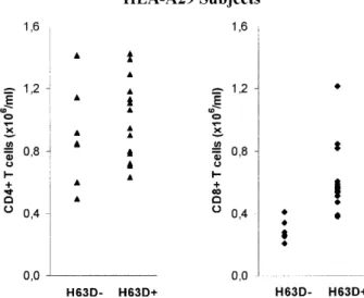
Analysis of total lymphocyte, and CD4
+and CD8
+T-cell counts based on the presence or absence
of the H63D mutation and the HLA-A29 allele
As a result of the strong linkage disequilibrium between
the two distant alleles HLA-A29 and H63D, we addressed
the question of a possible interaction between HFE and
HLA by examining lymphocyte numbers based on the
presence or absence of the relevant alleles. The results
are summarized in Table 4. Statistically significant, higher
average numbers of CD8
+T lymphocytes were observed
in subjects carrying both HLA-A29 and the H63D
muta-tion (0.61±0.22
×
10
6cells/ml) when compared with
sub-jects carrying HLA-A29 without an HFE mutation
(0.29±0.17
×
10
6cells/ml) (Table 4). No effect was seen on
CD4
+T cell numbers (Table 4). This result is illustrated
in Fig. 1.
Discussion
The conjecture that some alleles would be associated
more frequently than expected based on chance was put
forward by Dausset some years ago (referred to in Dausset
1998). Many examples of linkage disequilibria have
since been described among the alleles within the HLA
system, apparently as normal polymorphisms. We
con-firm the linkage disequilibrium between the HLA-A3-B7
and the C282Y mutation of the HFE gene, and
demon-strate in a large sample of normal chromosomes the
link-age disequilibrium between the HLA-A29 allele and the
H63D mutation. In the case of the C282Y-A3-B7 linkage
disequilibrium, a founder effect and a genetic drift are
presently the most widely accepted explanations,
assum-ing a recent age for the C282Y mutation (Ajioka et al.
1997; Thomas et al. 1998). In contrast, the H63D
muta-tion is thought to be older in origin based on the high
allele frequencies reported and a wider geographical
dis-tribution (reviewed in Merryweather-Clarke et al. 2000;
Porto and De Sousa 2000). Thus, the maintenance of its
linkage disequilibrium with HLA-A29 needs a more
elab-orate explanation. In general, linkage disequilibrium is
expected to diminish with recombination map distance
(Abecasis et al. 2001; Cargill et al. 1999; Kruglyak
1999; Reich et al. 2001). Strong linkage disequilibrium
at a large physical distances could be explained by: (1) a
low recombination rate in the chromosomal region; (2) a
strong founder effect by a recent allele/mutation; or (3)
selection. The MHC-class I region has approximately
one fifth of the expected recombination rate under the
usual rule that 1 Mb corresponds to approximately 1 cM
(Martin et al. 1995). This has been confirmed for a 6 Mb
region containing HFE (Malfroy et al. 1997). However,
in our view, it is improbable that the low recombination
rate of the region could suffice to explain the strong
link-age disequilibrium observed with this particular allele,
knowing that both the H63D and HLA-A29 alleles are
common and distributed worldwide. This also argues
against a recent founder effect. Thus, an additional
alter-haplotypes (0.435, P=0.00003), particularly in those
haplotypes carrying HLA-A29 without B44 (0.515,
P=0.00037). No differences were seen in H63D
frequen-cies in relation to the HLA-B44 allele. A more detailed
description of HLA alleles and haplotypes in
chromo-somes grouped according to the presence or absence of
the H63D mutation is given in Table 3. No significant
differences were observed for the HLA-B44 frequencies
in the two groups of chromosomes. A strong and
signifi-cant enrichment of the HLA-A29 allele and, consequently,
all HLA-A29-carrying haplotypes, was observed in
chro-mosomes carrying the H63D mutation (P<0.00001). The
most common single haplotype is HLA-A29-B44, as
expected by the linkage disequilibrium that exists between
HLA-A29 and HLA-B44 in the general population, and
also in this particular population (see Materials and
methods). Therefore, the difference between the two
groups of chromosomes in terms of the frequency of
HLA-A29-B44 is not so marked as for other HLA-A29
containing haplotypes, which are uncommon in
chromo-somes without H63D (frequency=0.050), and
sig-nificantly enriched in H63D-carrying chromosomes
(frequency=0.207). In those haplotypes, no specific HLA-B
allele was seen to be more significantly associated with
HLA-A29 (see Table 3). No differences were observed
between the two groups of chromosomes for other
HLA-B44 containing haplotypes. Altogether, the results
demonstrate the strong linkage disequilibrium between
the H63D mutation and the HLA-A29 allele itself
(D=0.036; D’=0.290; P=0.00037) in a control Portuguese
population, and do not confirm the hypothesis that the
H63D mutation influences the linkage disequilibrium
between the HLA-A29 and B44 alleles.
Fig. 1 CD4+and CD8+T cell subpopulations in HLA-A29 subjects according to the presence (H63D+) or absence (H63D--) of the
H63D HFE mutation. Significantly higher CD8+T cell numbers but not CD4+T cell numbers were observed in HLA-A29 subjects which were carriers of the H63D mutation. No significant differences in CD4+and CD8+T cell numbers were observed among subjects based on the presence or absence of any of the alleles alone (data not shown)
native explanation could be the co-selection of this
particular combination of alleles imposed by some
bio-logical advantage. In this context, the present finding of
significantly higher numbers of CD8
+T lymphocytes in
HLA-A29 subjects carrying the H63D mutation is of
con-siderable interest. The influence of MHC-class I antigens
on the setting of CD4:CD8 ratios and T-lymphocyte
numbers is documented in mice (van Meerwijk et al.
1998), rats (Damoiseaux et al. 1999) and in humans (for
review see Price et al. 1999). To our knowledge, this is
the first demonstration of the impact of an HLA-A allele
(HLA-A29) and a mutation in a non-classical MHC-class
I gene located 4 Mb away (H63D) on lymphocyte
num-bers. The consistent finding of a phenotypic association
between low lymphocyte numbers and high hepatic iron
storage in HH patients (Porto et al. 1997, 1998, 2001)
and in lymphocyte-defective knockout mice (De Sousa
et al. 1994; Santos et al. 1996, 2000), led us to the
pres-ent finding of significantly higher numbers of CD8
+T
cells in HLA-A29 normal subjects carrying the H63D
mutation. This observation may give us some insight
into the mechanism whereby the lymphocytes could
con-tribute to the regulation of iron metabolism. The
postu-lated influence of the H63D mutation on the regulation
of the transferrin receptor-mediated iron uptake (Feder et
al. 1998) occurring in a specific MHC class I
back-ground could contribute both to the setting of CD8
+numbers and to the regulation of transferrin iron loading.
Activated T lymphocytes express transferrin receptors
(Pattanapanyasat and Hoy 1991). Both activated and
non-activated T lymphocytes synthesize ferritin but do
not secrete (Dorner et al. 1980; Pattanapanyasat and Hoy
1991; Pollack et al. 1983). Lymphocytes could therefore
act as a “mobile” and easily “mobilizable” iron-storage
compartment protecting from iron-mediated toxicity, as
originally postulated by De Sousa (1978). Finally, we
could speculate that in human evolution individuals with
higher numbers of CD8
+T lymphocytes might have been
better equipped to survive life-threatening viral
epi-demics. Further studies are currently being done to
clarify the expression of -related proteins and genes in
lymphocytes.
Acknowledgements We gratefully acknowledge Jorge Vieira for many valuable discussions. We also acknowledge the technical staff from the North Histocompatibility Centre for HLA typing. This work was funded in part by FCT grant POCTI/32986/ MGI/2000, the Gulbenkian/FCT Hemochromatosis project and a grant from Innova/APRF (USA). C.S.C. is a recipient of a National Foundation for Science and Technology (FCT) grant (PRAXIS XXI BD/13383/97). We thank Vienna Lab (Austria) for their support with kits for HFE genotyping.
References
Abecasis GR, Noguchi E, Heinzmann A, Traherne JA, Bhattacharyya S, Leaves NI, Anderson GG, Zhang Y, Lench NJ, Carey A, Cardon LR, Moffatt MF, Cookson WO (2001) Extent and distribution of linkage disequilibrium in three genomic regions. Am J Hum Genet 68:191–197
Ajioka RS, Jorde LB, Gruen JR, Yu P, Dimitrova D, Barrow J, Radisky E, Edwards CQ, Griffen LM, Kushner JP (1997) Haplotype analysis of hemochromatosis: evaluation of different linkage-disequilibrium approaches and evolution of disease chromosomes. Am J Hum Genet 60:1439–1447
Alves H, Dias M, Mendes F, Herrero D, Calvo I, Gonçalves R, Adion I, Mendes A, Charron D (2001) Allele and haplotype frequencies for HLA-A, B, C, DRB1 and DQB in random Portuguese families from the north population. Eur J Immuno-genet 28:282 (Abstract 169)
Arosa FA, Porto G, Cabeda JM, Lacerda R, Resende D, Cruz E, Cardoso C, Fonseca M, Simões C, Rodrigues P, Bravo F, Oliveira JC, Alves H, Fraga J, Justiça B, De Sousa M (2000) Expansions of CD8+CD28– and CD8+TcRVβ5.2+ T cells in peripheral blood of alcohol heavy drinkers. Alcoholism Clin Exp Res 24:519–527
Cardoso CS, Oliveira P, Oberkanins C, Mascarenhas M, Rodrigues P, Miranda C, Kury F, De Sousa M, Porto G (2001) HFE mutations in the normal Portuguese population: regional differences for the frequency of C282Y mutation. Eur J Immuno-genet 28:290 (Abstract 185)
Cargill M, Altshuler D, Ireland J, Sklar P, Ardlie K, Patil N, Shaw N, Lane CR, Lim EP, Kalyanaraman N, Nemesh J, Ziaugra L, Friedland L, Rolfe A, Warrington J, Lipshutz R, Daley GQ, Lander ES (1999) Characterization of single-nucleotide poly-morphisms in coding regions of human genes. Nat Genet 22: 231–238
Cullen LM, Gao X, Easteal S, Jazwinska EC (1998) The hemo-chromatosis 845 G-A mutations: prevalence in non-Caucasian populations. Am J Hum Genet 62:1403–1407
Damoiseaux JG, Cautain B, Bernard I, Mas M, van Breda-Vriesman PJ, Druet P, Fournie G, Saoudi A (1999) A dominant role for the thymus and MHC genes in determining the peripheral CD4/CD8 T cell ratio in the rat. J Immunol 163:2983–2989 Dausset J (1998) Clin d’oeil à la vie – la grande aventure HLA.
Odile Jacob, Paris, France
De Sousa M (1978) Lymphoid cell positioning: a new proposal for the mechanism of control of lymphoid cell migration. Symp Soc Exp Biol 32:393–410
De Sousa M, Reimao R, Lacerda R, Hugo P, Kaufmann SH, Porto G (1994) Iron overload in beta 2-microglobulin-deficient mice. Immunol Lett 39:105–111
De Sousa M, Porto G, Arosa F, Cardoso C, Cabeda JM, Lacerda R, Fraga J (2000) T-lymphocyte expression and function in hemochromatosis. In: Barton JC, Edwards CQ (eds) Hemo-chromatosis: genetics, pathophysiology, diagnosis and treatment. Cambridge University Press, Cambridge, pp 396–407
Dorner MH, Silverstone A, Nishiya K, de Sostoa A, Munn G, de Sousa M (1980) Ferritin synthesis by human T lymphocytes. Science 209:1019–1021
Feder JN, Gnirke A, Thomas W, Tsuchihashi Z, Ruddy DA, Basava A, Dormishian F, Domingo R Jr, Ellis MC, Fullan A, Hinton LM, Jones NL, Kimmel BE, Kronmal GS, Lauer P, Lee VK, Loeb DB, Mapa FA, McClelland E, Meyer NC, Mintier GA, Moeller N, Moore T, Morikang E, Wolff RK (1996) A novel MHC class I like gene is mutated in patients with hereditary haemochromatosis. Nat Genet 13:399–406 Feder JN, Penny DM, Irrinki A, Lee VK, Lebron JA, Watson N,
Tsuchihashi Z, Sigal E, Bjorkman PJ, Schatzman RC (1998) The hemochromatosis gene product complexes with the trans-ferrin receptor and lowers its affinity for ligand binding. Proc Natl Acad Sci U S A 95:1472–1477
Fleming RE, Holden CC, Tomatsu S, Waheed A, Brunt EM, Britton RS, Bacon BR, Roopenian DC, Sly WS (2001) Mouse strain differences determine severity of iron accumulation in Hfe knockout model of hereditary hemochromatosis. Proc Natl Acad Sci U S A 98:2707–2711
Imanishi T, Akaza T, Kimura A, Tokunaga K, Gojobori T (1992) HLA 1991: Proceedings of the Eleventh International Histo-compatibility Workshop and Conference (vol 1). In: Tsuji K, Aizaga M, Sasazuki T (eds) Oxford University Press, Oxford, pp 1065–1074
Porto G, Alves H, Rodrigues P, Cabeda JM, Portal C, Ruivo A, Justica B, Wolff R, De Sousa M (1998) Major histocompatibility complex class I associations in iron overload: evidence for a new link between the HFE H63D mutation, HLA-A29, and non-classical forms of hemochromatosis. Immunogenetics 47: 404–410
Porto G, Cardoso CS, Gordeuk V, Cruz E, Fraga J, Areias J, Oliveira JC, Bravo F, Gangaidzo IT, MacPhail AP, Gomo ZAR, Moyo VM, Melo G, Silva C, Justiça B, De Sousa M (2001) Clinical and genetic heterogeneity in hereditary hemo-chromatosis: association between lymphocyte counts and expression of iron overload. Eur J Haematol 67:110-118 Price P, Witt C, Allcock R, Sayer D, Garlepp M, Kok CC, French
M, Mallal S, Christiansen F (1999) The genetic basis for the association of the 8.1 ancestral haplotype (A1, B8, DR3) with multiple immunopathological diseases. Immunol Rev 167: 257–274
Reich DE, Cargill M, Bolk S, Ireland J, Sabeti PC, Richter DJ, Lavery T, Kouyoumjian R, Farhadian SF, Ward R, Lander ES (2001) Linkage disequilibrium in the human genome. Nature 411:199–204
Sanchez M, Bruguera M, Bosch J, Rodes J, Ballesta F, Oliva R (1998) Prevalence of Cys282Tyr and His63Asp HFE gene mutations in Spanish patients with hereditary hemochromatosis and in controls. J Hepatol 29:725–728
Santos M, Schilham MW, Rademakers LH, Marx JJ, De Sousa M, Clevers H (1996) Defective iron homeostasis in beta 2-micro-globulin knockout mice recapitulates hereditary hemochroma-tosis in man. J Exp Med 184:1975–1985
Santos MM, De Sousa M, Rademakers LH, Clevers H, Marx JJ, Schilham MW (2000) Iron overload and heart fibrosis in mice deficient for both beta2-microglobulin and Rag1. Am J Pathol 157:1883–1892
Simon M, Bourel M, Fauchet R, Genetet B (1976) Association of HLA-A3 and HLA-B14 antigens with idiopathic haemochro-matosis. Gut 17:332–334
Sproule TJ, Jazwinska EC, Britton RS, Bacon BR, Fleming RE, Sly WS, Roopenian DC (2001) Naturally variant autosomal and sex-linked loci determine the severity of iron overload in beta 2-microglobulin-deficient mice. Proc Natl Acad Sci USA 98:5170–5174
Ten Elshof AE, Brittenham GM, Chorney KA, Page MJ, Gerhard G, Cable EE, Chorney MJ (1999) Gamma delta intraepithelial lymphocytes drive tumor necrosis factor-alpha responsiveness to intestinal iron challenge: relevance to hemochromatosis. Immunol Rev 167:223–232
Thomas W, Fullan A, Loeb DB, McClelland EE, Bacon BR, Wolff RK (1998) A haplotype and linkage disequilibrium analysis of the hereditary hemochromatosis gene region. Hum Genet 102: 517–525
Van Meerwijk JP, Bianchi T, Marguerat S, MacDonald HR (1998) Thymic lineage commitment rather than selection causes genetic variations in size of CD4 and CD8 compartments. J Immunol 160:3649–3654
Jaswinska EC (2000) The ancestral haplotype in hemochromatosis. In: Barton JC, Edwards CQ (eds) Hemochromatosis: genetics, pathophysiology, diagnosis and treatment. Cambridge University Press, Cambridge, pp 91–98
Kruglyak L (1999) Prospects for whole-genome linkage disequi-librium mapping of common disease genes. Nat Genet 22: 139–144
Levy JE, Montross LK, Andrews NC (2000) Genes that modify the hemochromatosis phenotype in mice. J Clin Invest 105: 1209–1216
Malfroy L, Roth MP, Carrington M, Borot N, Volz A, Ziegler A, Coppin H (1997) Heterogeneity in rates of recombination in the 6-Mb region telomeric to the human major histocompatibility complex. Genomics 43:226–231
Martin M, Mann D, Carrington M (1995) Recombination rates across the HLA complex: use of microsatellites as a rapid screen for recombinant chromosomes. Hum Mol Genet 4: 423–428
Merryweather-Clarke AT, Pointon JJ, Shearman JD, Robson KJH (1997) Global prevalence of putative haemochromatosis muta-tions. J Med Genet 34:275–278
Merryweather-Clarke AT, Pointon JJ, Jouanolle AM, Rochette J, Robson KJ (2000) Geography of HFE C282Y and H63D muta-tions. Genet Test 4:183–198
Mullighan CG, Bunce M, Fanning GC, Marshall SE, Welsh KI (1998) A rapid method of haplotyping HFE mutations and linkage disequilibrium in a Caucasoid population. Gut 42: 566–569
Murphy S, Curran MD, McNougall N, Callender ME, O’Brien CJ, Middleton D (1998) High incidence of the Cys282Tyr mutation in the HFE gene in the Irish population – implications for haemochromatosis. Tissue Antigens 52:484–488
Oberkanins C, Kazemi-Shirazi L, Kury F, et al (1998) Genotyping of common hereditary haemochromatosis mutations in microtiter plates. Eur J Hum Genet 6 [Suppl 1]:62
Pattanapanyasat K, Hoy TG (1991) Expression of cell surface transferrin receptor and intracellular ferritin after in vitro stimulation of peripheral blood T lymphocytes. Eur J Haematol 47:140–145
Pollack MS, da Silva BM, Moshief RD, Groshen S, Bognacki J, Dupont B, De Sousa M (1983) Ferritin secretion by human mononuclear cells: association with HLA phenotype. Clin Immunol Immunopathol 27:124–134
Porto G, De Sousa M (2000) Variation of hemochromatosis preva-lence and genotype in national groups. In: Barton JC, Edwards CQ (eds) Hemochromatosis: genetics, pathophysiology, diagnosis and treatment. Cambridge University Press, Cambridge, pp 51–62
Porto G, Vicente C, Teixeira MA, Martins O, Cabeda JM, Lacerda R, Goncalves C, Fraga J, Macedo G, Silva BM, Alves H, Justica B, De Sousa M (1997) Relative impact of HLA pheno-type and CD4/CD8 ratios on the clinical expression of hemo-chromatosis. Hepatology 25:397–402