RESUMO.- [Caracterização histomorfológica e imuno--histoquímica de 172 neoplasias cutâneas de células redondas em cães.]Este trabalho descreve o uso de um painel de anticorpos (CD117, CD3, CD79a, CD45, citoque-ratina, vimentina e e-caderina em tecidos formalizados e parainizados para o diagnóstico de neoplasias de
Histomorphological and immunohistochemical characterization
of 172 cutaneous round cell tumours in dogs
1Marina Rios Araújo2, Ingred Sales Preis2, Gleidice Eunice Lavalle3, Geovanni Dantas Cassali4
and Roselene Ecco2*
ABSTRACT.- Araújo M.R., Preis I.S., Lavalle G.E., Cassali G.D. & Ecco R. 2012. Histomor-phological and immunohistochemical characterization of 172 cutaneous round cell tumours in dogs. Pesquisa Veterinária Brasileira 32(8):772-780. Setor de Patologia Vete-rinária, Escola de VeteVete-rinária, Universidade Federal de Minas Gerais, Av. Antônio Carlos 6627, Belo Horizonte, MG 31270-901, Brazil. E-mail: [email protected]
This paper describes the use of a panel of antibodies (CD117, CD3, CD79a, CD45, cy-tokeratin, vimentin and E-cadherin) on formalin-ixed, parafin-embedded sections of ca-nine cutaneous round cell tumours. Neoplastic tumours were diagnosed by histology and histochemical stains and included 107 mast cell tumours, 31 cutaneous histiocytomas, two localized histiocytic sarcomas, 21 cutaneous lymphomas, three plasma cell tumours, one transmissible venereal tumour and seven unclassiied round cell tumours. The histologic diagnosis was modiied in 39.5% of the total 172 neoplasms. The staining for CD45 and E-cadherin were variable, and therefore, the inal diagnoses of cutaneous histiocytoma and localized histiocytic sarcoma were made based on histology in association with negative results for CD3, CD79a, CD117 and cytokeratin. The cellular origin of unclassiied round cell tumours was deined in all cases. Cutaneous B-cell lymphoma and plasma cell tumours were CD79a-positive and could be distinguished from each other by the morphological characteristics. Mast cell tumours and T cell lymphoma were CD117and CD3 positive, re-spectively. The positive staining for vimentin and the negative staining for CD3, CD79a, CD117 and cytokeratin favoured the diagnosis of transmissible venereal tumours. Thus, the inal diagnosis of cutaneous round cell tumours should be based on the interpretation of immunohistochemical results together with the cellular morphology observed by histol-ogy. Therefore, more studies to optimize the speciic markers in formalin-ixed, parafin-embedded tissues (especially for histiocytes) are required for deinitive diagnosis of round cell tumours in dogs.
INDEX TERMS: Dogs, skin, round-cells tumours, immunohistochemistry, CD117, CD3, CD79a.
¹ Received on December 12, 2011. Accepted for publication on May 15, 2012.
2 Departamento de Clínica e Cirurgia Veterinária, Escola de Veterinária,
Universidade Federal de Minas Gerais (UFMG), Avenida Antônio Carlos 6627, Pampulha, Belo Horizonte, MG 31270-901, Brazil. *Corresponding author: [email protected]
3 Hospital Veterinário, Escola de Veterinária, UFMG, Belo Horizonte, MG. 4 Laboratório de Patologia Comparada, Instituto de Ciências Biológicas,
UFMG, Belo Horizonte, MG.
foram distinguidos entre si pelas características morfológi-cas. Marcação positiva para vimentina e negativa para CD3, CD79a, CD117 e citoqueratina favoreceram o diagnóstico dos tumores venereos transmissíveis. Assim, o diagnósti-co inal dos tumores de células redondas foram baseados na interpretação dos resultados da imuno-histoquímica em conjunto com a avaliação das características morfoló-gicas observadas na histologia. Finalmente, mais estudos em relação à padronização de marcadores especíicos para tecidos parainizados (especialmente para histiócitos) são necessários para o diagnóstico deinitivo das neoplasias de células redondas em cães.
TERMOS DE INDEXAÇÃO: Cães, pele, neoplasias, celulas redon-das, imunoistoquimica, CD117, CD3 e CD79a .
INTRODUCTION
The skin is one of the most common sites of neoplasms and accounts for 9.5% to 51% of all tumours in dogs (Bronden et al. 2010). Cutaneous round cell tumours may have a simi-lar morphological appearance, and a diagnosis based only on routine histopathology is often challenging (Fernandez et al. 2005). Among these neoplasms are mast cell tumours (MCT) with intermediate differentiation, or grade II, mast cell tumours poorly differentiated, or grade III, cutaneous T-cell lymphoma (CTL), cutaneous B-cell lymphoma (CBL), plasma cell tumours (PCT), cutaneous histiocytomas (HCT) and transmissible venereal tumours (TVT) (Goldschimidt & Hendrick 2002). Accurate diagnosis is essential to deter-mine a prognosis and treatment (Fernandez et al. 2005). Immunohistochemistry (IHC) has been proven to be one of the most important ancillary techniques in the charac-terization of neoplastic diseases in veterinary medicine, as oncologists demand more speciic diagnoses (Ramos-Vara et al. 2008).
MCTs are one of the most common neoplasms in dogs, comprising 7% to 21% of all skin tumours (Rothwell et al. 1987, Yager & Wilcock 1994).These tumours are routinely graded for malignancy, based on histomorphological cha-racteristics (Patnaik et al. 1984). However, the typical in-tracytoplasmic granules of mast cells are more dificult to identify using histology and histochemical stains (Giemsa or toluidine blue) in tumours with intermediate differen-tiation (grade II) and poor differendifferen-tiation (grade III), whi-ch often makes the diagnosis more dificult (Patnaik et al. 1984). Recently, CD117 antibody, or c-KIT, has been used as an immunohistochemical speciic marker for mast cells, supporting the diagnosis of less differentiated MCTs (Re-guera et al. 2000). Now, three KIT-staining patterns have been proposed: membrane-associated staining (KIT I), fo-cal-to-stippled cytoplasmic staining with decreased mem-brane-associated staining (KIT II), and diffuse cytoplasmic staining, (KIT III) (Kiupel et al. 2004). Apparently, tumours with the most aberrant expression (cytoplasmic staining/ KIT II or KIT III) have a worse prognosis (Kiupel et al. 2004, Webster et al. 2006).
Skin neoplasms involving T or B lymphocytes are un-common in dogs (Day 1995). Cutaneous lymphomas (CLs) are classiied into epitheliotropic or non-epitheliotropic
according to their morphological appearance (Gross et al. 2006). Cutaneous epitheliotropic lymphomas have a wide variety of clinical presentations and are characterized by the iniltration of neoplastic T lymphocytes with a speciic tropism for the epidermis and adnexal structures (Moo-re et al. 2009, Fontaine et al. 2010). Non-epitheliotropic forms of cutaneous lymphoma are less common than epi-theliotropic forms. The vast majority of cutaneous canine non-epitheliotropic lymphomas have T cell origin (Gross et al. 2006). Cutaneous B-cell non-epitheliotropic lympho-mas are extremely rare (Day 1995). Similar to what occurs in humans, the determination of the immunophenotype of lymphoma in dogs has become essential because of the re-lationship to biological behavior and response to therapy (Fontaine et al. 2010, Fournel-Fleury et al. 2002). Curren-tly, the immunohistochemical markers most commonly used to characterize T and B lymphocytes in formalin--ixed, parafin-embedded tissues include CD3 (Fournel--Fleury et al. 2002) and CD79a (Milner et al. 1996), res-pectively.
The extramedullary PCTs constitute a neoplastic proli-feration of plasma cells without bone marrow involvement. Cutaneous PCTs are uncommon and are usually benign (Ja-cobs et al 2002). Immunohistochemical markers such as CD79a (Schrenzel et al. 1998) and immunoglobulins (Platz et al. 1999) have been used as ancillaries in the diagnosis of this neoplasm. Variable immunohistochemical expression of the CD79a antigen has been found in PCTs, and positive staining CD79a was present in 56.2% to 80% of cases stu-died (Schrenzel et al. 1998, Vara et al. 1998, Ramos--Vara et al. 2007). The CBL also can be stained by CD79a and immunoglobulin markers. However, cutaneous PCTs and CBL can be distinguished by histomorphologic appe-arance and clinical course (Brunnert & Altman 1991). Cur-rently, the antibody MUM-1 has been indicated for speciic diagnostic of plasma cell tumours (Ramos-Vara et al. 2007). HCT is a benign neoplasm derived from Langerhans cells (Baines et al. 2008).The immunophenotypic markers of Langerhans cells include MHC-I, MHC-II, CD1a, CD1c, CD11b, CD11c, CD18, CD44, CD45, CD45RA, CD49d, ICAM-1 and E-cadherin (Baines et al. 2008, Ginn et al. 2007, Ramos--Vara & Miller 2011). However, most of these antibodies must be used in fresh or frozen tissue, which is not often available to the veterinary. Only the CD45, CD45RA, CD11d, CD18 and E-cadherin antibodies can be used in formalin--ixed, parafin-embedded tissues (Affolter & Moore 2002, Gross et al. 2006, Fulmer & Mauldin, 2007).
This study evaluated a panel of immunohistochemical stains as an additional technique for the diagnosis of 172 cutaneous round cell tumours in dogs.
MATERIALS AND METHODS
One hundred and seventy-two archival canine cutaneous round cell tumours were selected from those diagnosed between 1996 and 2010 at the Laboratory of Veterinary Pathology, Veterinary School, Universidade Federal de Minas Gerais, Brazil. All the tu-mours had been diagnosed using histopathology, but in suspected cases of mast cell tumours, histochemical (Giemsa) stains were also used. The tumours selected included 107 MCTs (35 of grade I, 29 of grade II and 43 of grade III, classiied according to a previous study)(Patnaik et al. 1984), 31 HCTs, two localized histiocytic sar-comas (LHS), 21 CLs, three PCTs, one TVT and seven unclassiied round cell tumours (URCTs).
Parafin blocks were selected, and consecutive three-micro-metre sections were obtained and mounted on silanised slides for immunohistochemical study. A panel of antibodies was used (Table 1), and the choice of antibody was based on the previous histological diagnosis. Heat-induced epitope retrieval using Dako antigen retrieval solution, pH 6.0 (Dako), was performed before dipping the sections in a water bath (20 minutes) for CD117, CD3, CD79a, CD45, cytokeratin AE1/AE3 and vimentin. For E-cadherin, the slides were incubated in an antigen retrieval solution (Dako) (pH =6.0) in a microprocessor-controlled pressure chamber for 2 minutes. The slides were then cooled at room temperature for 20 minutes in the antigen retrieval buffer. The sections were in-cubated at room temperature in 3% (vol/vol) H2O2 for 15 min, in
primary antibodies for 16 h at 4°C (CD117, CD79a, CD45, CD3)
or for 1h at room temperature (cytokeratin AE1/AE3, E-cadherin and vimentin), in a reagent containing secondary antibodies (Ad-vance HRP link, Dako, Carpinteria-CA, USA) for 30 min and a poly-mer reagent (Advance HRP enzyme, Dako, Carpinteria-CA, USA) for 30 minutes. Between incubations, the slides were washed for 2×5 minutes in 10% phosphate-buffered saline solution. The immunoreactivity was visualized using diaminobenzidine (DAB substrate chromogen system, Dako, Carpinteria-CA, USA). The slides were then counterstained with Harris haematoxylin. Posi-tive and negaPosi-tive control slides were included in each batch. As positive controls, the lung (CD45), the lymph node (CD79a) and the thymus (CD3) were used. For CD117, cytokeratin AE1/AE3, E-cadherin and vimentin, the self-tissue was used as an internal control. Negative controls were assessed by using normal serum (Ultra V block, Labvision, Fremont, CA, USA) to replace the prima-ry antibody.
The immunostains for CD79a, CD45, CD3, cytokeratin AE1/ AE3, E-cadherin and vimentin were evaluated qualitatively as po-sitive (+) or negative (-). The MCTs were classiied according to their KIT-staining patterns (Kiupel et al. 2004): membrane-asso-ciated staining (KIT I), focal-to-stippled cytoplasmic staining with
Table 1. Panel of antibodies used for immunohistochemistry
Target antigen Clone Dilution Manufacturer
CD117 (c-KIT) --- 1:800 Dako (Carpinteria/CA, USA) CD79a HM47/A9 1:500 DBS (Pleasanton/CA, USA)
CD45 CA1210C12 1:200 University of California, Davis/
CA, USA
CD3 CD3-12 1:500 University of California, Davis/
CA, USA
Cytokeratin AE1/ AE1/AE3 1:100 Dako (Carpinteria/CA, USA) AE3
E-caderin 4A2C7 1:60 Invitrogen (Camarillo/CA, USA) Vimentin V9 1:50 Dako (Carpinteria/CA, USA)
Table 2. Expected immunohistochemical results for canine cutaneous round cell tumours
CD117 CD3 CD79a CD45 CK E-cad Vim
MCT + - - + - - +
HCT - - - +/- - +/-
PCT - - +/- - - - +
CTL - + - +/- - -
CBL - - + +/- - -
TVT - - - +/- - - +
LHS - - - +/- - +/- +
MCT = mast cell tumour; HCT = cutaneous histiocytoma; PCT = plasma cell tumour; CTL = cutaneous T cell lymphoma; CBL = cutaneous B cell lymphoma; TVT = transmissible venereal tumour; LHS = localized his-tiocytic sarcoma; + positive; - negative; +/- variable; CK = cytokeratin AE1/AE3; E-cad = E-cadherin; Vim = vimentin
RESULTS
The stains for CD45 and E-cadherin were variable, and the-refore, the inal diagnosis of HCT and LHS were made ba-sed on histology in association with the negative results for CD3, CD79a, CD117 and cytokeratin AE1/AE3. Thus, CTL, CBL, PCT, MCT and tumour or basal cell carcinoma were ruled out based on the negative staining for CD3, CD79a, CD117 and cytokeratin AE1/AE3, respectively. The staining for cytokeratin AE1/AE3 was used in the neoplasms in whi-ch cords of cells were observed arranged near and perpen-dicular to the epidermis, similar to the tumour or basal cell carcinoma. The results of the panel of antibodies for all the tumours and the inal diagnoses are summarized in Table 3.
Mast cell tumours
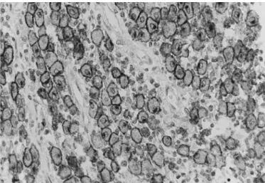
Of the 107 tumours that were diagnosed histologically as MCTs, 78 (72.9%) were conirmed by IHC. All except one (34/35) of the MCTs in grade I, (27/29) grade II and (17/41) grade III were positive for CD117 and were clas-siied as pattern KIT I (42/78, 53.85%) (Figure 1), pat-tern KIT II (24/78, 30.77%) (Figure 2) and patpat-tern KIT III (12/78, 15.38%).
Nine HCTs (Figure 3) and one CL that had been diag-nosed histologically were positive for CD117 and deined as MCTs. These tumours were negative for CD45, CD3 and CD79a.
Histiocytoma
Of the 31 tumours that were diagnosed histologically as HCT, eight (25.81%) were conirmed. Seventeen tumours not diagnosed by histology as HCT were classiied as HCT using CD45 and E-cadherin (Figure 4) as markers, in asso-ciation with morphological characteristics (especially large decreased membrane-associated staining (KIT II) and diffuse cytoplasmic staining (KIT III). Each MCT was assigned one of the-se three staining patterns bathe-sed on the highest staining pattern (staining patterns I versus II versus III) present in at least 10% (estimated based on 100 neoplastic cells in a high-power ield) of the neoplastic cell population or present in large clusters of neo-plastic cells within the tumour. Expected results for each tumour type are summarized in Table 2.
Table 3. Immunohistochemical stain panel with results for 172 canine cutaneous round cell tumours
Histologic Nº Final CD117 CD3 CD79a CD45 CK E-cad Vim diagnosis tumours diagnosis
MCT I 24 MCT KIT I ND ND ND ND ND ND MCT I 10 MCT KIT II ND ND ND ND ND ND MCT II 16 MCT KIT I ND ND ND ND ND ND MCT II 7 MCT KIT II ND ND ND ND ND ND MCT II 4 MCT KIT III ND ND ND ND ND ND MCT III 2 MCT KIT I ND ND ND ND ND ND MCT III 7 MCT KIT II ND ND ND ND ND ND MCT III 8 MCT KIT III ND ND ND ND ND ND
MCT III 6 HCT - - - - - + ND
MCT III 1 HCT - - - - - - ND
MCT III 1 LHS - - - + - ND ND
MCT III 1 LHS - - - - - ND ND
MCT II 1 CTL - + - + ND ND ND MCT III 3 CTL - + - + ND ND ND MCT III 9 CTL - + - - ND ND ND MCT I 1 PCT - - - - ND ND ND MCT II 1 PCT - - + - ND ND ND MCT III 1 PCT - - + - ND ND ND MCT III 1 PCT - - - - ND ND ND
MCT III 1 TVT - - - - - ND +
MCT III 1 TVT - - - + ND ND + MCT III 1 TVT - - - - ND ND +
HCT 4 HCT - - - + - + ND
HCT 3 HCT - - - + ND
HCT 1 HCT - - - ND
HCT 1 MCT KIT I - - - ND ND ND HCT 3 MCT KIT II - - - ND ND ND HCT 5 MCT KIT III - - - ND ND ND
HCT 1 LHS - - - - ND - ND
HCT 4 CTL ND + - + ND ND ND
HCT 4 CTL ND + - - ND ND ND
HCT 1 PCT - - - - ND ND ND
HCT 1 PCT - - + - ND ND ND
HCT 1 PCT ND - - - ND ND ND
HCT 1 TVT ND - - - - ND +
HCT 1 TVT - - - - - ND +
CL 7 CTL ND + - + ND ND ND
CL 5 CTL ND + - - ND ND ND
CL 1 CBL ND - + - ND ND ND
CL 2 HCT - - - - - + ND
CL 1 HCT - - - + - - ND
CL 3 HCT - - - - - - ND
CL 1 LHS - - - - ND ND ND
CL 1 MCT KIT III - - - ND ND ND
LHS 1 LHS - - - + ND ND ND
LHS 1 LHS - - - - ND ND ND
PCT 2 PCT ND - + - ND ND ND
PCT 1 HCT - - - - - - ND
TVT 1 TVT ND - - + ND ND +
URCT 3 HCT - - - - - - ND
URCT 1 CBL ND - + - ND ND ND
URCT 1 CBL - - + + ND ND ND
URCT 1 CTL - + - + ND ND ND
URCT 1 TVT - - - - - ND +
MCT I = mast cell tumour grade I; MCT II = mast cell tumour grade II; MCT III = mast cell tumour grade III HCT = cutaneous histiocytoma; LHS = localised histiocytic sarcoma; CL = cutaneous lymphoma; CTL = cutaneous T-cell lymphoma; CBL = cutaneous B-cell lymphoma; PCT = plasma cell tumour; TVT = transmissible venereal tumour; URCT = un-classiied round cell tumour; ND = not done; + positive; - negative; KIT I = membrane-associated staining; KIT II = focal-to-stippled cytoplasmic staining with decreased membrane-associated staining; KIT III = diffuse cytoplasmic staining; CK = cytokeratin AE1/AE3; E-cad = E-cadherin; Vim = vimentin
Fig.3. Skin, Dog 12: Mast cell tumour. Neoplastic mast cells charac-terized by diffuse cytoplasmic staining, KIT-staining pattern III. Chromogen DAB; Mayer’s Haematoxylin counter stain, 600x.
Fig.1. Skin, Dog 55: Mast cell tumour. Neoplastic mast cells cha-racterized by membrane-associated staining with little or no cytoplasmic staining, KIT-staining pattern I. Chromogen DAB, Mayer’s Haematoxylin counter stain, 600x.
round cells with abundant amphophilic cytoplasm and nu-cleus round to indented): seven (41.2%) MCTs, six (35.3%) CLs, one (5.9%) PCT and three (17.6%) URCTs.
Localized histiocytic sarcoma
The two tumours that were diagnosed histologically as LHS were conirmed. Two MCTs (50.0%), a CL (25%) and a HCT (25%) that had been diagnosed histologically were deined as LHS using IHC (negative for CD117, CD3 and CD79). The differentiation between the HCT and LHS was performed by observation of the morphology and cellular characteristics of malignancy.
Cutaneous lymphoma
Of the 21 tumours that were diagnosed histologically as CL, 13 (61.9%) were conirmed by IHC. CTLs were the most common (12/13, 92.3%) based on positive CD3 stai-ning, and only one CBL (1/13, 7.69%) was observed and characterized by staining positive for CD79a. Of the 12 tu-mours diagnosed as CTL, seven (58.33%) were classiied
as epitheliotropic T-cell lymphoma (Figure 5 and 6) and were characterized by the presence of tumour cells stai-ning positive for CD3 in the epidermis, arranged diffusely or in small groups (Pautrier microabscesses) and also in the epithelium of hair follicles and the apocrine and seba-ceous glands. Of the 12 tumours diagnosed as CTL, three (25.0%) were classiied as non-epitheliotropic T-cell lym-phoma. The tumour cells positive for CD3 were located in the supericial dermis, near the epidermis but with no close contact between them, or were located in the deep dermis. Two (16.67%) of the 12 tumours diagnosed as CTL could not be classiied as epitheliotropic or non-epitheliotropic due to ulceration of the epidermis and the hair follicles and the fact that the glands were not visible.
Twenty-two tumours diagnosed histologically as MCTs (13/22, 59.09%), HCTs (8/22, 36.36%) and URCT (1/22, 4.5%) were deined as CLT based on positive CD3 staining. Of these 22 tumours, eight (36.36%) were classiied as lym-phoma T-cell epitheliotropic, 11 (50.0%) were classiied as lymphoma T-cell non-epitheliotropic, and three (13.64%) Fig.4. Skin, Dog 41: Cutaneous histiocytoma. (a) Magniication 200x, immunostaining positive for E-cadherin in the epidermis (internal control) and in the histiocytes of the supericial dermis. The dense deep portion lacks expression for E-cadherin. (b) Magniication 600x, details of positive stain of the histiocytes in the supericial dermis. Chromogen DAB, Mayer’s Haematoxylin counter stain.
Fig.5. Skin, Dog 22: Lymphoma T-cell epitheliotropic. Neoplastic lymphocytes stained positive for CD3 invading the apocrine gland epithelium (epitheliotropic). Chromogen DAB, Mayer’s Haematoxylin counter stain, 600x.
were not classiied. Two URCTs were deined as CBLs based on positive CD79a staining. The differentiation between CBL and PCT, which can also stain positive for CD79a, was performed by evaluation of cellular morphology.
Plasma cell tumours
Of the three tumours that were diagnosed histologically as PCT, two (61.67%) were conirmed by IHC based on stai-ning positive for CD79a (Figure 7). Seven tumours diagnosed histologically as MCTs (4/7, 57.14%) and HCTs (3/7, 42.86%) were deined as PCT based on the IHC results (CD117 and CD3 negative) in association with histological characteristics.
Transmissible venereal tumours
One tumour that was diagnosed histologically as TVT was conirmed by IHC based on staining positive for vi-mentin (Figure 8). Six tumours classiied histologically as MCTs (3/6, 50.0%), HCTs (2/6, 33.33%) and URCT (1/6, 16.67%) were determined as TVT based on staining positi-ve for vimentin and staining negatipositi-ve for CD3, CD79, CD117 and cytokeratin AE1/AE3.
Unclassiϐied round cell tumours
The diagnosis was obtained for all cases (5/5, 100.0%) of histologically unclassiied round cell tumours with the support of IHC: three (42.86%) HCTs, two (28.57%) CBLs, one (14.29%) CTL and one (14.29%) TVT.
DISCUSSION
In this study, the frequency of all the cutaneous round cell tumours classiied by IHC was different from the frequency of these tumours categorized by histology. The MCTs were the most commonly observed but in different propor-tions, 51.2% (88/172) by IHC versus 62.2% (107/172) by histology. The high prevalence of MCTs was expected be-cause this neoplasm represents 7% to 21% of all skin tu-mours (Rothwell et al. 1987, Yager & Wilcock 1994). The frequency of CL diagnosed by IHC was 1.8 times higher (21.5%, 37/172) compared to histology (12.2%, 21/172). The frequency of CTL (19.8%, 34/172) was more common than CBL (1.7%, 3/172). The frequency of HCT decreased when diagnosed with the support of IHC (14.5%, 25/172) compared with histology (18.02%, 31/172). In this stu-dy, the frequency of CLs was higher than HCTs. Studies on the prevalence of skin tumours in dogs show that the frequency of HCTs is greater than CLs, but in these cases, only histology was used as a diagnostic tool (Kaldrymidou et al. 2002, Pakhrin et al. 2007). The diagnosis of PCT, LHS and TVT using IHC (5.2%, 9/172, 3.5%, 6/172 and 4.1%, 7/172, respectively) was greater than the diagnoses using only histology (1.74%, 3/172, 1.17%, 2/172 and 0.06%, 1/172, respectively).
The antibody CD117 (c-KIT), as expected, proved to be extremely useful for the diagnosis of MCTs, especially for grade III. In this study, the three patterns of KIT-staining described by Kiupel et al. (2004) were observed. Of the tu-mours diagnosed histologically as CL, 4.8% (1/21) were po-sitive for CD117 and were re-diagnosed as MCTs. Similarly, 2.9% (9/31) of the tumours previously diagnosed as HCT were positive for CD117 and were re-diagnosed as MCTs. In 90% (9/10) of these cases, cytoplasmic staining for CD117 (KIT II or KIT III) was present, indicating that the tumours were less differentiated. Histologically, the intracytoplas-mic granules typical of mast cells are more dificult to visu-alize in tumours with intermediary differentiation (grade II) and poor differentiation (grade III) (Patnaik et al. 1984). Special stains such as toluidine blue and Giemsa are used to improve the visibility of these granules. In many cases, the granules are not seen even with the use of the histochemi-cal stains. Thus, IHC becomes a vital tool to help histology provide a more deinitive diagnosis of round cell tumours, especially in cases of poorly differentiated MCTs (Reguera et al. 2000).
CD45 is a common leukocyte antigen, and Moore et al. (1996)and Affolter & Moore (2002)report the expression of this marker in HCTs and LHSs, respectively. In this stu-dy, approximately 20% (5/25) and 33.33% (2/6) of the tumours deined as HCTs and LHSs, respectively, were po-sitive for CD45. The expression of CD45 is also reported in CTLs (Moore et al. 1994), CBLs (Gross et al. 2006), PCTs (Schrenzel et al. 1998), TVTs (Sharkey & Willman 2011) Fig.8. Skin, Dog 90: Transmissible venereal tumour. Round cells
with large nucleus showing positive staining for vimentin. Chromogen DAB, Mayer’s Haematoxylin counter stain, 600x Fig.7. Skin, Dog 4: Plasma cell tumour. Neoplastic plasma cells
and MCTs (Gross et al. 2006, Jakab et al. 2009). In this stu-dy, 47% (16/34) of the CTLs, 33.33% (1/3) of the CBLs and 28.6% (2/7) of the TVTs were positive for CD45. The lack of expression or weak expression of the common leukocyte antigen CD45 has been described in cases of CTLs (Affol-ter et al. 2009). Therefore, the most likely hypothesis that could explain the lack of positive staining for CD45 obser-ved in this study would be that the loss of differentiation in the cells would not permit the expression of the antigen on the cell surface.
In 60% (15/25) of all tumours with a inal diagnosis of HCT, positive staining for E-cadherin was observed. In all these cases, immunostaining was observed in tumour cells near and arranged perpendicular to the epidermis. According to Ramos-Vara & Miller (2011), the intensity of labelling for E-cadherin in HCTs decreases with increa-sing distance from the epidermis due to the maturation of Langerhans cells or the immune response during tumour regression. According to Baines et al (2008), although the immunostaining for E-cadherin in HTCs is well known, Gross et al. (2006) reported that the lack of expression of this marker can be observed. In this study, 40% (10/25) of the tumours were negative for E-cadherin, probably due to the differentiation of the tumour cells. The deinitive diag-nosis of tumours of histiocytic origin is challenging given the lack of speciic cell markers in formalin-ixed, parafin--embedded tissues. In a study in humans, Vos et al. (2005)37 demonstrated that the antibody CD163 could be an appa-rently speciic marker for histiocytes.
The single case of LHS submitted to IHC for E-cadherin was negative. Information about immunostaining for E--cadherin in LHS is controversial. According to Gross et al (2006), these tumours do not express E-cadherin, but re-cently Ramos-Vara & Miller (2011)described positive stai-ning for E-cadherin in ive histiocytic sarcomas analysed.
The cytokeratin AE1/AE3 is expressed in all tumours of epithelial origin. Basal cell tumour and basal cell carcinoma (tumours originating from the basal layer of the epidermis) have morphological characteristics similar to HCT, that is, a growth pattern near the epidermis in cords of cells ar-ranged perpendicular to the epidermis (Gross et al. 2006). In this study, the cytokeratin was used in tumours that had those morphological characteristics and that were negati-ve for CD45 to rule out a possible epithelial origin. None of the tumours showed reactivity for cytokeratin and CD3, CD79, CD117 as well. Thus, the inal diagnosis of HCT was made by the association of the combined results of the IHC analysis and the morphological characteristics of the neo-plastic cells. HCTs are constituted by proliferation of large round cells with abundant amphophilic cytoplasm and nu-cleus round to indented. A growth pattern is characterized by cord of cells arranged perpendicular to the epidermis (Gross et al. 2006).
Vimentin is a marker of mesenchymal cells and is ex-pressed in the TVT (Pereira et al. 2000), in the MCT (Jakab et al. 2009, Lin et al. 2010), in the PCT (Baer et al. 1989, Schrenzel et al. 1998) and in the LHS (Azakami et al. 2006). In this study, the tumours deined as TVT by IHC were po-sitive for vimentin in 100% (6/6) of the cases and
positi-ve for CD45 in 28.6% (2/7) of the cases. All of them were negative for CD117, CD3 and CD79. The tumours submit-ted to immunostaining for cytokeratin AE1/AE3 also were negative. This panel of antibodies rule out the possibilities of poorly differentiated MCT, CTL, CBL, PCT or poorly diffe-rentiated carcinoma. Thus, the positive staining for vimen-tin favours the diagnosis of TVT. The possibility of HCT and amelanotic melanoma was also ruled out by the evaluation of the morphological characteristics of the tumour cells. The TVT neoplastic cells had large nuclei, scant cytoplasm, a high mitotic index and cellular monotony, were someti-mes vacuolated, and their predominant location was in the dermis and not near and perpendicular to the epidermis, as observed in HCTs (Gross et al. 2006).
Although the reactivity of CD79a in canine PCT and CBL is well established, there are large variations in the percen-tage of cases and number of cells stained with this marker, with up to 80% of PCTs expressing CD79 (Schrenzel et al. 1998, Ramos-Vara et al. 1998). Other authors observed po-sitive staining for CD79a in 59 (56.2%) of 105 cases of PCT (Ramos-Vara et al. 2007).In the present study, the tumours deined as PCTs were positive for CD79a in 66.66% (6/9) of the cases, and the labelling was predominantly cytoplasmic diffuse. The differentiation between the PCT and CBL, whi-ch also expressed CD79 (3/3, 100%), was performed by evaluation of the cellular morphology because all the PCTs had, in different proportions, well-differentiated cells cha-racteristic of plasma cells, a perinuclear halo (Golgi zone), a peripheral nucleus, and abundant amphophilic cytoplasm, including cells with Russel’s corpuscle (Mott cell), to sup-port the diagnosis (Ginn et al. 2007).
The CD3 is a speciic marker for T lymphocytes (Four-nel-Fleury et al. 2002). In this study, 92% (34/37) of the CLs were positive for CD3. The frequency of T lympho-mas, T-cell epitheliotropic and non-epitheliotropic, were very close. Approximately 44% (15/34) were classiied as lymphoma T-cell epitheliotropic and 41% (14/34) as lymphoma T-cell non-epitheliotropic. According to Gross et al (2006) 12, lymphoma T-cell non-epitheliotropic is much less common than lymphoma T-cell epitheliotropic, and the lymphoma B-cell non-epitheliotropic is still more rare.
select the antibodies and support the interpretation of the results from IHC. Because IHC is a relatively expensive diag-nostic procedure, pathologists tend to use only one or two antibodies to conirm diagnostic suspicions in some cases. Considering these conditions, CD117 is very useful to dis-card or conirm the diagnostic of undifferentiated mast cell tumours.
Finally, more studies on the standardization of speciic markers in formalin-ixed, parafin-embedded tissues (es-pecially for histiocytes and transmissible venereal tumor) are required for deinitive diagnosis of cutaneous round cell tumours in dogs.
Acknowledgments.- This study was supported by the Pró-Reitoria de Pesquisa da Universidade Federal de Minas Gerais (PRPq-UFMG), Con-selho Nacional de Desenvolvimento Cientíico e Tecnológico (CNPq) and Fundação de Amparo à Pesquisa (FAPEMIG), Brasil.
REFERENCES
Affolter V.K. & Moore P.F. 2002. Localized and disseminated histiocytic sar-coma of dendritic cell origin in dogs. Vet. Pathol. 39(1):74-83.
Affolter V.K., Gross T.L. & Moore P.F. 2009. Indolent cutaneous T-cell lym-phoma presenting as cutaneous lymphocytosis in dogs. Vet. Dermatol. 20(5/6):577-85.
Azakami D., Bonkobara M., Washizu T., Iida A., Kondo M., Kato R., Niikura Y., Iwaki S., Tamahara S., Matsuki N. & Ono K. 2006.Establishment and biological characterization of canine histiocytic sarcoma cell lines. J. Vet. Med. Sci. 68(12):1343-1346.
Baer K.E., Patnaik A.K., Gilbertson S.R. & Hurvitz A.I. 1989. Cutaneous plas-macytomas in dogs: A morphologic and immunohistochemical study. Vet. Pathol. 26(3):216-221.
Baines S.J., McInnes E.F. & McConnell I. 2008. E-cadherin expression in ca-nine cutaneous histiocytomas. Vet. Rec. 162(16):509-513.
Bronden L.B., Eriksen T. & Kristensen A.T. 2010. Mast cell tumours and other skin neoplasia in Danish dogs: Data from the Danish Veterinary Cancer Registry. Acta Vet. Scand. 52(6). Available at <http://www.acta-vetscand.com/content/ 52/1/6> Accessed on Jan. 3, 2011.
Brunnert S.R. & Altman N.H. 1991. Identiication of immunoglobulin light chains in canine extramedullary plasmacytomas by thiolavine T and immunohistochemistry. J. Vet. Diagn. Invest. 3(3):245-251.
Day M.J. 1995. Immunophenotypic characterization of cutaneous lym-phoid neoplasia in the dog and cat. J. Comp. Pathol. 112(1):79-96. Fernandez N.J., West K.H., Jackson M.L. & Kidney B.A. 2005.
Immunohisto-chemical and HistoImmunohisto-chemical stains for differentiating canine cutaneous round cell tumors. Vet. Pathol. 42(4):437-445.
Fontaine J., Heimann M. & Day M.J. 2010. Canine cutaneous epitheliotropic T-cell lymphoma: A review of 30 cases. Vet. Dermatol. 21(3):267-275. Fournel-Fleury C., Ponce F., Felman P., Blavier A., Bonnefont C., Chabanne
L., Marchal T., Cadore J.L., Goy-Thollot I., Ledieu D., Ghernati I. & Magnol J.P. 2002. Canine T-cell lymphomas: A morphological, immunological, and clinical study of 46 new cases. Vet. Pathol. 39(1):92-109.
Fulmer A.K. & Mauldin G.E. 2007. Canine histiocytic neoplasia: An over-view. Can. Vet. J. 48(10):1041-1050.
Ginn P.E., Mansell J.E.K.L. & Rakich P.M. 2007. The skin and appendages, p.553-781. In: Maxie M.G. (Ed.), Jubb, Kennedy, and Palmer’s Pathology of Domestic Animals. Vol.1. Saunders Elsevier, Philadelphia.
Goldschmidt M.H. & Hendrick M.J. 2002. Tumors of the skin and soft tis-sues, p.44-117. In: Meuten J.D. (Ed.), Tumors in Domestic Animals. Iowa State University Press, Ames.
Gross T.L., Ihrke P., Walder E.J. & Affolter V.K. 2009. Skin diseases of the dog and cat: clinical and histopathologic diagnosis. 2nd ed. Blackwell
Pu-blishing, Ames. 932p.
Jacobs R.M., Messick J.B. & Valli V.E. 2002. Tumors of the hemolymphatic system, p.119-198. In: Meuten J.D. (Ed.), Tumors in Domestic Animals. Iowa State University Press, Ames.
Jakab C., Szász A.M., Kulka J., Schaff Z., Rusvai M., Németh T. & Gáli P.2009. Cutaneous mast cell tumour within a lipoma in a boxer.Acta Vet. Hung. 57(20):263-274.
Kaldrymidou H., Leontides L., Koutinas A.F., Saridomichelakis M.N. & Ka-rayannopoulou M. 2002. Prevalence, distribution and factors associated with the presence and the potential for malignancy of cutaneous neo-plasms in 174 dogs admitted to a clinic in northern Greece. J. Vet. Med. A, Physiol. Pathol. Clin. Med. 49(2):87-91.
Kiupel M., Webster J.D., Kaneene J.B., Miller R. & Yuzbasiyan-Gurkan V. 2004. The use of KIT and Tryptase expression patterns as prognostic tools for canine cutaneous mast cell tumors. Vet. Pathol. 41(4):371-377. Lin T.Y., Hamberg A., Pentecost R., Wellman M. & Stromberg P. 2010. Mast
cell tumors in a llama (Lama glama). J. Vet. Diagn. Invest. 22(5):808-811. Milner R.J., Pearson J., Nesbit J.W. & Close P. 1996.Immunophenotypic
classiication of canine malignant lymphoma on formalin-mixed para-fin wax-embedded tissue by means of CD3 and CD79a cell markers. On-derstepoort J. Vet. Res. 63(4):309-313.
Moore P.F., Affolter V.K., Graham P.S. & Hirt B. 2009. Canine epitheliotropic cutaneous T-cell lymphoma: An investigation of T-cell receptor immu-nophenotype, lesion topography and molecular clonality. Vet. Dermatol. 20(5/6):569-576.
Moore P.F., Olivry T. & Naydan D. 1994. Canine cutaneous epitheliotropic lymphoma (mycosis fungoides) is a proliferative disorder of CD8+ T cells. Am. J. Pathol. 144(2):421-429.
Moore P.F., Schrenzel M.D., Affolter V.K., Olivry T. & Naydan D. 1996. Ca-nine cutaneous histiocytoma is an epidermotropic Langerhans cell his-tiocytosis that expresses CD1 and speciic f32-integrin molecules. Am. J. Pathol. 148(5):1699-1708.
Pakhrin B., Kang M.S., Bae I.H., Park M.S., Jee H., You M.H., Kim J.H., Yoon B.I., Choi Y.K. & Kim D.Y. 2007. Retrospective study of canine cutaneous tumors in Korea. J. Vet. Sci. 8(3):229-236.
Park M.S., Kim Y., Kang M.S., Oh S.Y., Cho D.Y., Shin N.S. & Kim D.Y. 2006. Disseminated transmissible venereal tumor in a dog. J. Vet. Diagn. In-vest. 18(1):130-133.
Patnaik A.K., Ehler W.J & Macewen E.G. 1984. Canine cutaneous mast cell tumor: Morphologic grading and survival time in 83 dogs. Vet. Pathol. 21(5):468-74.
Pereira J.S., Silva A.B. & Martins A.L. 2000. Immunohistochemical charac-terization of intraocular metastasis of a canine transmissible venereal tumor. Vet. Ophthalmol. 3(1):43-47.
Platz S.J., Breuer W., Pleghaar S., Minkus G., Hermanns W. 1999. Prognos-tic value of histopathological grading in canine extramedullary plas-macytomas. Vet. Pathol. 36(1):23-27.
Ramos-Vara J.A. & Miller M.A. 2011. Immunohistochemical expression of E-cadherin does not distinguish canine cutaneous histiocytoma from other canine round cell tumors. Vet. Pathol. 48(3):758-763.
Ramos-Vara J.A., Kiupel M., Baszler T., Bliven L., Brodersen B., Chelack B., Czub S., Del Piero F., Dial S., Ehrhart E.J., Graham T., Manning L., Paulsen D., Valli V.E. & West K. 2008. Suggested guidelines for immunohistoche-mical techniques in veterinary diagnostic laboratories. J. Vet. Diagn. In-vest. 20(4):393-413.
Ramos-Vara J.A., Miller M.A. & Valli V.E. 2007. Immunohistochemical de-tection of multiple myeloma 1/interferon regulatory factor 4 (MUM1/ IRF-4) in canine plasmacytoma: Comparison with CD79a and CD20. Vet. Pathol. 44(6):875-884.
Ramos-Vara J.A., Miller M.A., Pace L.W., Linke R.P., Common R.S. & Wat-son G.L. 1998. Intestinal multinodular Al-amyloid deposition associated with extramedullary plasmacytoma in three dogs: Clinicopathological and immunohistochemical studies. J. Comp. Pathol. 119(3):239-249. Reguera M.J., Rabanal R.M., Puigdemont A. & Ferrer L. 2000. Canine mast
Rothwell T.L., Howlett C.R., Middleton D.J., Grifiths D.A. & Duff B.C. 1987. Skin neoplasms of dogs in Sydney. Aust. Vet. J. 64(6):161-164.
Schlafer D.H. & Miller R.B. 2007. Female genital system, p.429-564. In: Ma-xie M.G. (Ed.), Jubb, Kennedy, and Palmer’s Pathology of Domestic Ani-mals. Vol.3. Saunders Elsevier, Philadelphia.
Schrenzel M.D., Naydan D.K. & Moore P.F. 1998. Leukocyte differentiation antigens in canine cutaneous and oral plasmacytomas. Vet. Dermatol. 9(1):33-41.
Sharkey L.C. & Wellman M.L. 2011. Diagnostic cytology in veterinary me-dicine: a comparative and evidence-based approach. Clin. Lab. Med. 31(1):1-19.
Vos J.A., Abbondanzo S.L., Barekman C.L., Andriko J.W., Miettinen M. & Aguilera N.S. 2005.Histiocytic sarcoma: A study of ive cases including the histiocyte marker CD163. Mod. Pathol. 18(5):693-704.
Webster J.D., Kiupel M. & Yuzbasiyan-Gurkan V. 2006. Evaluation of the kinase domain of c-KIT in canine cutaneous mast cell tumors. BMC. Can-cer. 6:85. Available at <http://www.biomedcentral.com/1471-2407/ 6/85> Accessed on Dec. 20, 2010.