The X-X-/E+E+ genotype of the
Xba
I
/Eco
RI polymorphisms of the
apolipoprotein B gene as a marker
of coronary artery disease in a
Brazilian sample
Departamentos de 1Patologia Médica, 2Genética, and
3Bioquímica, Universidade Federal do Paraná, Curitiba, PR, Brasil
4Departamento de Clínica Médica, Faculdade de Medicina de Ribeirão Preto, Universidade de São Paulo, Ribeirão Preto, SP, Brasil
M. Scartezini1, M.A. Zago4, E.A. Chautard-Freire-Maia2, A. Pazin-Filho4, J.A. Marin-Neto4, J.K.S. Hotta4, A.J. Nascimento3 and J.E. Dos-Santos4
Abstract
Studies that consider polymorphisms within the apolipoprotein B (apo B) gene as risk factors for coronary artery disease (CAD) have reported conflicting results. The aim of the present study was to search for associations between two DNA RFLPs (XbaIand EcoRI) of the
apo B gene and CAD diagnosed by angiography. In the present study we compared 116 Brazilian patients (92 men) with CAD (CAD+) to 78 control patients (26 men) without ischemia or arterial damage (CAD-). The allele frequencies at the XbaI(X) and EcoRI (E) sites did
not differ between groups. The genotype distributions of CAD+ and CAD- patients were different (c2(1) = 6.27, P = 0.012) when assigned
to two classes (X-X-/E+E+ and the remaining XbaI/EcoRIgenotypes).
Multivariate logistic regression analysis showed that individuals with the X-X-/E+E+ genotype presented a 6.1 higher chance of developing CAD than individuals with the other XbaI/EcoRI genotypes,
inde-pendently of the other risk factors considered (sex, tobacco consump-tion, total cholesterol, hypertension, and triglycerides). We conclude that the X-X-/E+E genotype may be in linkage disequilibrium with an unknown variation in the apo B gene or with a variation in another gene that affects the risk of CAD.
Correspondence
M. Scartezini
Setor de Ciências da Saúde Sede Botânico, UFPR
Rua Pref. Lothario Meissner, 3400 80210-170 Curitiba, PR Brasil
Fax: +55-41-360-4127 E-mail: [email protected]
Research supported by FAPESP, CAPES and CNPq.
Received April 12, 2002 Accepted December 16, 2002
Key words
·Apo B XbaI/EcoRI
polymorphisms
·Coronary artery disease ·CAD risk
Introduction
Two restriction fragment length polymor-phisms detected with the restriction enzymes
XbaI and EcoRI represent single base
alter-ations in the coding region of the apolipopro-tein B (apo B) gene (1).
The XbaI RFLP in exon 26 of the apo B
gene involves the 2,488th nucleotide (ACC®ACT). The presence of thymine cre-ates a restriction site for the XbaI enzyme
characterizing the X+ allele, whereas its ab-sence determines the X- allele. These are syn-onymous variations and so they do not affect the amino acid sequence of apo B (2-4). The
ap-pears in exon 29 (GAA®AAA; 4,154th nucleotide) (1,2), resulting in an amino acid change (Glu®Lys) (5). A restriction site for the EcoRI enzyme is present with the guanine
(E+ allele); otherwise it is lost (E- allele). The allele lacking the XbaI site (X-) and/
or its homozygous genotype (X-X-) have been reported as more common in survivors of myocardial infarction and in patients with coronary artery disease (CAD) than in con-trols (6-10).
In the present prospective, cross-sectional study, the apo B alleles detected with the XbaI
and EcoRI restriction enzymes were examined
for association with CAD by evaluating their frequency distributions in patients with CAD (CAD+) as compared to patients with the con-firmed absence of this disease (CAD-).
Material and Methods
Study population
A total of 116 (92 men and 24 women) CAD+ patients and 78 control patients (26 men and 52 women) without ischemia or arterial damage (CAD-) were diagnosed by angiography at the University Hospital of Ribeirão Preto, SP, Brazil. Mean age was 44 ± 7 years (ascertainment below 56 years) for both groups. The CAD+ group comprised consecutive patients with acute ischemic syn-dromes and angiographically proven CAD (with at least one coronary stenosis with
³50% of narrowing of the luminal diameter). The CAD- group comprised consecutive pa-tients being catheterized for clinical reasons and presenting non-cyanogenic congenital cardiopathy or valvulopathies. All patients in the CAD- group had angiographically nor-mal coronaries. All coronary angiograms were analyzed visually by two experienced interventional cardiologists according to con-ventional American Heart Association meth-ods. No exclusion criteria, other than not signing the informed consent form, were applied to the consecutively enrolled
pa-tients in either group. This study was ap-proved by the Ethics Committee of the Hos-pital das Clínicas, USP, Ribeirão Preto, SP, Brazil.
Blood (10 ml) was collected without an-ticoagulant after a 12-h fast and the serum was used for the determination of lipids, lipoproteins and apolipoproteins. The levels of total cholesterol (TC) and triglycerides (TG) were obtained by an enzymatic proce-dure (Roche Diagnostic GmbH, Mannheim, Germany) using a Cobas Mira S autoanalyzer (Roche). HDL-cholesterol (HDL-C) was measured by an enzymatic method (Roche) in the supernatant after precipitation with phosphotungstate-MgCl2 (Roche). LDL-C
levels were estimated by the method of Friedewald et al. (11). Apo A-I, A-II, B, E and Lp(a) were measured by immunoneph-elometry (Behring, Marburg, Germany).
Determination of DNA polymorphism
Leukocyte genomic DNA was extracted from 10 ml of whole blood collected with EDTA by the Super Quick Gene procedure (Analytical Genetic Testing Center, Inc., Denver, CO, USA). The desired segments were amplified by PCR (12) using the apo B
XbaI and apo B EcoRI protocols (13) with
the respective primers (Gibco BRL, Rock-ville, MD, USA): 5'(5’GGAGACTATTCAG AAGCTAA3') and 3'(5’GAAGAGCCTG AAGACTGACT3'); 5'(5’CTGAGAGAAGT GTCTTCGAAG3') and 3'(5’CTCGAAAGG AAGTGTAATCAC3'). The final amplifica-tion products were submitted to digesamplifica-tion with the respective restriction enzymes (XbaI
and EcoRI) and the variations were
visual-ized after electrophoresis on 1.5% agarose gel and 6% polyacrylamide gel, respectively, with ethidium bromide under UV light, fol-lowed by photographic documentation.
Statistical analysis
-test and logarithmic transformation was used when the data did not fit a normal distribu-tion. Allelic frequencies were calculated by gene counting. Comparison of observed and expected genotypes under Hardy-Weinberg equilibrium, as well as comparison of the CAD+ and CAD- groups were made by the
c2 test. Haplotype frequencies and linkage disequilibrium were estimated with the Arlequin program (14). A multivariate logis-tic regression (Statislogis-tica for Windows, ver-sion 4.2, Statsoft Inc., 1993) was performed considering CAD as the dependent variable and the following independent variables:
XbaI/EcoRI genotypes, sex, hypertension,
tobacco consumption, TC, and TG. For this analysis numbers were assigned to the vari-ables (CAD- = 0, CAD+ = 1; female = 0, male = 1; absence of hypertension = 0, pres-ence = 1; abspres-ence of tobacco consumption = 0, presence = 1). The XbaI/EcoRI genotypes
were classified as X-X-/E+E+ (= 1) and other genotypes as 0. TC and TG were clas-sified as 0 when below the median (195 mg/ dl and 122 mg/dl, respectively) and as 1 when equal or above the median. The odds ratios were calculated as explained in Hosmer Jr. and Lemeshow (15).
Results
Comparisons concerning the frequency distributions of ethnic origin and risk factors in the CAD+ and CAD- groups are shown in Table 1.
The genotype and allele frequency distri-butions for the CAD+ and CAD- groups with respect to XbaI and EcoRI
polymor-phisms were compared by c2 tests (Table 2).
None of the differences in allele or genotype frequencies between the CAD+ and CAD-groups were statistically significant. In both groups, these genotype frequencies for XbaI
and EcoRI were distributed according to
Hardy-Weinberg equilibrium.
Table 3 shows haplotype frequencies and estimates of linkage disequilibrium for XbaI/
Table 1. Frequency distribution of ethnic origin and risk factors for coronary artery disease found in the CAD+ (N = 116) and CAD- (N = 78) groups.
Variable CAD+ (%) CAD- (%) P
Ethnic origin
Euro-Brazilians 78 81 >0.05
Risk factors
Male sex 79 33 <0.0001
Hypertension 62 38 <0.01
Diabetes mellitus 16 8 >0.05
Tobacco consumption 70 31 <0.01
Familial hypercholesterolemia 31 24 >0.05
Obesity (BMI >30) 25 21 >0.05
Data were compared statistically by the c2 test. The complementary ethnic group
consisted of Afro-Brazilians. BMI = body mass index.
Table 2. Genotype and allele frequencies of XbaI and EcoRI (apo B gene) in the CAD+ (N = 114) and CAD- (N = 78) groups.
Apo B Genotype c2 (P) Allele c2 (P)
X+X+ X+X- X-X- X+
X-XbaI
CAD+ 15.8 45.6 38.6 38.6 61.4
CAD- 18.0 56.4 25.6 3.54 (0.17) 46.2 53.8 2.18 (0.14)
E+E+ E+E- E-E- E+
E-EcoRI
CAD+ 62.0 34.5 3.50 79.3 20.7
CAD- 60.0 36.0 4.00 0.07 (0.96) 78.2 21.8 0.07 (0.79)
Data are reported as percent and the two groups were compared by the c2 test.
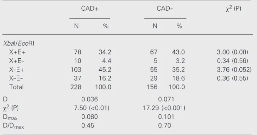
Table 3. Absolute and relative frequencies of the apo B (XbaI/EcoRI) haplotypes and results of linkage disequilibrium analyses in the CAD+ and CAD- groups.
CAD+ CAD- c2(P)
N % N %
XbaI/EcoRI
X+E+ 78 34.2 67 43.0 3.00 (0.08)
X+E- 10 4.4 5 3.2 0.34 (0.56)
X-E+ 103 45.2 55 35.2 3.76 (0.052)
X-E- 37 16.2 29 18.6 0.36 (0.55)
Total 228 100.0 156 100.0
D 0.036 0.071
c2 (P) 7.50 (<0.01) 17.29 (<0.001)
Dmax 0.080 0.101
D/Dmax 0.45 0.70
Haplotypes were estimated by the Arlequin program (14). X+E+ and X-E- were considered to be the cis conformations. The c2 test was used to compare the two
ing the CAD+ (N = 22) and CAD- (N = 6) groups, only HDL-C showed a statistically significant difference. When only the other
XbaI/EcoRI genotypes were considered
to-gether and the means of these same variables were compared in the CAD+ (N = 87) and CAD- (N = 72) groups, all means were higher in the CAD+ than in the CAD- group, except for HDL-C and apo A-I that did not differ statistically between these two groups. Com-paring the X-X-/E+E+ genotype with the class formed by all the other XbaI/EcoRI
genotypes, no statistically significant differ-ence was detected for the means of these variables in the CAD+ group (22 and 87 patients, respectively) or in the CAD- group (6 and 72 individuals, respectively).
Calcu-Table 4. Frequency distributions of two apo B genotype classes (XbaI/EcoRI) compared by the c2 test in the
CAD+ and CAD- groups.
Genotypes CAD+ CAD- c2(P)
N % N %
X-X-/E+E+ 24 21.1 6 7.7
Other genotypes 90 78.9 72 92.3 6.27 (0.012)
Total 114 100.0 78 100.0
EcoRI and the results of comparisons
be-tween the CAD+ and CAD- groups. When the CAD+ and CAD- haplotype frequencies were compared (Table 3), the difference closest to the significance limit was shown by the X-E+ haplotype. This result led to a comparison between the CAD+ and CAD- groups, as classified on the basis of only two genotype classes, i.e., X-X-/ E+E+ and all the other genotypes (Table 4). The distributions of these genotypes dif-fered in the CAD+ and CAD- groups (c2
(1) =
6.27, P = 0.012).
With respect to the mean values of serum lipids, lipoproteins, apoproteins and of the apo B/apo A-I ratio (Table 5), considering only the X-X-/E+E+ genotype and
compar-Table 5. Serum levels of lipids, lipoproteins, apolipoproteins (apo) and of the apo B/apo A-I ratio of the X-X-/ E+E+ genotype and of the other XbaI/EcoRI genotypes in the CAD+ and CAD- groups, as compared by the Student t-test.
Variable X-X-/E+E+ P Other XbaI/EcoRI genotypes P
CAD+ CAD- CAD+
CAD-(N = 22) (N = 6) (N = 87) (N = 72)
Total Cholesterol 215.0 ± 15.2 182.8 ± 10.8 >0.20 205.2 ± 4.9 176.2 ± 5.1 <10-4
Triglycerides 170.8 ± 27.9 88.8 ± 14.0 >0.10 161.3 ± 10.5 119.4 ± 8.3 <0.01 HDL-Cholesterol 37.4 ± 3.0 53.3 ± 7.3 =0.027 38.9 ± 1.4 42.9 ± 1.7aaaaa >0.05
LDL-Cholesterol 138.9 ± 11.3bbbbb 111.8 ± 12.3 >0.20 134.9 ± 4.1ccccc 107.9 ± 4.1aaaaa <10-5
Lipoprotein (a) 47.4 ± 9.5 47.2 ± 11.8 >0.90 52.6 ± 5.2 32.9 ± 3.6 <0.01 Apoprotein B 154.5 ± 12.2 109.0 ± 10.0 >0.05 143.5 ± 3.8 109.0 ± 3.9 <10-6
Apoprotein A-I 135.0 ± 7.6 146.8 ± 10.2 >0.40 137.4 ± 3.7 138.3 ± 4.3 >0.80 Apoprotein A-II 31.1 ± 2.0 30.5 ± 1.2 >0.80 31.7 ± 1.0 27.1 ± 0.9 <0.001 Apoprotein E 5.0 ± 0.5 3.6 ± 0.5 >0.10 4.1 ± 0.2 3.5 ± 0.2 <0.01 apo B/apo A-I 1.2 ± 0.1 0.8 ± 0.1 >0.10 1.1 ± 0.04 0.83 ± 0.04 <10-4
Data are reported as means ± SEM in mg/dl.
lations using the data from Table 5 showed that the CAD+ group (N = 109) presented significantly higher means than the CAD-group (N = 78) for TC, TG, LDL-C, Lp(a), apo A-II, apo B, apo E, apo B/apo A-I, whereas mean HDL-C concentration was significantly higher in the CAD- than in the CAD+ group. Only mean apo A-I concentra-tions did not differ significantly between these groups.
Table 6 shows the results of multivariate logistic regression analysis which consid-ered CAD as the dependent variable and the following independent variables: XbaI/EcoRI
genotypes, sex, hypertension, tobacco con-sumption, TC, and TG levels. The table also shows the odds ratios obtained for each of these statistically significant risk factors for CAD. The other variables that differed be-tween the CAD+ and CAD- groups (HDL-C, LDL-C, Lp(a), apo B, apo A-II, apo E, and apo B/apo A-I ratio) were not included in the analysis presented in Table 6, since they did not present statistically significant values of ß when considered in the regression analy-sis.
Discussion
The allele frequencies found in the pres-ent study referring to the XbaI and EcoRI
sites do not differ from those reported for
Brazilian CAD patients and controls (16). Bohn and Berg (17) cited significant posi-tive associations between CAD and the X-allele and/or the X-X- genotype in five stud-ies (6-10), and reported that this association was not observed in seven other studies (18-24). The X-X- genotype was more frequent in Brazilian women with CAD than in con-trol women (25). Studies on Caucasians from Europe (26) and on Chinese subjects (27) did not detect significant associations be-tween XbaI variability and CAD.
Bohn et al. (10), applying multivariate logistic regression analysis, obtained an odds ratio of 2.16 (P<0.01) for the X-X- homozy-gotes having myocardial infarction compared to the combined group of X+X+ and X+X-genotypes. This increased risk for the X-X-genotype was not apparently conferred by higher levels of TC, LDL-C or apo B, but by some mechanism not closely related to the traditional risk factors. However, the X-X-genotype was associated with higher serum concentrations of TC and LDL-C in Brazil-ian women classified as presenting a high risk lipid profile for CAD (28).
In the present study the difference in the frequencies of the X-E+ haplotype between CAD+ and CAD- individuals (Table 3) was close to the significance limit and the fre-quency of the X-X-/E+E+genotype was sig-nificantly higher in the CAD+ group when
Table 6. Results of the Student t-test and the odds ratios obtained by multivariate logistic regression analysis, considering CAD as the dependent variable and XbaI/EcoRI genotypes, sex, hypertension, tobacco consump-tion, total cholesterol (TC) and triglycerides (TG) as independent variables in CAD+ (N = 108) and CAD-(N = 78) individuals.
Constant ßo XbaI/EcoRI Sex Hypertension Tobacco TC TG
Estimate of ß -3.748 1.803 1.970 1.414 1.740 1.432 0.950
SE of ß 0.626 0.683 0.437 0.435 0.445 0.456 0.467
t(179) -5.982 2.640 4.502 3.250 3.908 3.138 2.035
P 0.0000 0.009 0.0000 0.0014 0.0001 0.0019 0.0433
X-X-/E+E+ Male Hypertension Tobacco TCb TGb
Odds ratio 6.1a 7.2 4.1 5.7 4.2 2.6
compared to the CAD- group (Table 4), sug-gesting that this genotype may be a risk factor for CAD. Data in Table 5 for the X-X-/E+E+genotype show a significantly lower mean HDL-C in the CAD+ group than in the CAD- group. At first we may assume the existence of an association between this geno-type and lower mean levels of HDL-C. How-ever, analysis of these data showed 73 and 17% of men with the X-X-/E+E+ genotype
in the CAD+ and CAD- groups, respectively. So, the reason for this difference in mean HDL-C may reside in the different sex pro-portions of these two groups. Possibly, a case-control study with matchingfor gender would have been more appropriate. How-ever, we set out to form a control group of patients being referred for cardiac catheter-ization due to clinical conditions other than ischemic heart disease.
The importance of the X-X-/E+E+ geno-type as an independent risk factor for CAD can be seen in the results of the regression analysis shown in Table 6. These results seem to exclude the possibility that the higher risk found for this genotype (Table 4) could be due to sample stratification caused by the differences between the two CAD groups (Table 1) in terms of the proportions of sex, hypertension and tobacco consumption. It is important to note that these three variables also appear to be independent risk factors in this analysis (Table 6). Among the six inde-pendent risk factors found in this study, the male sex and the X-X-/E+E+ genotype pre-sented the highest odds ratios (7.2 and 6.1,
respectively).
Two limitations could be pointed out in relation to the present study. One is the fact that the CAD+ group was composed of pa-tients with a diagnosis of acute coronary syndrome reflecting their tendency to coro-nary thrombotic complications. However, all patients were studied after the acute phase, and the angiography clearly showed signifi-cant atherosclerotic disease in all of them. The second limitation refers to the relatively small number of patients that may lead to type II errors. However, the patient number in the present study is comparable to those reported in several similar investigations.In spite of these limitations, the present study is the first in which the X-X-/E+E+ genotype is compared to all the other XbaI/EcoRI
geno-types, suggesting this sort of comparison for further studies.
Since the X- and X+ variations are syn-onymous, it is assumed that the X-X-/E+E+ genotype may be in linkage disequilibrium with an unknown variation in the apo B gene or with a variation in another gene that influ-ences the risk for CAD. The chance of de-tecting the effect of this putative variation would be two times higher if the homozy-gous genotype rather than the haplotype were considered in the analyses.
Acknowledgments
The authors gratefully acknowledge Marly Tavela and Amélia G. Araújo for excellent technical collaboration.
References
1. Barni N, Talmud PJ, Carlsson P et al. (1986). The isolation of genom-ic recombinants for the human apolipoprotein B gene and the map-ping of three common DNA polymorphisms of the gene - a useful marker for human chromosome 2. Human Genetics, 73: 313-319. 2. Priestley L, Knott T, Wallis S, Powell L, Pease R, Brunt H & Scott J
(1985). RFLP for the human apolipoprotein B gene: I, BamHI; II,
EcoRI; III, EcoRV; IV, MspI; V, XbaI. Nucleic Acids Research, 13: 6789-6793.
3. Law A, Powell LM, Brunt H et al. (1986). Common DNA polymor-phism within coding sequence of apolipoprotein B gene associated
with altered lipid levels. Lancet, 1: 1301-1303.
4. Blackhart BD, Ludwig EM, Pierotti VR et al. (1986). Structure of the human apolipoprotein B gene. Journal of Biological Chemistry, 261: 15364-15367.
5. Shoulders CC, Myant NB, Sidoli A, Rodriguez JC, Cortese C, Baralle FE & Cortese R (1985). Molecular cloning of human LDL-apolipopro-tein B cDNA. Evidence for more than one gene per haploid genome.
Atherosclerosis, 58: 277-292.
polymor-phisms associated with myocardial infarction. New England Journal of Medicine, 315: 1509-1515.
7. Monsalve MV, Young R, Jobsis J, Wiseman SA, Dhamu S, Powell JT, Greenhalgh RM & Humphries SE (1988). DNA polymorphisms of the gene for apolipoprotein B in patients with peripheral arterial disease. Atherosclerosis, 70: 123-129.
8. Myant NB, Gallagher J, Barbir M, Thompson GR, Wile D & Humphries SE (1989). Restriction fragment length polymorphism in the apo-B-gene in relation to coronary-artery disease. Atherosclero-sis, 77: 193-201.
9. Tybjaerg-Hansen A, Nordestgaard BG, Gerdes LU & Humphries SE (1991). Variation of apolipoprotein B gene is associated with myocar-dial infarction and lipoprotein levels in Danes. Atherosclerosis, 89: 69-81.
10. Bohn M, Bakken A, Erikssen J & Berg K (1993). XbaI polymorphism in DNA at the apolipoprotein B locus is associated with myocardial infarction (MI). Clinical Genetics, 44: 241-248.
11. Friedewald WT, Levy RI & Fredrickson DS (1972). Estimation of the concentration of low-density lipoprotein cholesterol in plasma, with-out use of the preparative ultracentrifuge. Clinical Chemistry, 18: 499-502.
12. Saiki RK, Gelfand DH, Stoffel S, Scharf SJ, Higuchi R, Horn GT, Mullis KB & Erlich HA (1988). Primer-directed enzymatic amplifica-tion of DNA with a thermostable DNA polymerase. Science, 239: 487-491.
13. Renges HH, Peacock R, Dunning AM, Talmud P & Humphries SE (1992). Genetic relationship between the 3’VNTR and diallelic apo-lipoprotein B gene polymorphisms: haplotype analysis in individuals of European and South Asian origin. Annals of Human Genetics, 56: 11-33.
14. Excoffier L, Schneider S, Kueffer JM & Roessli D (1997). Arlequin: version 1.1. Geneva. Download in 1998. http://anthropologie. unige.ch/arlequin.
15. Hosmer Jr DW & Lemeshow S (1989). Applied Logistic Regression. John Wiley & Sons, Inc., New York, NY, USA, 44.
16. Machado MO, Hirata MH, Bertolami MC & Hirata RDC (2001). Apo B gene haplotype is associated with lipid profile of higher risk for coronary heart disease in Caucasian Brazilian men. Journal of Clini-cal Laboratory Analysis, 15: 19-24.
17. Bohn M & Berg K (1994). The XbaI polymorphism at the apolipopro-tein B locus and risk of atherosclerotic disease. Clinical Genetics, 46: 77-79.
18. Deeb S, Failor A, Brown BG, Brunzell JD, Albers JJ & Motulsky AG (1986). Molecular genetics of apolipoprotein and coronary heart
disease. Cold Spring Harbor Symposia on Quantitative Biology, 56: 403-409.
19. Ferns GAA, Robinson D & Galton DJ (1988). DNA haplotypes of the human apolipoprotein B gene in coronary atherosclerosis. Human Genetics, 81: 76-80.
20. Wiklund O, Darnfors C, Bjursell G, Nilsson J, Linden T, Olofsson SO, Wilhelmsen L & Bondjers G (1989). XbaI restriction fragment length polymorphism of apolipoprotein B in Swedish myocardial infarction patients. European Journal of Clinical Investigation, 19: 255-258. 21. Genest JJ, Ordovas JM, McNamara JR et al. (1990). DNA
polymor-phisms of the apolipoprotein B gene in patients with premature coronary artery disease. Atherosclerosis, 82: 7-17.
22. Paulweber B, Frield W, Krempler F, Humphries SE & Sandhofer F (1990). Association of DNA polymorphism at the apolipoprotein B gene locus with coronary heart disease and serum very low density lipoprotein levels. Arteriosclerosis, 10: 17-24.
23. Peacock R, Dunning A, Hamsten A, Tornvall P, Humphries SE & Talmud P (1992). Apolipoprotein B gene polymorphism, lipoproteins and coronary atherosclerosis: a study of young myocardial infarction survivors and healthy population-based individuals. Atherosclerosis, 92: 151-164.
24. Nieminen MS, Mattila KJ, Aalto-Setälä K, Kuusi T, Kontula K, Kauppinen-Mäkelin R, Ehnholm C, Jauhiainen M, Valle M & Taskinen M-R (1992). Lipoproteins and their genetic variation in subjects with and without angiographically verified coronary artery disease. Arte-riosclerosis and Thrombosis, 12: 58-69.
25. Salazar LA, Hirata MH, Giannini SD, Forti N, Diament J, Lima TM & Hirata RD (2000). Seven DNA polymorphisms at the candidate genes of atherosclerosis in Brazilian women with angiographically docu-mented coronary artery disease. Clinica Chimica Acta, 300: 139-149. 26. Turner PR, Talmud PJ, Visvikis S, Ehnholm C & Tiret L (1995). DNA polymorphisms of the apoprotein B gene are associated with altered plasma lipoprotein concentrations but not with perceived risk of cardiovascular disease: European Atherosclerosis Research Study.
Atherosclerosis, 116: 221-234.
27. Pan J, Chiang A, Tai JJ, Wang S & Chang M (1995). Restriction fragment length polymorphisms of apolipoprotein B gene in Chi-nese population with coronary heart disease. Clinical Chemistry, 41: 424-429.