Ultrastructural description of rabies virus infection in cultured
sensory neurons
Myriam L Velandia*, Rosalía Pérez-Castro*, Hernán Hurtado*/**,
Jaime E Castellanos/
+Instituto de Virología, Universidad El Bosque, Transv. 9A Bis 132–55, Edificio de Rectoría, Laboratorio 205, Bogotá, Colombia *Laboratorio de Neurociencias, Instituto Nacional de Salud, Bogotá, Colombia **Facultad de Biología, Universidad Militar
Nueva Granada, Bogotá, Colombia
Primary cultures were made from adult mouse spinal ganglia for depicting an ultrastructural description of rabies virus (RABV) infection in adult mouse sensory neuron cultures; they were infected with rabies virus for 24, 36, and 48 h. The monolayers were processed for transmission electron microscopy and immunochemistry studies at the end of each period. As previously reported,sensory neurons showed great susceptibility to infec-tion by RABV; however, in none of the periods evaluated were assembled virions observed in the cytoplasm or seen to be associated with the cytoplasmic membrane. Instead, fibril matrices of aggregated ribonucleoprotein were detected in the cytoplasm. When infected culture lysate were inoculated into normal animals via intra-cerebral route it was observed that these animals developed clinical symptoms characteristic of infection and transmission electron microscopy revealed assembled virions in the cerebral cortex and other areas of the brain. Sensory neurons infected in vitro by RABV produced a large amount of unassembled viral ribonucle-oprotein. However, this intracellular material was able to produce infection and virions on being intra-cere-brally inoculated. It can thus be suggested that the lack of intracellular assembly in sensory neurons forms part of an efficient dissemination strategy.
Key words:rabies virus - sensory neurons - viral assembly
Rabies virus (RABV) is a negative, non-segmented, single-strand RNA virus which infects animals and the human nervous system (Mattos et al. 2001). RABV en-try to cells is due to viral glycoprotein (G) binding to some of the membrane proteins proposed as being viral receptors (the nicotinic acetylcholine receptor, the neu-ral cell adhesion molecule, and the p75 low affinity neurotrophin receptor) (Lafon 2005). Once within the cytoplasm, the ribonucleoprotein (RNP) complex is re-leased; this is formed by the RNA strand and non-struc-tural proteins N, P, and L (nucleoprotein, phosphopro-tein, and viral polymerase, respectively). Viral transcrip-tion and replicatranscrip-tion begin on this complex, following processes culminating in new viral progeny being re-leased (Finke & Conzelmann 2005). Viral capture and transport in a rabid accident is produced by endocytic process, reaching the soma of sensory neurons located in the spinal ganglia or the motoneurons of the spinal cord’s (SC) ventral horn. Sensory neurons’ ability to cap-ture, transport, and replicate RABV has been described
in sensory neuron cultures from different animal spe-cies. Our group has developed an in vitro RABV infec-tion model using adult mouse sensory neurons which has confirmed and evaluated susceptibility to infection and the antiviral and anti-replication effect of some drugs on these cells (Castellanos et al. 2005). Such suscepti-bility has been observed in models in vivo and it has been confirmed that these neurons are fundamentally involved in viral transport from the periphery to the central ner-vous system (CNS) via retrograde and anterograde ax-onal transport (Tsiang et al. 1983, 1989, 1991, Coulon et al. 1989, Dietzschold et al. 2005).
Even though the virus’ tropism towards neurons is widely known, it has been observed that some non-neu-ron cells may support viral entry and replication. Labo-ratory-adapted “fixed strain” viruses are frequently used in in vitro studies for infecting cells or tissue having different origin to the neurons. It has been found that baby hamster kidney fibroblast cells present high sus-ceptibility to infection and produce assembled viral par-ticles, recuperating in culture medium 12 to 18 h post-infection (pi) (Madore & England, 1977). On the con-trary, even though high susceptibility to infection has been observed in myocyte primary cultures, no as-sembled viral particles have been detected or released to the extracellular space. Just a large amount of viral in-clusions can be observed in cytoplasm, even up to 72 h pi (Tsiang et al. 1986).
On the other hand, it has been observed in both in vitro and in vivo neuron infection studies that even though marked viral neurotropism is maintained, there are differences in infection ability and viral production between different neuron types. Sensory neurons
pro-Partial financial support: Instituto Colombiano para la Ciencia y la Tecnología, Colciencias grant 2104-04-11724
+ Corresponding author: [email protected]
duce between 10- to 100-fold less viral particles respect-ing cortical neurons (Mrak & Young 1994). Such results suggest that each neuronal type has particular metabolic and biochemical characteristics which may interfere in different ways in the main processes of the viral cycle, just the same as that observed in fibroblasts and myocytes. It is thus particularly interesting to evaluate the ultra-structural characteristics of RABV infection in sensory neuron cultures and adult mouse cortex neurons by im-munochemistry [immunoperoxidase (IP), immunofluo-rescence (IF), and transmission electron microscopy (TEM)] techniques.
MATERIALS AND METHODS
Sensory neuron cultures - Four independent adult ICR strain mouse spinal ganglia sensory neuron cultures were done according to previously reported protocol (Castellanos & Hurtado 1999). The animals were sacrificed by cervical dislocation; the complete vertebral column was extracted and the ganglia dissected. They were then me-chanically and enzymatically dissociated in a 200 U/ml collagenase solution in complete medium (CM, DMEM supplemented with 10% bovine foetal serum, 100 U/ml penicillin, and 100 µg/ml streptomycin). The cell suspen-sion was centrifuged at 200 × g for 5 min. Cell viability was then evaluated with Trypan blue and 2500 cells seeded on Thermanox coverslips pre-treated with poly-L-lysine (10 µg/ml) in 24-well dishes and kept at 37ºC with 5% CO2. The medium was changed each 48 h.
In vitro infection - Sensory neuron cultures (consist-ing of sensory neurons, Schwann cells, and fibroblasts) were infected with a 1:10 dilution of CVS-11 strain rabies virus (Challenge Virus Standard), produced in mouse brain (102.43 DL50) diluted in complete medium. The inoculum was removed 1 h pi, complete medium was introduced and the cultures were taken to 24, 36, and 48 h pi. Once these periods had elapsed, half of the infected cultures were fixed with 4% paraformaldehyde in PBS, pH 7.4, for later processing by IF. The rest of the samples were processed by IP and then fixed with Karnowsky’s modi-fied solution (0.5% glutaraldehyde, 2% paraformaldhyde in 0.1M phosphate buffer, pH 7.4). A total of four cul-tures were processed with three replicas per culture.
In vivo infection - Three independent sensory neu-ron cultures were processed; they were infected at 120 h pi (according to the previously described protocol) and lysed in CM by mechanical pipetting. This cell lysate was then inoculated by intracerebral route into five ICR adult mice; these animals were observed daily and sacri-ficed on day 5. Six-week-old ICR mice were used as in-fection controls and intra-cerebrally inoculated with a 1/10 dilution of CVS-11 strain RABV and the mock-in-fected mice were inoculated with CM. Both groups of animals were anaesthetised with a mixture of ketamine (90 mg/kg) and xylazine (15 mg/kg) on the fourth day pi. They were then intra-cardially perfused with a washing solution (0.8 % NaCl, 0.1% procaine, 0.1% glucose) and fixed by perfusion with modified Karnowsky’s solution. The brain and cerebellum were extracted and each
hemi-sphere was independently processed for later immu-nochemistry (IF and IP) or TEM. Tissue processed by immunochemistry was post-fixed in the same fixing so-lution overnight at 4ºC and then vibratome sectioned into 100 µm thick sections (Castellanos et al. 1996). All the procedures described above had been previously ap-proved by the Instituto Nacional de Salud’s Ethics’ Com-mittee, in line with Colombian Ministry of Health Reso-lution 8430/1993.
Immunochemistry - The samples (cells or tissue) were permeabilised with 50% ethanol. Endogenous per-oxidase was inactivated by using a 0.05% NaN3 and 0.04% H2O2 solution in PBS. Non-specific binding sites were blocked with 10% horse serum. Viral antigen was detected by using a hamster anti-rabies antibody at 1:900 dilution (diagnostic antibody produced by the Instituto Nacional de Salud, Colombia) incubated in Tris buffer, pH 8.6, for 1 h at 37ºC (Castellanos et al. 1996). The respective biotinylated anti-hamster secondary antibody was then added (2.5 µg/ml). Samples were then incubated with biotinylated avidin-peroxidase (ABC Vector Inc) complex diluted 1:100, a 0.05% diaminobenzidine so-lution in 50 mM HCl Tris buffer pH 7.2 and 0.02% H2O2 used as chromogen. The reaction was interrupted by suc-cessive washing with tap water. Sensory neuron cultures fixed with PFA were processed by IF. They were thus permeabilised with Triton X-100 and incubated with ham-ster anti-rabies antibody coupled to FITC (1:30) in PBS at 37°C for 30 min in a wet chamber. The cells were then counterstained with Evans’ blue (1/10.000) and washed with PBS and distilled water. The cells were then mounted in aqueous medium (buffered glycerine, pH 9.0) and observed by epifluorescence microscopy.
Electron microscopy - Once immunoperoxidase de-veloping time had elapsed; three samples by duplicate per culture were processed for TEM. Both kinds of sample were then post-fixed with 1% osmium tetroxide and dehydrated in increasing ethanol gradients (70, 80, 90, 95, and 100%). The monolayers were infiltrated with a mixture of 100% ethanol and Epon-812 Polybed resin in 3:1, 1:1, 1:3 ratios and then embedded in a mixture of Epon-Araldite resins. Slices of nerve tissue were infil-trated with a mixture of propylene oxide and Epon-Araldite resin in the ratios described above. They were finally embedded in Epon-Araldite and polymerised at 67ºC for 18 h. Semi-thin and ultra-thin sections were ob-tained by using a Sorvall MT1 ultramicrotome. Semi-thin sections were stained with toluidine blue and specific ar-eas were defined for obtaining the ultra-thin sections. These were contrasted with uranyl acetate and lead citrate and observed using a Carl Zeiss EM 109 electron microscope.
RESULTS
Morphological description - The sensory neurons cultures presented a typical heterogeneous aspect. Neu-ronal and non-neuNeu-ronal cells were identified. The neu-rons were seen to be refringent, spherical, having an ec-centric nucleus; neuritic processes were observed in most of them which increased in length during culture time. The non-neuronal cell population (Schwann cells and fibroblasts) appeared to be non-refringent cells which were strongly adhered to the surface of the plate. No appreciable differences were observed between infected culture morphology compared to that of non-infected ones. When evaluating the number of neurons and non-neuronal cells infected with RABV at 24 h pi, it was ob-served that 39.8% of the neurons had become infected whilst the population of non-neuronal cells (fibroblasts and Schwann cells) in cultures presented 2.1% infec-tion (Table).
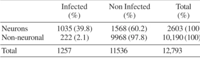
Immunochemistry - Viral antigen was observed in sensory neuron cultures at three periods pi, evaluated by both IF and IP techniques. Immunoreactivity (IR) was low at 24 h pi and was located mainly in the perinuclear re-gion and in sensory neuron neurites. IR was also observed in some non-neuronal cells. A large amount of cytoplas-mic vesicles which were IR for viral antigen 36 and 48 h pi were observed in the cytoplasm of infected neurons; these vesicles were located in the cytoplasm’s central area. An increase in the number of these vesicles was also observed 48 h pi; they were located very close to the neuronal membrane (Fig. 1A). Infection in the brain
TABLE
Percentage of neuronal and non-neuronal cells from sensory neuron cultures infected with rabies virus
Infected Non Infected Total
(%) (%) (%)
Neurons 1035 (39.8) 1568 (60.2) 2603 (100) Non-neuronal 222 (2.1) 9968 (97.8) 10,190 (100)
Total 1257 11536 12,793
was observed in different areas of the cortex and in the hippocampus’ pyramidal cell layer. IR was observed in the cerebellum in the molecular and granular layers of the cortex. However, it was preferentially detected in Purkinje neuron soma in which cytoplasmic inclusions were identified close to the cell membrane (Fig. 1B).
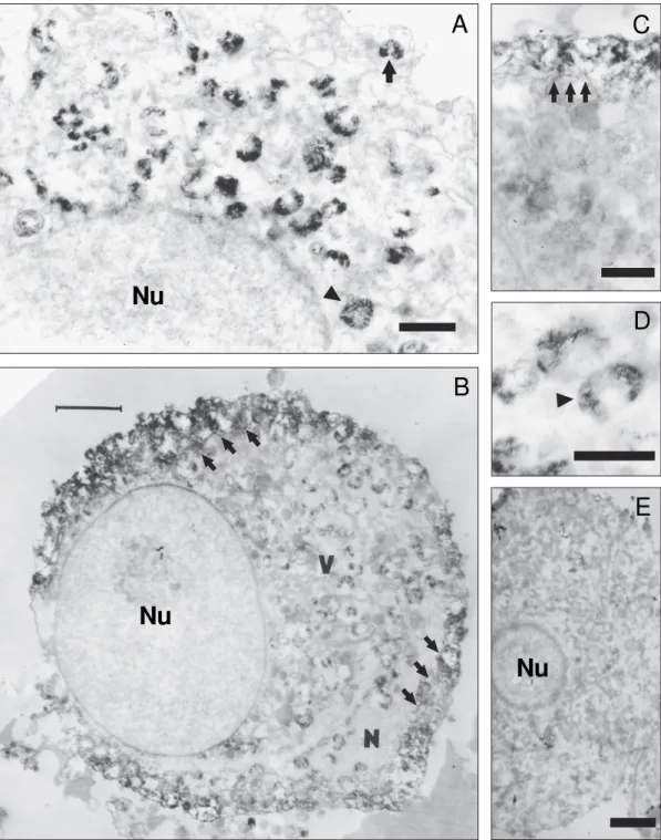
Infected cell ultrastructure - Infected neuron cyto-plasm presented multi-vesicular and lamellar bodies. It was possible to identify subcellular structures such as lysosomes, lipofuscin granules, mitochondria, and Golgi apparatus. Type B neurons (dark neurons) were most found in cultures. Cytoskeleton structures, such as mi-crotubules and neurofilaments, could only be identified in some neurons (i.e. being more evident in neuritic pro-cesses). Granular electron-dense aggregates (fibrillar matrices) were observed during all pi periods around the nucleus or very close to the plasma membrane. Such ag-gregates were more evident at 36 and 48 h pi (Figs 2A, B). TEM revealed that these electron-dense structures were half-moon shaped and were IR for viral antigens. This mor-phological pattern was maintained during the periods ana-lyzed; however, a larger number of these aggregates was found at 48 h pi, located very close to the plasma mem-brane or fused to it (Figs 2C, D). No assembled viral par-ticles were found at any time analyzed or in the cytoplasm or close to the cell membrane. Aspect of mock infected sensory neuron cultures is showed in Fig 2E.
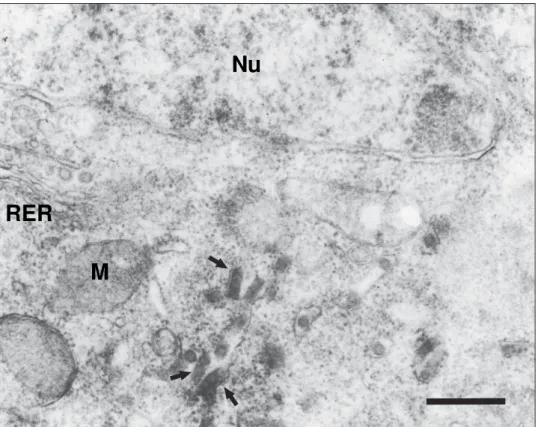
Infected cell lysates were inoculated by the intra-cerebral route into mice at 48 h pi to confirm the infec-tious ability of fibril matrices present in the sensory neuron culture cytoplasm. The animals presented symp-toms characteristic of RABV infection 5 days pi. The sections presented a positive reaction in the hippocam-pus, cerebral cortex, and the cerebellar Purkinje neu-rons when evaluated by IFA and/or IP. TEM revealed vi-ral particles in pyramidal cells from the hippocampus, some of them presenting degeneration and dilated mito-chondrial matrix. Each infected neuron cell presented 2 to 8 cytoplasmic inclusions which seemed to be Negri body-like (viral RNP) structures, observed as being a strongly electron-dense matrix; 75 nm diameter, vari-able length (approximately 200 nm), assembled viral
Fig. 1:immunochemical staining of rabies virus. A: sensory neurons culture processed by immunofluorescence. Infected sensory neurons (arrow-heads); note the immunoreactive neurites (arrows); B: cerebellum sections processed by immunoperoxidase technique. Purkinje cell bodies are positive to viral antigen (arrowheads) and dendrites are shown with immunoreactive vesicles (arrow). Bars = 50 µm.
particles were observed around such structures. These viral particles were also observed to be sometimes as-sociated with vesicles close to the endoplasmic reticule (ER) or Golgi apparatus; they were only observed in these neuron cells budding through the cytoplasmic membrane of soma or some synaptic buttons. Electron immuno-microscopy revealed granular, rounded precipitates as-sociated with ER. Assembled viral particles were ob-served close to these structures in some cases (Fig. 3).
DISCUSSION
Sensory ganglia are peripheral nervous system struc-tures, mainly consisting of pseudomonopolar neurons and support tissue provided by Schwann cells and fibro-blasts. Sensory neurons have been described and characterised both in vivo and in vitro according to their physiology, biochemistry, and morphology (Thomas et al. 1993). Ultrastructurally, and according to their den-sity and ER distribution in the cytoplasm, these cells Fig. 2:electron micrographs of cultured sensory neurons. A: infected neuron 36 h pi. Many viral inclusions (arrow and arrowhead) were detected by using immunoperoxidase inside the cytoplasm near the nucleus (Nu). Bar = 1 µm; B: infected sensory neuron 48 h pi; viral inclusions (V) are concentrated near the plasma membrane (arrowhead). Bar = 5 µm; C: detail of cytoplasmic immunoreactive vesicles (arrows) and membrane fusion. Bar = 1 µm. D: detail of immunoreactive viral inclusion (arrows); note the electron-dense diaminobenzidine crystals. Bar = 0.5 µm. E: electron micrograph of sensory neuron from a non-infected culture; Bar = 1 µm.
Nu
A
C
D
E
Nu
Nu
can be classified into clear (type A) and dark neurons (type B) (Martinez et al. 2002). Our culture model favoured the presence of type B neurons, corresponding to intermediate and small nociceptive-associated neu-rons. On the other hand, the loss of large neurons (type A) could have been due to the technique employed for obtaining them and the culture’s senescence. These find-ings coincide with those previously described by Sommer et al. (1986) in in situ models and by Martínez et al. (2002) and Rambourg et al. (1983) in in vitro models.
Viral antigen distributed in infected sensory neurons’ soma and neurites was detected during the present work, confirming marked RABV neurotropism. High rabies’ virus neurotropism was confirmed by the percentage of infected cells present in cultures where 39.8% of sen-sory neurons were infected, whilst only 2.1% of non-neuronal cells were infected.
A particular cytoplasmic IR structure was observed near the nucleus, associated or not with ER during all evaluated periods in infected neurons. These aggregates were dense fibril structures, suggesting that they were viral ribonucleoprotein (RNP), being free inside the cy-toplasm or accumulated forming irregular-shaped struc-tures. The absence of assembled viral particles in these cell cultures coincided with that preliminarily reported in rat sensory neurons and myocytes (Tsiang et al. 1986, Martinez et al. 2000). Both cell types can be considered to be primary viral capture and replication sites, suggest-ing that the virus initially enters these cells to avoid im-mune system attack and/or undergo some adaptation
pro-cesses (Iwasaki et al. 1973, Watson et al. 1981, Lycke & Tsiang 1987). Low assembled virus production in these types of cell could possibly be attributed to differences in the machinery of neurons from the sensory neurons and brain, these being transport and immune response evasion strategies.
The cells’ biochemical and metabolic differences could be implicated in viral replication and transcrip-tion; they could also modulate interaction between viral and cellular proteins, concluding in correct viral assem-bly (Nakamichi et al. 2005). Rhabdovirus assemassem-bly de-pends on the correct interaction of RNP with protein M and that of the latter with the plasma membrane. A direct interaction between RABV matrix protein N-terminal re-gion L domain and the WW domain present in the pro-teasome-associated membrane protein cytoplasmic region has been previously demonstrated (Charlton & Casey 1979, Lenard 1999, Mebatsion et al. 1999, Harty et al. 1999, Harty et al. 2001). It is possible that alterations are presented in the proteasome-associated membrane proteins domains’ sensory neurons, thereby impeding correct M interaction with these neurons’ membrane.
Regarding a transport strategy, the same line of thought as that reported by Mc Quaid et al. (1998) in neuronal models of infection with measles’ virus could be adopted. They observed a large amount of viral nucleo-capsids accumulating without assembled particles being produced in the syncytia formed during infection by the virus in Vero cells, suggesting that the measles virus uses different forms of dispersion, without the need to
as-Nu
RER
M
semble viral particles. Viral dispersion and immune eva-sion would thereby become increased. It is possible that RABV may not be assembled in typical viral particles within the cytoplasm of sensory neurons by as yet un-known biochemical or metabolic mechanisms. By con-trast, it may produce a large amount of RNP aggregates, these being constantly stored and transported during pro-longed infection; such mechanism thereby guaranteeing the efficiency of spreading, silencing and persistence of infection in sensory neurons. These neurons could then transport RNP complexes to the CNS where the vi-rus could conventionally assemble and disperse itself by using genetic information and a different cellular machinery. The latter may be suggested as intra-cere-brally inoculating supernatants and homogenised prod-ucts from sensory neurons cultures into adult mice has led to the animals developing symptoms characteristic of infection. Assembled viral particles have also been observed in cortical neurons.
Our results suggest that sensory neurons support in-fection and offer RABV an excellent scenario for viral replication and transcription; however, a large amount of non-assembled RNP are produced which are able to produce the characteristic signs of paralysis and death in infected animals due to their biochemical and func-tional characteristics. These RNP also produce viral cor-tical neuron progeny which are assembled and released. Such phenomenon offers a strategy for the mass and si-lent production of viral RNP in sensory neurons which might be transported and then released in the dorsal horn of spinal cord so that RABV can then start on a new in-fectious cycle. The virus might possibly encounter suit-able metabolic, biochemical, and functional conditions in the latter neurons, allowing correct and efficient as-sembly of new viral progeny. The present evidence leads to suggesting that RABV adopts different mechanisms in each type of neuron which it infects to increase its ability to disperse itself within the organism and avoid the immune system’s attack. How and why the virus uses such types of strategy is still to be determined. Further studies are also needed for a much more precise defini-tion of the sensory neurons’ role during viral capture, transport, and spreading within the nervous system.
ACKNOWLEDGEMENTS
To Jason Garry for translating this paper.
REFERENCES
Castellanos J, Hurtado H 1999. Viral infection studied in adult sensory neurons. In L Haynes, The Neuron in Tissue Cul-ture, John Wiley & Sons Ltd., Chichester, p. 289-293.
Castellanos JE, Castañeda D, Velandia A, Hurtado H 1996. Inmunodetección por peroxidasa de células de ganglio sen-sorial infectadas por virus de rabia. Biomedica16: 214-218.
Castellanos JE, Martinez-Gutierrez M, Hurtado H, Kassis R, Bourhy H, Acosta O, Lafon M 2005. Studying neurotrophin antiviral effect on rabies-infected dorsal root ganglia cultures.
J Neurovirol 11: 403-410.
Charlton K, Casey G 1979. Experimental rabies in Skunks: im-munofluorescence light and electron microscopic studies.
Lab Invest 41: 36-44.
Coulon P, Dervin C, Kucera P, Lafay F, Prehaud C, Flamand A 1989. Invasion of the peripheral nervous systems of adult mice by the CVS strain of rabies virus and its avirulent derivate. J Virol 63: 3550-3554.
Dietzschold B, Schnell M, Koprowski H 2005. Pathogenesis of rabies. Curr Top Microbiol Immunol 292: 45-56.
Finke S, Conzelmann K 2005. Replication strategies of rabies virus. Virus Res 111: 120-131.
Harty R, Brown M, Mc Gettigan J, Wang G, Jayakar H, Huibregtse J, Whitt M, Schnell M 2001. Rhabdoviruses and the cellular ubiquitin-proteasome system: a budding interaction. J Virol 75: 10623-10629.
Harty R, Paragas J, Sudol M, Palese P 1999. A prolin-rich motif within the matrix protein of vesicular stomatitis virus and ra-bies virus interacts with WW domains of cellular proteins: implications for viral budding. J Virol 73: 1921-1929.
Iwasaki Y, Wiktor T, Koprowski H 1973. Early events of rabies virus replication in tissue cultures: an electron microscopy study. Lab Invest 28: 142-148.
Lafon M 2005. Rabies virus receptors. J Neurovirol 11: 82-87.
Lenard J 1999. Negative-strand virus M and retrovirus MA pro-teins: all in a family? Virology 216: 289-298.
Lycke E, Tsiang H 1987. Rabies virus infection of cultures rat sensory neurons. J Virol 61: 2733-2741.
Madore HP, England J 1977. Rabies virus protein synthesis in infected BHK-21 cells. J Virol 22: 102-112.
Martínez M, Quiroga N, Castellanos J, Hurtado H 2000. Subpoblaciones neuronales presentes en el ganglio de la raíz dorsal. Biomédica 20: 248-260.
Martínez M, Velandia M, Quiroga N, Castellanos JE 2002. Dif-ferential susceptibility in sensory cultured neurons to rabies virus infection. J Neurovirol 8 (Suppl.1): 109.
Mattos C, Mattos C, Rupprecht C 2001. Rhabdoviruses. In DM Knipe, PM Howley, L Williams (eds), Fields Virology,
Lippincott, Williams & Wilkins, Philadelphia, p. 1245-1277.
McQuaid S, Campbell S, Wallace I, Kirk J, Cosby S 1998. Measles virus infection and replication in undifferentiated and differ-entiated human neuronal cells in culture. J Virol 72: 5245-5252.
Mebatsion T, Weiland F, Colzemann K 1999. Matrix protein or rabies virus is responsible for the assembly and budding of bullet-shaped particles and interacts with the transmembrane spike glycoprotein G. J Virol 73: 242-250.
Mrak R, Young L 1994. Rabies encephalitis in humans: pathol-ogy, pathogenesis and pathophysiology. J Neuropathol Exp Neurol 53: 1-10.
Nakamichi K, Saiki M, Sawada M, Takayama-Ito M, Yamamuro Y, Morimoto K, Kurane I 2005. Rabies virus-induced activa-tion of mitogen-activated protein kinase and NF-kB signaling pathways regulates expression of CXC and CC chemokine ligands in microglia. J Virol 79: 11801-11812.
Rambourg A, Clermont Y, Beaudet A 1983. Ultrastructural fea-tures of six types of neurons in rat dorsal root ganglia. J Neurocytol 12: 47-66.
Sommer E, Kazimeierczak J, Droz B 1986. Neuronal subpopula-tions in the dorsal root ganglion of the mouse as characterised by combinations of ultrastructural and cytochemical features.
Thomas PK, Berthold C, Ochoa C 1993. Microscopic anatomy of the peripheral nervous system. In P Dyck, Peripheral Neuropathy, WB Sauders, p. 230-263.
Tsiang H, De la Porte S, Ambroise D, Derer M, Koenig J 1986. Infection of cultured rat myotubes and neurons from the spinal cord by rabies virus. J Neuropathol Exp Neurol 45: 28-42.
Tsiang H, Koulakoff A, Bizzini B, Berwald-Netter Y 1983. Neu-rotropism of rabies virus: an in vitro study. J Neuropath Exp Neurol 42: 439-452.
Tsiang H, Lycke E, Ceccaldi P, Ermine A, Hirardot X 1989. The anterograde transport of rabies virus in rat sensory dorsal root ganglia neurons. J Gen Virol 70: 2075-2085.
Tsiang H, Ceccaldi PE, Lycke E 1991. Rabies virus infection and transport in human sensory dorsal root ganglia neurons.
J Gen Virol 72: 1191-1194.