D e te rm inatio n o f m acro m o le cular
exchange and PO
2
in the microcirculation:
a sim ple syste m fo r
in vivo
fluo re sce nce
and pho spho re sce nce vide o m icro sco py
Departamento de Ciências Fisiológicas, Instituto de Biologia Roberto Alcântara Gomes,
Universidade do Estado do Rio de Janeiro, Rio de Janeiro, RJ, Brasil L.N. Torres and
I.P. Torres Filho
Abstract
We have developed a system with two epi-illumination sources, a DC-regulated lamp for transillumination and mechanical switches for rapid shift of illumination and detection of defined areas (250-750 µm2) by fluorescence and phosphorescence videomicroscopy. The
system permits investigation of standard microvascular parameters, vascular permeability as well as intra- and extravascular PO2 by
phosphorescence quenching of Pd-meso-tetra (4-carboxyphenyl) por-phine (PORPH). A Pechan prism was used to position a defined region over the photomultiplier and TV camera. In order to validate the system for in vivo use, in vitro tests were performed with probes at
concentrations that can be found in microvascular studies. Extensive
in vitro evaluations were performed by filling glass capillaries with
solutions of various concentrations of FITC-dextran (diluted in blood and in saline) mixed with different amounts of PORPH. Fluorescence intensity and phosphorescence decay were determined for each mix-ture. FITC-dextran solutions without PORPH and PORPH solutions without FITC-dextran were used as references. Phosphorescence de-cay curves were relatively unaffected by the presence of FITC-dextran at all concentrations tested (0.1 µg/ml to 5 mg/ml). Likewise, fluores-cence determinations were performed in the presence of PORPH (0.05 to 0.5 mg/ml). The system was successfully used to study macromo-lecular extravasation and PO2 in the rat mesentery circulation under
controlled conditions and during ischemia-reperfusion.
Co rre spo nde nce I.P. Torres Filho Departamento de Ciências Fisiológicas, IBRAG, UERJ 20551-030 Rio de Janeiro, RJ
Brasil
Fax: + 55-21-254-5442 E-mail: ivo@ uerj.br
Presented at the XV Annual Meeting of the Federação de Sociedades de
Biologia Experimental, Caxambu, MG, Brazil, August 23-26, 2000.
Received April 13, 2000 Accepted August 14, 2000
Ke y wo rds ·O xygen ·Permeability ·Microcirculation ·Rat mesentery ·Intravital microscopy ·Phorphyrin
Intravital microscopy has become a stan-dard technique for investigating microvas-cular phenomena. In the past, most studies measured only variables such as internal di-ameter and red blood cell velocity. More recently, investigators have been using fluo-rescence intravital microscopy for studying microvascular permeability to small and large molecules (1-3), leukocyte-endothelium
(8). Using a new investigating tool, Torres Filho et al. (9,10) measured intravascular and extravascular PO2 in small tissue areas. This PO2 measurement system has been implemented also in other laboratories (6,11). Due in part to the specific nature of light sources and collection devices, it is difficult to perform physiological studies using the same set-up for bright-field microscopy as well as fluorescence and phosphorescence. While there have been previous reports of systems capable of measuring time-resolved luminescence (12,13), there are no reports of such a system using FITC and metallopor-phyrins in the microcirculation. Therefore, the aim of the present study was to imple-ment a simple system that allows a rapid shift for illumination and capture of well-defined areas using fluorescence and phos-phorescence microscopy as well as transillu-mination. The system allows quantitative on-line evaluation of intra- and extravascu-lar PO2, macromolecuextravascu-lar extravasation as well as blood vessel diameter and length. In order to allow in vivo evaluations, in vitro
and in vivo tests were performed with fluo-rescence and phosphofluo-rescence probes in con-centration ranges that can be found in the microvascular studies. We also investigated whether fluorescence determinations were affected by the presence of the phosphores-cence probe and whether phosphoresphosphores-cence evaluations were affected by the presence of FITC.
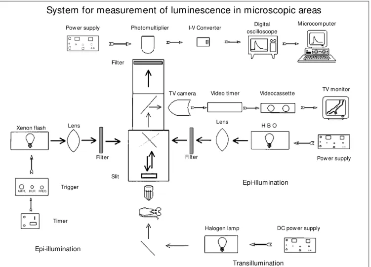
Figure 1 schematically illustrates the sys-tem used to measure fluorescence and phos-phorescence in small regions. All studies were performed using a Leitz Orthoplan mi-croscope equipped with an epi-illumination system (Ploemopak; Leitz, Wetzlar, Ger-many), a tungsten-halogen source for trans-illumination and a bright-field substage con-denser. The epi-illumination light sources were a high-pressure mercury vapor lamp (HBO50, Aus Jena, Jena, Germany) and a xenon strobe arc (EG&G Electro Optics, Salem, MA, USA). A mechanical switch
allowed rapid shifts of epi-illumination sources. The epi-illumination head included three rapidly interchangeable cubes: one for observations during transillumination and two additional cubes with appropriate di-chroic mirrors as well as excitation and emis-sion filters for fluorescence and phosphores-cence microscopy (see below).
A specially designed optical adapter al-lowed the image magnified by the objective to be projected either to a charge coupled device (CCD) camera (model STPM314, Sony) or to a photomultiplier (model 77344, Oriel, Stratford, CT, USA). The camera had a sensitivity of 0.5 lux (F1.2) A Pechan prism image rotation unit (Hoptik, Tucson, AZ, USA) was inserted into the optical path, allowing better positioning of the region of interest. Observations were made using wa-ter immersion objectives (Zeiss 20X, NA = 0.50; Zeiss 40X, NA = 0.75; Leitz 55X, NA = 0.84) yielding final magnifications of 1230-3080 times on the screen of the video moni-tor (model WV5400BN, National, Tokyo, Ja-pan). The horizontal resolution of the monitor was 700 lines at center. These magnifications were equivalent to 2-5 raster lines per µm.
lens in the optical path allowed the slit to be placed on a conjugate plane with the image. The phosphorescence emission from the epi-illuminated area passed through a light filter with cut off at 630 nm before being captured by a photomultiplier (model 77344, Oriel), with a peak radiant sensitivity (approximately 70 mA/W) in the 600-800 nm range. The signals from the photomultiplier were sent to an oscilloscope (model 54603B, Hewlett-Packard). The oscilloscope averaged 10-40 curves, yielding a single smoothed curve after 1-4 s that was then digitized and stored
for later analysis. Each smoothed phospho-rescence decay curve was computer pro-cessed for the calculation of PO2.
The PO2 of a given area was calculated by mathematically fitting the decay of phos-phorescence to a single exponential using a conventional least squares method. The time constant t of the fitted exponential curve was used to calculate PO2 according to the Stern-Volmer equation:
t0/t = 1 + kq x t0 x PO2
where t0 and t are the phosphorescence
lifetimes in the absence of O2 and in the area
Pow er supply Photomultiplier Digital oscilloscope
Videocassette TV monitor
Pow er supply I-V Converter M icrocomputer
Filter
TV camera Video timer
Lens
H B O
Filter Filter
Slit
Epi-illumination
Epi-illumination
Transillumination
Trigger
Timer Xenon flash Lens
DC pow er supply Halogen lamp
System for measurement of luminescence in microscopic areas
AM PL DUR FREQ
dextran solutions without porphyrin and por-phyrin solutions without FITC-dextran were used as references. Background measure-ments were also obtained with glass capillar-ies filled only with blood or saline. Two capillaries were prepared for each solution and all capillaries were evaluated twice in random order.
In vivo studies were performed on 4 male Wistar rats (150-200 g) acclimated to an animal care facility and maintained on stan-dard rat chow and water ad libitum. After 12 h of fasting, the animals were anesthetized
with sodium pentobarbital (40 mg/kg, ip)
and the trachea was cannulated to insure a patent airway. The left and right femoral veins were cannulated using polyethylene catheters (PE 50). The superior mesenteric artery was isolated and a snare was placed around it using a polyethylene tube. The rat mesentery was prepared for intravital mi-croscopy as described in detail earlier (14). Briefly, after abdominal midline incision, the mesentery was carefully spread over a Lucite pedicle of a specially designed acrylic board and covered with thin plastic film to avoid drying and exposure to atmospheric gases. The Lucite pedestal consisted of a chamber through which thermostatically con-trolled warm water was circulated continu-ously to maintain the temperature at 37oC. Observations were performed in animals spontaneously breathing room air.
The board was attached to the micro-scope stage and the mesenteric tissue image was projected onto the TV camera connected to a videotape recorder (model SLV88HFBR, Sony), a video timer (model VTG33, For A) and a video monitor. A schematic drawing of the microvascular anatomy in the area under observation was made to facilitate later anal-ysis. Each field was recorded under trans-mitted light for at least 1 min before or after each PO2 determination. The temperature of the area being studied was periodically meas-ured using a thermocouple placed over the pedestal (YSI Co., Inc., Yellow Springs, OH, being analyzed, respectively, and kq is the
quenching constant. Appropriate values for
t0 and kq (8,9) were used according to the
probe and to the temperature of the area under study.
Photometric evaluation of the fluores-cence emitted by extravasated FITC allows estimation of microvascular permeability. To elicit fluorescent signals, the area was illuminated with a 50-W mercury lamp. Photobleaching was avoided by keeping the light exposure time to 1-3 s. The filter system for fluorescence evaluation included a band pass filter (420-490 nm) and an emission filter with cut off at 515 nm. The excitation light passed through the same adjustable rectangular slit used for the phosphorescence studies. The fluorescence emission from the epi-illuminated area was also captured by the photomultiplier. The signals were sent to an oscilloscope, digitized and stored for later computer analysis. Since fluorescent light intensity is proportional to FITC-dextran con-centration, the integrated optical density of the illuminated area after background sub-traction would be proportional to the total amount of tracer molecules within the area. Background levels were obtained by meas-uring the light intensity in a given epi-illumi-nated area before the addition of the fluores-cent compound.
FITC-USA). Microvessel inner diameters were measured off-line from videotape record-ings with a calibrated digital caliper (Mitutoyo).
In order to evaluate the influence of FITC on PO2 measurements, each animal received a slow (3-5 min) intravenous injection of Pd-meso-tetra (4-carboxyphenyl) porphine pre-viously bound to albumin. The dose used was 15 mg/kg body weight at a concentra-tion of 18 mg/ml. A series of intravascular PO2 determinations were performed and the animals then received FITC-dextran (50 mg/ kg body weight) intravenously at a concen-tration of 50 mg/ml. After 5 min, PO2 deter-minations were repeated at the same loca-tions. In order to evaluate the influence of porphyrin on fluorescence measurements, each rat received an intravenous injection of FITC-dextran (50 mg/kg body weight) at a concentration of 50 mg/ml. After a period of at least 20 min, a series of fluorescence determinations were performed in intravas-cular and perivasintravas-cular areas before, during and after ischemia. Changes in microvascu-lar permeability were provoked by subject-ing the mesentery to an ischemic period of 20 min followed by reperfusion. Ischemia was induced by occluding the previously isolated superior mesenteric artery. The ani-mals then received the phosphorescence probe (15 mg/kg body weight at a concentra-tion of 18 mg/ml). After a period of 5 min, fluorescence determinations were repeated at the same locations.
The switch for illumination and emission between fluorescence and phosphorescence set-ups was mechanically simple, took about 10-15 s, but precluded simultaneous meas-urements. The in vitro calibration showed that the system was linear for fluorescence measurements and FITC-dextran concentra-tions ranging from 0.1 µg/ml to 5 mg/ml yielded average light intensities (which were
measured as voltages) from 101
to 104 mV. The analysis of 80 capillaries filled with test solutions (320 luminescence measurements)
showed that phosphorescence decay meas-urements were unaffected by the presence of FITC-dextran at all concentrations tested (0.1 µg/ml to 5 mg/ml). Likewise, in vitro fluo-rescence determinations were unaffected by the presence of porphyrin at all concentra-tions tested (0.05 to 0.5 mg/ml).
Similar findings were obtained using the mesentery in vivo. Intravascular and extravas-cular measurements of fluorescence were unaffected by the injection of porphyrin (Fig-ure 2, top panel). Ischemia-reperfusion caused the extravasation of FITC-dextran to perivascular spaces. This increase in perme-ability could be monitored by measuring the fluorescence in well-determined regions. Since optical characteristics of in vivo prepa-rations are different from the in vitro set-up, absolute values of light intensity cannot be directly translated into absolute values of FITC-dextran levels in vivo. However, the linearity of the system is preserved and fluo-rescent light intensities measured in vivo can be readily used to express relative changes in intra- and extravascular FITC-dextran con-centrations.
Intravascular and extravascular measure-ments of phosphorescence were unaffected by the injection of FITC-dextran. An ex-ample of an intravascular PO2 measurement under control conditions is presented in Fig-ure 2 (lower panel). In addition, we noted that in some poorly perfused areas, late in-jection of porphyrin would not allow meas-urements of interstitial PO2 since the leakage of the probe under these conditions would be too small.
There have been previous reports on sys-tems capable of measuring fluorescence and phosphorescence (12,13) but only a few have been applied to microvascular preparations (6,15,16). These systems utilize more so-phisticated approaches, ranging from acousto-optic tunable filters and phase-locked mechanical choppers to slow-scan CCD cameras and gated multichannel plate image intensifiers. A major advantage of the present system is that quantitative fluores-cence and phosphoresfluores-cence may be per-formed in the same precise microregion us-ing a microscope normally found in most microcirculation research laboratories. Therefore, on-line determinations of PO2 can be done in regions following changes in microvessel permeability. Assembly, cali-bration, data collection and analysis are rather straightforward, basically requiring the com-putation of signals produced by a photomul-tiplier inserted into the optical path.
Previous investigators have used
rectan-gular fields to determine integral optical den-sity (1). A similar procedure has been em-ployed in our study, since only a particular region was excited by epi-illumination. This is advantageous also because the total tissue area exposed to strong illumination is mini-mized. In the case of PO2 measurements, this procedure of limiting illumination has been successfully implemented earlier (11).
Once injected into the circulation, fluo-rescent and phosphorescence probes reach plasma and interstitial concentrations that have been previously estimated (17). In or-der to validate the system for in vivo studies,
in vitro tests were performed with probes within the concentration ranges that can be found in blood and interstitium. We ob-served that phosphorescence decay curves were relatively unaffected by the presence of FITC-dextran at all tested concentrations (0.1 µg/ml to 5 mg/ml). Likewise, fluorescence determinations could be performed in the presence of porphyrin (0.05 to 0.5 mg/ml). This is not surprising since the decay time of the porphyrin phosphorescence is long enough to avoid the prompt fluorescence while maintaining sufficient data for PO2 determination.
A complete evaluation of mesenteric microvessel permeability was beyond the scope of the present study. However, follow-ing appropriate calibrations and adequate fluorescence determinations at precise times and locations, we showed that the system can be readily used to estimate microvessel permeability. Macromolecular extravasation following ischemia-reperfusion, as used in the present study, has been used by several authors (18,19).
The system may be further improved in several ways. A three-wavelength labeling method could be used, for instance, by em-ploying porphyrin for PO2 determinations, rodamine labeling for white cell visualiza-tion, and fluorescein markers for blood plasma. Since a closed circuit TV system is available, video-image digital processing may
L ig h t in te n s it y ( m V ) S Venule Interstitium Porphyrin Ischemia B
0 10 20 30 40 50 60 70 Time (min) 100 80 60 40 20 0 V e n u la r P O2 ( m m H g ) 40 35 30 25 20 15 10
-10 0 10 20 30 Time (min)
S
FITC-dextran Figure 2 - Examples of
lumines-cence measurements from tw o
in vivo experiments in the rat m esent ery preparat ion. Top panel, Typical curves of fluores-cence measurements obtained before, during and after mesen-teric ischem ia in one experi-ment. B represents background measurements before FITC-dex-tran injection. The curve on top w as obtained from m easure-ments in a 40-µm venule. The bottom curve presents fluores-cence from a tissue area as a result of increased levels of ex-travasated FITC-dextran. The ar-row indicates the iv injection of Pd-meso-tetra (4-carboxyphenyl) porphine (15 m g/m l). Bottom panel, Typical curve show ing PO2 levels from a 45-µm venule
also be employed, as described previously (1,7,20). Once appropriate analysis is per-formed, a single illumination source could be used for all luminescence measurements and a multiple wavelength filter cube could be developed to detect both fluorescence and phosphorescence. These improvements would lead to a faster detection system since the switch between fluorescence and phos-phorescence light sources and filter cubes
would be eliminated.
In summary, a system is described which permits in vitro and in vivo quantitative flu-orescence and phosphflu-orescence in well-de-fined microregions. On-line determinations of PO2 and macromolecular extravasation can be made after changes in microvessel permeability. Preliminary experiments us-ing the rat mesentery confirmed this versatil-ity.
Re fe re nce s
1. Bekker AY, Ritter AB & Durán WN (1989). Analysis of microvascular permeability to macromolecules by video-image digital processing. M icrovascular Research, 38: 200-216.
2. Gerlow ski LE & Jain RK (1986). M icrovas-cular permeability of normal and neoplas-tic tissues. M icrovascular Research, 31: 288-305.
3. Svensjö E, Arfors K-E, Arturson G & Rutili G (1978). The ham ster cheek pouch preparation as a model for studies of mac-romolecular permeability of the microvas-culature. Upsala Journal of M edical Sci-ences, 83: 71-79.
4. Costa JJ, Harris AG, Delano FA, Zw eifach BW & Schmid-Schonbein GW (1999). M ast cell degranulation and parenchymal cell injury in the rat mesentery. M icrocir-culation, 6: 237-244.
5. Johnston B, Gaboury JP, Suematsu M & Kubes P (1999). Nitric oxide inhibits mi-crovascular protein leakage induced by leukocyte adhesion-independent and ad-hesion-dependent inflammatory media-tors. M icrocirculation, 6: 153-162. 6. Richmond KN, Shonat RD, Lynch RM &
Johnson PC (1999). Critical PO2 of
skel-etal muscle in vivo. American Journal of Physiology, 277: H1831-H1840. 7. Suzuki S, Sw ei A, Zw eifach BW &
Schmid-Schonbein GW (1995). In vivo evidence for microvascular oxidative stress in spon-taneously hypertensive rats. Hydroethi-dine microfluorography. Hypertension,
25: 1083-1089.
8. Lo LW, Koch CJ & Wilson DF (1996). Cali-bration of oxygen-dependent quenching of the phosphorescence of Pd-meso-tetra (4-carboxyphenyl) porphine: a phosphor w ith general application for measuring oxygen concentration in biological sys-tems. Analytical Biochemistry, 236: 153-160.
9. Torres Filho IP & Intaglietta M (1993). M icrovessel PO2 measurements by
phos-phorescence decay method. American Journal of Physiology, 265: H1434-H1438. 10. Torres Filho IP, Leunig M , Yuan F, Inta-glietta M & Jain R (1994). Non-invasive measurement of microvascular and inter-stitial PO2 profiles in a human tumor in
SCID mice. Proceedings of the National Academy of Sciences, USA, 91: 2081-2085.
11. Zheng L, Golub AS & Pittman RN (1996). Determination of PO2 and its
heterogene-ity in single capillaries. AmericanJournal of Physiology, 271: H365-H372. 12. Hennink EJ, de Haas R, Verw oerd NP &
Tanke HJ (1996). Evaluation of a time-resolved fluorescence microscope using a phosphorescent Pt-porphine model sys-tem. Cytometry, 24: 312-320.
13. M arriott G, Clegg RM , Arndt-Jovin DJ & Jovin TM (1991). Time resolved imaging microscopy. Phosphorescence and de-layed fluorescence imaging. Biophysical Journal, 60: 1374-1387.
14. Zw eifach BW (1973). The microcirculation
in the intestinal mesentery. M icrovascu-lar Research, 5: 363-367.
15. Helmlinger G, Yuan F, Dellian M & Jain RK (1997). Interstitial pH and PO2
gradi-ents in solid tumors in vivo: high-resolu-tion measurements reveal a lack of corre-lation. Nature M edicine, 3: 177-182. 16. Shonat RD, W achm an ES, Niu W &
Koretsky AP (1997). Near-simultaneous hemoglobin saturation and oxygen ten-sion maps in mouse brain using an AOTF m icroscope. Biophysical Journal, 73: 1223-1231.
17. Tsai AG, Friesenecker B, M azzoni M C, Kerger H & Intaglietta M (1998). M icro-vascular and tissue oxygen gradients in the rat mesentery. Proceedings of the Na-tional Academy of Sciences, USA, 95: 6590-6595.
18. Liao L, Harris NR & Granger DN (1996). Oxidized low -density lipoproteins and mi-crovascular responses to ischemia-reper-fusion. American Journal of Physiology, 271: H2508-H2514.
19. Panés A, Kurose I, Rodrigues-Vaca D, Anderson DC, M iyasaka M , Tso P & Granger DN (1996). Diabetes exacerbates inflammatory responses to ischemia-re-perfusion. Circulation, 93: 161-167. 20. Ohshima N & Sato M (1987). M ass
trans-fer kinetics from blood to lymph in the mesenteric microcirculation studied by fluorescent intravital microscope method.