Objective: The aim of this study was to perform a systematic review on the morphological characteristics of the skull base (flexion, anterior length and posterior length) and the concomitant development of malocclusions, by comparing
differences in dimorphism, ethnicity and age. Methods: The articles were selected by means of electronic search on
BBO, MEDLINE and LILACS databases from 1966 to 2016. A qualitative evaluation of the methodologies used on the articles was also performed. Results: Although the literature on this topic is abundant, only 16 articles were selected for the present systematic review. The cranial base angle itself does not seem to play a significant role in the development of malocclusions. In fact, the cranial base angle is relatively stable at the ages of 5 to 15 years. Conclusions: A more obtuse angle at the skull base, in association or not with a greater anterior length of the cranial base, can contribute to the development of Class II division 1 malocclusions. On the other hand, a more acute angle at the skull base can con-tribute to a more anterior positioning of the mandible and to the development of Class III malocclusions.
Keywords: Malocclusion. Cephalometry. Growth. Skull base.
1 Universidade Estadual Paulista, Faculdade de Odontologia de Araraquara, Departamento de Ortodontia e Odontopediatria (Araraquara/SP, Brazil). 2 Private practice (Araraquara/SP, Brazil).
» The authors report no commercial, proprietary or financial interest in the products or companies described in this article.
Influence of the cranial base flexion on Class I, II and III
malocclusions: a systematic review
Kélei Cristina Mathias de Almeida1, Taísa Boamorte Raveli1, Camila Ivini Viana Vieira2,
Ary dos Santos-Pinto1, Dirceu Barnabé Raveli1
DOI: https://doi.org/10.1590/2177-6709.22.5.056-066.oar
How to cite: Almeida KCM, Raveli TB, Vieira CIV, Santos-Pinto A,
Rav-eli DB. Influence of the cranial base flexion on Class I, II and III malocclu-sions: a systematic review. Dental Press J Orthod. 2017 Sept-Oct;22(5):56-66. DOI: https://doi.org/10.1590/2177-6709.22.5.056-066.oar
Submitted: June 02, 2016 - Revised and accepted: August 27, 2017
Contact address: Dirceu Barnabé Raveli
Rua Humaitá, 1680, Araraquara/SP – CEP: 14.801-903 E-mail: [email protected]
Objetivo: o objetivo desse estudo foi realizar uma revisão sistemática sobre as características morfológicas da base do crânio (flexão, comprimento anterior e comprimento posterior) e o desenvolvimento concomitante da má oclusão, com-parando as diferenças do dimorfismo, etnia e idade. Métodos: os artigos foram selecionados por meio de busca eletrônica nas bases de dados BBO, MEDLINE e LILACS, de 1966 a 2016. Uma avaliação qualitativa da metodologia dos artigos também foi executada. Resultados: ainda que a literatura seja abundante nesse assunto, somente 16 artigos foram sele-cionados para a presente revisão sistemática. O ângulo da base do crânio, por si só, não parece desempenhar papel signi-ficativo no desenvolvimento das más oclusões. De fato, o ângulo da base do crânio é relativamente estável dos 5 aos 15 anos. Conclusões: um ângulo mais obtuso na base do crânio, associado ou não a um comprimento maior, pode contribuir para o desenvolvimento da má oclusão de Classe II, divisão 1. Por outro lado, um ângulo mais agudo na base do crânio pode contribuir para um posicionamento mais anterior da mandíbula e para o desenvolvimento da má oclusão de Classe III.
INTRODUCTION
The skull base plays a key role in the craniofacial growth, thus helping to integrate diferent growth pat-terns both spatially and functionally, regarding several regions adjacent to the skull — such as brain
compo-nents, nasal cavity, oral cavity and pharynx.1In this way,
the skull base supports the brain and allows the neuro-cranium and visceroneuro-cranium to adapt and develop
dur-ing growth.2,3 It is reported that the irst growth spurt
of the skull base occurs between 14 and 32 weeks of intra-uterine life, and the second one occurs during the irst year of life.4
The development of the skull base is closely related to both middle region of the face and mandibular po-sitioning, with its anterior-posterior growth playing an important role in mandibular and nasomaxillary growth, thus directly contributing to the degree of
facial prognathism.5 Based on geometrical
relation-ships, it would be reasonable to say that any change in the skull base flexion might affect the positioning of maxilla and mandible, thus influencing skeletal
pat-tern and occlusion as well.6
The literature is abundant, but controversial, re-garding the influence of the skull base flexion on the
development of malocclusions.6 Although there are
studies supporting that the skull base flexion is not a factor in the etiology of malocclusions, others suggest
the contrary.2,7 In fact, several authors2,8-12 showed
ev-idence that the skull base has a considerable influence in the inter-maxillary relationships.
Ricketts9 reported that the skull base area has
an important influence on total facial prognathism and development of anteroposterior relationship between maxilla and mandible. According to the same author, the Class II malocclusion worsens
with age. To Hopkin et al,10 the skull base angle was
lower in individuals with Class III malocclusions
(males = 122.4o and females = 122.2o) and higher in
those with Class II division 1 (males = 126.7o and
females = 128.8o). By analyzing the craniofacial
relationships in the mandibular prognathism and comparing them to Class I malocclusions, Horowitz
and Converse11 found mean values of 119.3o for the
BaSN angle in Class III malocclusions with
hori-zontal growth pattern, 116.6o in Class III
maloc-clusions with vertical growth pattern, and 124.1o in
Class I malocclusions.
Among studies showing no influence of the skull
base on malocclusions, Hildwein et al13 found no
sig-nificant difference in the BaSN angle in individuals
with Class II and Class I malocclusions. Kasai et al14
investigated the relationship between skull base and maxillofacial morphology in Japanese subjects and found no difference between Class I and Class II
malocclusions. Similarly, Wilhelm et al15 observed no
difference in the measurements of the skull base
re-garding Class I and Class II malocclusions.1
There are many studies corroborating the inding that skull base lexion has an inluence on malocclusions, whereas others show no such evidence. Diferent factors can contribute to these divergent indings, such mixed samples — in terms of age and gender — as well as the use of chronological age instead of skeletal age. Other factors possibly contributing to these divergent indings include the following: lack of radiographic standardization, small sample size, and number of inter-group comparisons. In this way, the inluence of the skull base lexion as an etiological factor inluencing inter-maxillary
relation-ships is still a matter of debate and investigation.16
The objective of the present study was to perform a systematic review on the relationship between skull base (flexion, anterior length and posterior length) and the development of malocclusions, by comparing differences in gender dimorphism, ethnicity and age.
METHODS Search strategy
The articles selected for this systematic review of the literature were found by means of electronic search on BBO, MEDLINE and LILACS databases, from 1966 to 2016. The keywords used for this bibli-ographic search were the following: skull base, cepha-lometry, malocclusion, Class I malocclusion, Class II malocclusion, Class III malocclusion.
Next, a manual search was also performed by ana-lyzing the bibliographic references of the articles ac-cording to the systematic review.
Criteria for study selection
The inclusion criteria were the following:
» Studies using lateral cephalometric radiography. » Meta-analysis, randomized clinical studies, ret-rospective and pret-rospective studies.
» Studies addressing non-treated Class I, Class II and Class II malocclusions.
» Studies with subjects aged between 6 and 12 years old (mixed dentition) and those aged between 12 to 18 years old (permanent dentition).
» Studies on skull base using linear (S-N, S-Ar, S-Ba) or angular (NSAr and NsBa) measurements.
» Articles written in Portuguese, English or Span-ish idioms.
The exclusion criteria were the following:
» Clinical cases, descriptive studies, opinion ar-ticles or abstracts.
» Case studies.
» Adult studies (subjects over 18 years old). » Animal studies or laboratory studies. » Craniofacial syndromes.
» Dissertations.
Article selection
Four researchers have independently examined titles, keywords and abstracts of the articles found in the databases, according to the inclusion and exclusion
criteria aforementioned. The articles were consensual-ly selected and integralconsensual-ly considered, and ater reading them a inal decision was made regarding their inclu-sion or not in the present study. The articles were clas-siied according to the criteria summarized in Table 1.
RESULTS
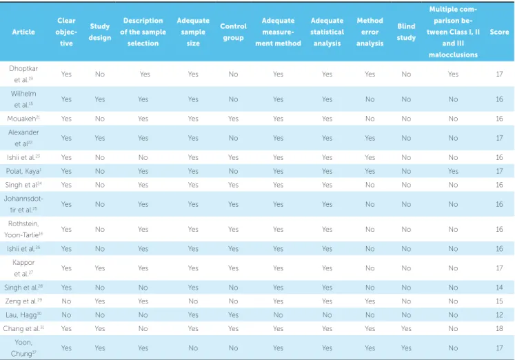
A total of 315 articles were initially identified, 8 from BBO, 12 from LILACS and 295 from MED-LINE. After reading the abstracts, only 55 were se-lected: no article from BBO, 2 from LILACS and 53 from MEDLINE. These were fully considered and after applying the inclusion and exclusion criteria, 39 articles were excluded and 16 remained, all from the MEDLINE database (Table 2). Next, another article was excluded due to lack of the measurements con-sidered for review.
A qualitative evaluation of the methodology used by these articles was performed according to previous stud-ies.17,18 The variables being considered for review are listed in Table 1, including corresponding values. Table 2 shows the articles presenting more accurate methodologies.
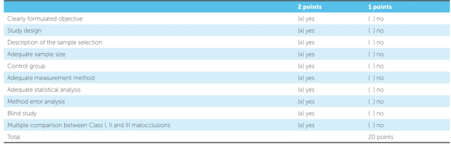
Table 1 - Qualification of the methodology used by the articles selected for review.
2 points 1 points
Clearly formulated objective (x) yes ( ) no
Study design (x) yes ( ) no
Description of the sample selection (x) yes ( ) no
Adequate sample size (x) yes ( ) no
Control group (x) yes ( ) no
Adequate measurement method (x) yes ( ) no
Adequate statistical analysis (x) yes ( ) no
Method error analysis (x) yes ( ) no
Blind study (x) yes ( ) no
Multiple comparison between Class I, II and III malocclusions (x) yes ( ) no
Each article was given points according to the items evaluated, as can be seen in Table 1. Each item had a maximum score of 2 points if there was a con-sensus on it, otherwise 1 point was given. This pro-cedure was applied to each article.
Therefore, each article could reach a maximum score of 20 points, thus allowing the study quality to be ranked as follows: ≤ 10 points = low score; > 10 and ≤15 = average score; > 15 and ≤ 18 = moderately high score; and > 18 points = high score.
Table 3 shows the sample characteristics, includ-ing the objective of each article, and Table 4 lists the
epitomes. In this last table, a study22 was excluded
because it did not present total measures, but rather growth increments.
Age
Seven studies have investigated the growth of the skull base by comparing the data obtained from
dif-ferent age groups16,20,22,23,24,28,37 with three22,24,28 of
them using samples of individuals with Class III mal-occlusion and whose age ranged from 5 to 18 years
old. These individuals were found22 to have a yearly
increment in the length of the anterior skull base in all age groups (6 to 18 years old), which was smaller than 1 mm for women and approximately equal to 1 mm for men, similar to the growth estimated for individuals
with Class I malocclusion. In another study24
com-paring Class III malocclusion to Class I malocclusion in individuals aged 5 to 11 years old, it was found that NSBa angle was more acute in the latter at ages of 5, 8, and 9 years old, and that NSAr angle had the same trend; but no statistically significant difference was
found at 8 years old. Researchers23 have reported that
the skull base of Japanese individuals with Class II di-vision 1 malocclusion had a significantly smaller an-terior length at 10 years and 10 months to 15 years and 10 months old compared to other age groups,
Table 2 - Evaluation of methodological quality of the 16 articles selected for review.
Article
Clear
objec-tive
Study
design
Description of the sample
selection
Adequate sample
size
Control
group
Adequate
measure-ment method
Adequate statistical
analysis
Method error
analysis Blind
study
Multiple
com-parison be-tween Class I, II
and III malocclusions
Score
Dhoptkar
et al.19 Yes No Yes Yes No Yes Yes Yes No Yes 17
Wilhelm
et al.15 Yes Yes Yes Yes No Yes Yes No No No 16
Mouakeh21 Yes No Yes Yes Yes Yes Yes No No No 16
Alexander
et al22 Yes Yes Yes Yes No Yes Yes Yes No No 17
Ishii et al.23 Yes No No Yes Yes Yes Yes Yes No No 16
Polat, Kaya1 Yes No Yes Yes No Yes Yes Yes No Yes 17
Singh et al24 Yes No Yes Yes Yes Yes Yes No No No 16
Johannsdot-tir et al.25 Yes No Yes Yes Yes Yes Yes No No No 16
Rothstein,
Yoon-Tarlie16 Yes No Yes Yes Yes Yes Yes No No No 16
Ishii et al.26 Yes No Yes Yes Yes Yes Yes No No No 16
Kappor
et al.27 Yes Yes Yes Yes Yes Yes Yes No No No 17
Singh et al.28 Yes No No Yes No Yes Yes No No No 14
Zeng et al.29 No Yes Yes No No Yes Yes Yes No No 15
Lau, Hagg30 No No No Yes Yes No No No No No 12
Chang et al.31 Yes Yes No Yes Yes Yes Yes Yes Yes No 18
Yoon,
Article Type of study (T or L) n Control group Malocclusion
Dhoptkar et al.19
(2002) Transversal 200 No
50 with Class I 50 with Class II div 1
50 with Class II div 2 50 with Class III
Wilhelm et al.15
(2001) Longitudinal 43 No
22 with Class I
21 with Class II
Mouakeh21
(2001) Transversal 138
Yes;
Paired by age, sex and ethnicity
69 with Class III 69 with Class I (control)
Alexander et al.22
(2009) Longitudinal 103 No 103 with Class III
Ishii et al.23
(2001) Transversal 279 Yes
190 with Class II div 1 89 with Class I (control)
Polat, Kaya1
(2007) Transversal 75 No
25 with Class I
25 with Class II 25 with Class III
Singh et al.24
(1997) Transversal 142 Yes
73 with Class III
69 with Class I (control)
Johannsdottir et al.25
(1999) Transversal 563 Yes
200 with Class I 363 with Class II
Rothstein, Yoon-Tarlie16
(2000) Transversal 613 Yes
278 with normal occlusion 335 with Class II div 1
Ishii et al.26
(2002) Transversal 124 No
Class II div 1 in Japanese females Classe II div 1 in Caucasian females
Kapoor et al.27
(2001) Transversal 100 Yes
50 with Class II
50 with normal occlusion
Singh et al.28
(1998) Transversal 141 No
72 with Class III, European/American 69 with Class III, Korean
Zeng et al.29
(1998) Transversal 160 No
40 with Class I Chinese; 40 with Class II Chinese (ch)
40 with Class I Swedish 40 with Class II Swedish (sw)
Lau e Hagg30
(1999) Transversal 416 No
105 Chinese with Class II div 1 107 Caucasians with Class II div 1 204 Chinese with normal occlusion
Chang et al.31
(2005) Transversal 200 Yes
100 with Class III 100 with normal occlusion (control)
Yoon, Chung37
(2015) Longitudinal 46 Yes
25 with Class I 21 with Class II
Sex % Ethnicity Age Objective
50% Caucasian
Class I: 10.4 yrs Class II div 1: 10.1yrs
Class II div 2: 11.1yrs Class III: 10.2 yrs
To compare the skull base
lexion to type of malocclusion as follows: Class I to Class II div 1 to Class II div 2 and Class I to Class III
50% European
1 month
2 yrs and 14 yrs old
To compare the longitudinal growth of the skull base (size and shape) to
Class I and Class II malocclusions)
66.6% female Syrian 5 - 12 yrs old
Mean 8.4 ± 2 yrs old
To identify morphological characteristics of the craniofacial complex in
Syrian patients with Class III malocclusion and compare them to controls
55 females
48 males Caucasian
6-7; 7-8; 8-9; 10-11; 11-12; 12-13 13-14; 15-16; 16-17; 17-18 yrs old
To investigate changes in the craniofacial growth in untreated individuals with Class III malocclusion
100% females Japanese
3 groups: Mixed dentition, Delayed mixed dentition, Young
permanent dentition
To compare the craniofacial characteristics of Japanese females with Class II division to those of controls
CIass I: 13 F, 11 M
Class II: 14 F, 11 M Class III: 13 M, 12 F
Not speciied (patients
treated at the Baskent University in Ankara, Turkey
15.74 yrs ± 4.28 To evaluate the diference in the skull base lexion between dental and
skeletal Class I, Class II, and Class III malocclusions
Approximately the same
number of males and females European/American 7 groups of 5 to 11 yrs
To diferentiate the skull base between individuals with Class I malocclusion
and those with Class III malocclusions
184 males
179 females Icelanders
5 yrs 7 mo to 7 yrs 8 mo Mean of 6 yrs 7 mo
To describe the craniofacial characteristics of Icelander children aged 6 years old with Class II malocclusion and compared them to Class II
malocclusion
T1: 47 F and 48 M
T2: 47 F and 43 M Caucasian
3 groups of subjects aged 10, 12 and 14 yrs old ± 6 years
To investigate whether Class II malocclusion is characterized by a poorly developed mandible or by its retropositioning
100% females 49 Japanese and
75 Caucasian females
Japanese: 11 yrs 8 mo British: 11 yrs 11 mo
To help deine the craniofacial morphology of the Japanese with Class II
malocclusion div 1 and to compare them with Caucasian individuals with the same malocclusion; and to elucidate craniofacial diferences
between ethnic groups
50% Indian
9 to 12 yrs old 10.64 yrs old, males
9.8 yrs old, females
To understand and diferentiate the skeletal-dental morphology of children
with straight terminal plane and those with Class II pattern
50% European/American Korean 5 to 11 yrs old
To compare Korean and European/American subjects with Class III malocclusion and provide data on ethnic diversity in individuals with
Class III malocclusion
50% Chinese
Swedish
8 to 10 yrs old 9.1 ± 0.9 males (M) ch Class II 9 ± 0.8 females (F) ch Class II 9.4 ± 0.8 M/F ch Class I
9.3±0.9 M sw Class I 9.3 ± 0.8 F sw Class I 8.7±0.8 M sw Class II 8.9±1.0 F sw Class II
To assess craniofacial structures in two ethnic groups and two malocclusions by comparing Chinese and Swedish individuals with Class I
and Class II malocclusions
Approx. 50% Chinese
Caucasians
10 to 15 yrs old 11 to 13 yrs old
12 yrs old
To assess the craniofacial pattern of Chinese and Caucasian individuals with
Class II div 1 malocclusion and those with normal occlusion; Chinese with Class II x Chinese with normal occlusion; Chinese with Class II div 1 x Caucasians with Class II div 1
50%
No speciication (radiographs from the Kaohsiung University,
Taiwan)
9.4 to 11.5 yrs old
To compare morphological characteristics of the skull base in children with malocclusion
Class III with normal children and provide database for subsequent Class III studies
100% females Caucasian ages 9, 14, and 18 To investigate and compare the craniofacial growth of untreated Class I and
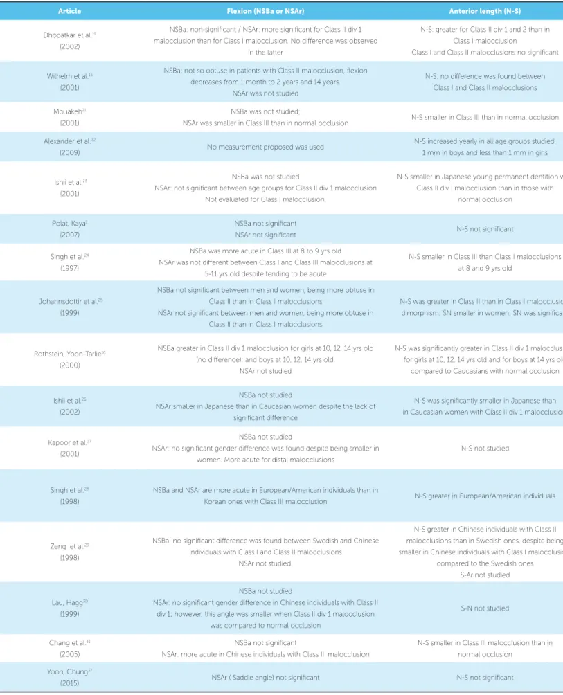
Article Flexion (NSBa or NSAr) Anterior length (N-S)
Dhopatkar et al.19
(2002)
NSBa: non-signiicant / NSAr: more signiicant for Class II div 1 malocclusion than for Class I malocclusion. No diference was observed
in the latter
N-S: greater for Class II div 1 and 2 than in
Class I malocclusion
Class I and Class II malocclusions no signiicant
Wilhelm et al.15 (2001)
NSBa: not so obtuse in patients with Class II malocclusion, lexion
decreases from 1 month to 2 years and 14 years. NSAr was not studied
N-S: no diference was found between Class I and Class II malocclusions
Mouakeh21 (2001)
NSBa was not studied;
NSAr was smaller in Class III than in normal occlusion N-S smaller in Class III than in normal occlusion
Alexander et al.22
(2009) No measurement proposed was used
N-S increased yearly in all age groups studied,
1 mm in boys and less than 1 mm in girls
Ishii et al.23 (2001)
NSBa was not studied
NSAr: not signiicant between age groups for Class II div 1 malocclusion
Not evaluated for Class I malocclusion.
N-S smaller in Japanese young permanent dentition with Class II div I malocclusion than in those with
normal occlusion
Polat, Kaya1
(2007)
NSBa not signiicant
NSAr not signiicant N-S not signiicant
Singh et al.24
(1997)
NSBa was more acute in Class III at 8 to 9 yrs old
NSAr was not diferent between Class I and Class III malocclusions at 5-11 yrs old despite tending to be acute
N-S smaller in Class III than Class I malocclusions
at 8 and 9 yrs old
Johannsdottir et al.25
(1999)
NSBa not signiicant between men and women, being more obtuse in Class II than in Class I malocclusions
NSAr not signiicant between men and women, being more obtuse in Class II than in Class I malocclusions
N-S was greater in Class II than in Class I malocclusion, dimorphism; SN smaller in women; SN was signiicant
Rothstein, Yoon-Tarlie16 (2000)
NSBa greater in Class II div 1 malocclusion for girls at 10, 12, 14 yrs old (no diference); and boys at 10, 12, 14 yrs old.
NSAr not studied
N-S was signiicantly greater in Class II div 1 malocclusions for girls at 10, 12, 14 yrs old and for boys at 14 yrs old
compared to Caucasians with normal occlusion
Ishii et al.26
(2002)
NSBa not studied
NSAr smaller in Japanese than in Caucasian women despite the lack of signiicant diference
N-S was signiicantly smaller in Japanese than
in Caucasian women with Class II div 1 malocclusions
Kapoor et al.27 (2001)
NSBa not studied
NSAr: no signiicant gender diference was found despite being smaller in
women. More acute for distal malocclusions
N-S not studied
Singh et al.28 (1998)
NSBa and NSAr are more acute in European/American individuals than in
Korean ones with Class III malocclusion N-S greater in European/American individuals
Zeng et al.29 (1998)
NSBa: no signiicant diference was found between Swedish and Chinese individuals with Class I and Class II malocclusions
NSAr not studied.
N-S greater in Chinese individuals with Class II
malocclusions than in Swedish ones, despite being smaller in Chinese individuals with Class I malocclusion,
compared to the Swedish ones S-Ar not studied
Lau, Hagg30
(1999)
NSBa not studied
NSAr: no signiicant gender diference in Chinese individuals with Class II
div 1; however, this angle was smaller when Class II div 1 malocclusion was compared to normal occlusion
S-N not studied
Chang et al.31
(2005)
NSBa not signiicant
NSAr: more acute in Chinese individuals with Class III malocclusion
N-S smaller in Class III malocclusion than in
normal occlusion
Yoon, Chung37
(2015) NSAr ( Saddle angle) not signiicant N-S not signiicant
Posterior length (S-Ba or S-Ar) Conclusion
S-Ba: greater in Class II div 1 and 2; than in Class I
Class I and Class III malocclusions no signiicant S-Ar was not studied
The skull base angle itself has no key inluence on the development of malocclusions
S-Ba: no diference between malocclusions
S-Ar was not used Growth in skull base is similar to that in Class I and Class II malocclusions
S-Ba was not studied
S-Ar smaller in Class III than in normal occlusion
Flexion, anterior and posterior lengths are signiicantly smaller in Class III malocclusions
S-Ba not studied
S-Ar not studied No measurement proposed was used
S-Ba not studied
S-Ar: not signiicant between age groups for Class I div 1 malocclusion
Not evaluated between Class II div 1 malocclusion and Class I malocclusion
Class II div 1 malocclusion had an increase in facial angle in association with a shortened mandibular ramus, compared to Class I malocclusion
SBa not diferent
S-Ar not signiicant
No diference was found in angle and dimensions of the skull base for
Class I, II and III malocclusions
S-Ba: no diference between age groups
S-Ar not studied
Skull base angle tending to be more acute in Class III malocclusion than in normal
occlusion
S-Ba and S-Ar smaller in women Dimorphism was found at the skull base at 6 yrs old in Class II malocclusion
S-Ba: no signiicant diference found in girls Signiicant diference was found in boys at 10 and 12 yrs old only
S-Ar not studied
Anterior base is more protrusive in Class II malocclusions, with length being excessive
at the skull base, maxillary and frontal sinuses increased (may contribute to Class II malocclusion increase)
Skull base lexion does not contribute to the retruded positioning of the mandible
S-Ba not studied
S-Ar: no signiicant diference Caucasian women have a more signiicantly longer anterior skull base
S-Ba not studied S-Ar not studied
NSAr is one of the factors indicating a Class II pattern in association with a distal mandibular positioning and skull base rotation
S-Ba and S-Ar greater in European/American individuals
For European/American individuals with Class III malocclusions, the craniofacial morphology is afected by an orthocephalization of the skull base, exacerbated by
prominent mandible and symphysis morphology
S-Ba greater in Chinese individuals with Class II malocclusions than in Swedish ones No signiicant diference was found in Class I malocclusion regarding both ethnic groups
S-Ar not studied
Anterior and posterior lengths of the skull base were greater in Chinese than in Swedish individuals
S-Ba not studied
S-Ar not studied
No dimorphism was found between Chinese individuals with Class II div 1 malocclusion
Skull base angle is more acute in Chinese individuals with Class II div 1 malocclusion than in those with normal occlusion
S-Ba smaller in Class III malocclusion, but with no signiicant diference
S-Ar smaller in Class III malocclusion than in normal occlusion
The decreased lexion and length in Class III malocclusion may be related to the
aetiology of this type of malocclusion
S-Ba not studied
involving individuals with same malocclusion and race (Group 1: 7 years and 6 months to 13 years and 6 months old; and Group 2: 9 years and 1 month to 13 years and 6 months old). On the other hand,
another study29 compared individuals with Class II
division 1 malocclusion to controls and found that the length of the anterior skull base was statistically greater in all six age groups studied (females aged 10, 12, 14 years old, and males aged 10, 12, 14 years
old). Two studies20,28 had samples with different age
groups, but it was not possible to use all data
be-cause the first study20 had compared age groups of
1 month, 2 years and 14 years old, whereas the
sec-ond study28 had assessed neither flexion nor the skull
base length at different age groups. For Yoon and
Chung,37 which compared Class I with Class II at
three different ages (9, 14 and 18 years — all female), no difference was observed in the flexion or length of the anterior skull base.
Ethnic group
Three studies have assessed different ethnic
groups,26,28,29 with Asian being compared to
Cauca-sian individuals in most cases, and significant differ-ences being found. The length of the anterior skull base was found to be greater for Japanese than for
Caucasian females.26 However, a study28 reported a
greater anterior skull base as well as a more acute NSAr angle in European/American than in Korean individuals. Similarly, with regard to the length of
the anterior skull base, a study29 showed this
mea-surement was smaller in Chinese than in Swedish individuals for both types of malocclusions (Class I and Class II), with the posterior length (S-Ba) be-ing also smaller in Chinese individuals with Class II
malocclusion. Another study24 evaluated two ethnic
groups but no comparison was possible because of the lack of data on skull base structures regarding Caucasian individuals.
Dimorphism
Six studies have evaluated the relationship be-tween sexual dimorphism and development of the
skull base,22,25,27,30,16,29 but no significant difference
was found in the angular measurements.
How-ever, one study25 showed that linear measurements
(S-N, S-Ar and S-Ba) were significantly greater in
Icelander male children compared to female ones with Class I and Class II malocclusions.
Malocclusion differences
Of the 16 articles selected, only one has not com-pared lexion or length of the skull base to some type
of malocclusion.22 Two studies1,19 compared lexion
or length of the skull base in individuals with Class I, Class II and Class III malocclusions. One of these
stud-ies19 found greater angular measurements for the skull
base in Class II division 1 malocclusion, compared to Class I malocclusion. In this same study, no diference was found between Class I malocclusion and other ones, regarding such angular measurements, whereas the length of the anterior and posterior skull base were greater in the cases of Class II malocclusion. However,
the other study1 reported signiicant diferences in the
skull base length between the three types of
malocclu-sion. Studies21,24,31 have also compared Class III
mal-occlusion to normal mal-occlusion, whereas another one28
compared this condition between Caucasian and Ko-rean individuals. All these studies found similar results, with the length of the anterior skull base being smaller in Korean individuals with Class III malocclusion and skull base lexion tending to be more acute in Caucasian individuals with the same condition.
Among these seven studies16,20,23,25,27,30,37 comparing
Class II malocclusion to Class I malocclusion, similar results have been reported. For example, individuals with Class II malocclusion had greater anterior and posterior lengths and more obtuse angular
measure-ments regarding the skull base. However, one study23
reported a smaller length of the skull base in Japanese girls with Class I malocclusion presenting permanent
dentition, whereas another30 found no significant
dif-ferences in the skull base flexion compared to normal occlusion. In the study, the compared groups (Class I and Class II) showed no differences in the length of the anterior skull base and flexion of the cranial base.
DISCUSSION
The growth of the skull base occurs by means of a complex balance between sutural growth, prolonga-tion of synchondroses, extensive cortical sliding, and remodelling. This combination allows an increase in the differential growth between base and vault of the skull, expansion of the contours of the various endo-cranial fossae, maintenance of vessel and nerve pathways, and prolongation of the processes, such as hypophysis. Prolongation of the skull base occurs with the growth of synchondroses and direct cortical growth. The cortical sliding of the skull loor produces several degrees of growth movement at diferent regions, usu-ally towards the ecto-cranial direction, with apposition
proportional to the external surface.5
Enlow32 has shown that maxilla growth is
influ-enced by the skull base, which in turn, is influinflu-enced by the growth of the brain. The mandible, because of its distant positioning, acts more independently, de-spite being articulated with glenoid fossa, thus being a potential factor influencing the skull base.
To better understand the cephalometric aspects, the skull base is divided into anterior and posterior lengths, the former extending anteriorly from sella turcica (S) to nasofrontal suture (N), and the latter extending from sella turcica to the anterior edge of
the foramen magnum, defined as Ba.21 There is a
consensus that the length of the anterior skull base corresponds to the linear N-S distance, but the same cannot be said about the posterior region, which
cor-responds to either S-Ba or S-Ar linear distances.19
Björk2 supported the use of the latter, as it is more
easily visualized, with most studies using this
mea-surement. Verjanne and Koski33 suggested the use of
Ba to measure the skull base angle as they considered
the S-Ar measurement too distant; Kerr and Adams34
used Ba to measure the skull base angle. Bhatia and
Leighton35 used N-S-Ba, N-S-Art as well as S-Ba
and S-Art, and found similar measurements.
According to the other authors,19 the skull base
fol-lows a neural (anterior region) and somatic (posterior region) growth pattern despite its cartilaginous origin (chondrocranium). Ater birth, especially in the early childhood, the growth of the anterior portion occurs mainly due to the increase in frontal sinus and remodel-ling of the nasal region, whereas the growth of the pos-terior region is related to the interstitial growth occur-ring in the spheno-occipital synchondrosis.
The two segments of the base of the skull form an flexion of 130-135 degrees at the angle formed at the Sela point (center of the sella turcica). This angle (NSBa) has approximately 142 degrees at birth, but
decreases to 130 degrees at 5 years old.19 From 5 to
15 years old, the skull base angle is relatively stable.23
Other studies suggest that there are no differences in this angle of the skull base during childhood,
pu-berty and adult phase.1,23
There is evidence showing that the skull base angle (N-S-Ba) is greater in Class II division 1 malocclusion than in Class I malocclusion or normal occlusion, with this angle not difering between Class II division 2 and
Class I malocclusions.19 In addition, studies comparing
Class II malocclusions to normal occlusion or Class I
malocclusion16,20,23,25,27,30,37 found similar results.16,25,27
However, two studies30,37 compared such
malocclu-sions and reported a smaller lexion of the skull base in individuals with Class II malocclusion. This can be explained by the fact that the posterior region of the skull base (S-Ba) forming the S-N-Ba angle might be anteriorly or posteriorly inclined, whereas the anterior region (S-N) might also be inclined anteriorly up-wards or downup-wards, thus causing a variation in either
S or N points vertically.36 Therefore, variable lengths
of the anterior and posterior regions of the skull base can compensate any cranial lexion, that is, a poste-rior acute angle anteposte-riorly positioned in relation to the mandible can neutralize the cranial lexion through the greater posterior length, thus positioning Ba and
man-dible posteriorly and vice-versa.20 However, the skull
base length was not assessed.30 Despite these indings,
the skull base lexion is thought to have no inluence
on malocclusions,1,19 whereas there is no consensus
among other studies regarding this issue.21,23-27,30-31
With regard to the Class III malocclusion, it has been reported that linear and angular measurements of the skull base are smaller when compared to other
types of malocclusion.1 These findings are
corrobo-rated by some studies,21,31 although one study1 had
found smaller angular measurements for Class III malocclusions, despite not being significant.
Asian individuals present a smaller anterior length and a more obtuse angle of the skull base, compared to Caucasians, with this finding compris-ing the both ethnic and morphological
CONCLUSIONS
Ater evaluating all these articles selected for the present systematic review, one can state the following:
1. The skull base angle itself does not seem to play a key role in the development of malocclusions.
2. The skull base angle is relatively stable at the ages of 5 to 15 years old.
3. A more obtuse skull base flexion, in association or not with a greater length of the anterior skull base, can contribute to the development of Class II divi-sion 1 maloccludivi-sion. A more acute skull base flexion
can contribute to a more anterior positioning of the mandible and to development of Class III malocclu-sion as well.
Author contributions
Conception or design of the study: KCMA, TBR, CIVV, ASP. Data acquisition, analysis or interpretation: KCMA, TBR, CIVV, ASP, DBR. Writing the article: KCMA, TBR, CIVV, ASP, DBR. Critical revision of the article: KCMA, TBR, ASP, DBR. Final approval of the article: KCMA, TBR, ASP.
1. Polat OO, Kaya B. Changes in cranial base morphology in diference malocclusions. Orthod Craniofac Res. 2007 Nov;10(4):216-21. 2. Björk A. Cranial base development. Am J Orthod. 1955 Mar;41(3):198-255.
3. Ford EHR. Growth of the human cranial base. Am J Orthod. 1958
July;44(7):498-506.
4. Scott JH. The cranial base. Am L Phys Anthropol. 1958 Apr;16(3):319-48. 5. Moyers RE. Ortodontia. 4ª ed. Rio de Janeiro: Guanabara Koogan; 1991. 6. Santos-Pinto A, Martins JCR, Uetanabaro T, Sakima T, Mendes AJD. Inluência
do grau de delexão da base craniana no relacionamento ântero-posterior dos maxilares. Ortodontia. 1983;16:5-9.
7. Björk A. The nature of facial prognathism and its relation to normal occlusion of the teeth. Am J Orthod. 1951 Feb;37(2):106-24.
8. Björk A. The face in proile. An Anthropological X-ray Investigation on Swedish Children and Conscripts. Svensk Tandläkare-Tidskrift. 1947;40(5B Suppl):1-180. 9. Ricketts RM. A foundation for cephalometric communication. Am J Orthod.
1960 May;46(5):330-57.
10. Hopkin GB, Houston WJ, James GA,et al. The cranial base as an aetiological, factor in malocclusion. Angle Orthod. 1968 July;38(3):250-5.
11. Horowitz SL, Converse JM. Craniofacial relationships in mandibular prognathism. Arch Oral Biol. 1969 Jan;14(1):121-31.
12. Dibbets JMH. Morphological associations between the Angle classes. Eur J Orthod. 1996 Apr;18(2):111-8.
13. Hildwein M, Bacon W, Turlot JC, Kuntz M. Spéciicités et discriminants majeurs dans une population de Classe II division 1. Revue d’Orthopédie Dento-Faciale. 1986 June;20(2):197-208.
14. Kasai K, Moro T, Kanazawa E, Iwasawa T. Relationship between cranial base and maxillofacial morphology. Eur J Orthod. 1995 Oct;17(5):403-10.
15. Wilhelm BM, Beck FM, Lidral AC, Vig KWL. A comparison of cranial base growth in Class I and Class II skeletal patterns. Am J Orthod Dentofacial Orthop. 2001 Apr;119(4):401-5.
16. Rothstein T, Yoon-Tarlie C. Dental and facial skeletal characteristics and growth of males and females with Class II, division 1 malocclusion between the ages of 10 and 14 (revisited)-Part I: characteristics of size form, and position. Am J Orthod Dentofacial Orthop. 2000 Mar;117(3):320-32.
17. Antczak AA, Tang J, Chalmers TC. Quality assessent of randomized control trials in dental research. I: Methods. J Periodontal Res. 1986 July;21(4):305-14. 18. Flores-Mir C, Major MP, Major PW. Search and selection methodology of
systematic reviews in orthodontics. Am J Orthod Dentofacial Orthop. 2006 Aug;130(2):214-7.
19. Dhoptkar A, Bhatia S, Rock P. An investigation into the relationship between the cranial base angle and malocclusion. Angle Orthodontist. 2002 Oct;72(5):456-63. 20. Andria LM, Leite LP, Prevatte TM, King LB. Correlation of the cranial base angle
and its components with other dental /skeletal variables and treatment time. Angle Orthod. 2004 June;74(3):361-6.
REFERENCES
21. Mouakeh M. Cephalometric evaluation of craniofacial pattern of Syrian children with Class III malocclusion. Am J Orthod Dentofacial Orthop. 2001 June;119(6):640-9.
22. Alexander AEZ, McNamara JA, Franchi L, Baccetti T. Semi longitudinal cephalometric study of craniofacial growth in untreated Class III malocclusion. Am J Orthod Dentofacial Orthop. 2009 June;135(6):701-14.
23. Ishii N, Deguchi T, Hunt N. Craniofacial morphology of Japanese girls with Class II division 1 malocclusion. J Orthod. 2001 Sept;28(3):211-5. 24. Singh GD, McNamara JA Jr, Lozanof S. Morphometry of the cranial base in
subjects with Class III malocclusion. J Dent Res. 1997 Feb;76(2):694-703. 25. Johannsdottir B, Thordarson A, Magnusson TE. Craniofacial morphology in
6-year-old Icelandic children. Eur J Orthod. 1999 June;21(3):283-90. 26. Ishii N, Deguchi T, Hunt NP. Morphological diferences in the craniofacial
structure between Japanese and Caucasian girls with Class II division 1 malocclusion. Eur J Orthod. 2002 Feb;24(1):61-7.
27. Kapoor S, Kapoor DN, Jaiswal JN. Cephalometric evaluation of Class II malocclusion in transitional dentition. J Indian Soc Pedo Dent. 2001 Dec;19(4):127-33.
28. Singh GD, McNamara JA Jr, Lozanof S. Craniofacial heterogeneity of prepubertal Korean and European-American subjects with Class III malocclusions: procrustes, EDMA, and cephalometric analyses. Int J Adult Orthod Orthognath Surg. 1998 Jan;13(3):227-40.
29. Zeng XL, Forsberg CM, Aronson SL. Craniofacial morphology in Chinese and Swedish children with Angle Class I and Class II occlusal relations. Australian Orthod J. 1998 Oct;15(3):168-76.
30. Lau JWP, Hagg U. Cephalometric morphology of Chinese with Class II division 1 malocclusion. Br Dental J. 1999 Feb;186(4):188-90.
31. Chang HP, Hsieh SH, Tseng YC, Chou TM. Cranial-base morphology in children with Class III malocclusion. Kaohsiung J Med Sci. 2005 Apr;21(4):159-65. 32. Enlow DH. Crescimento facial. 3ª ed. Rio de Janeiro: Artes Médicas; 1993. 33. Varjanne I, Koski K. Cranial base, sagittal jaw relationship and occlusion.
A radiological: craniometric appraisal. Proc Finn Dent Soc. 1982 Jan;78(4):179-83. 34. Kerr WJS, Adams CP. Cranial base and jaw relationship. Am J Phys Anthropol.
1988 Oct;77(2):213-20.
35. Bathia SN, Leighton BC. A manual of facial growth. Oxford: Oxford University Press; 1993.